

 PDF
PDF ePub
ePub Citation
 Print
Print


Abstract
The aim of this study was to evaluate the marginal and internal gaps in CEREC3 CAD/CAM inlays of three different preparation designs. CEREC3 Inlays of three different preparation designs (n = 10) were fabricated according to Group I-conventional functional cusp capping/shoulder preparation, Group II-horizontal reduction of cusps and Group III-complete reduction of cusps/shoulder preparation. After cementation of inlays, the bucco-lingual cross section was performed through the center of tooth. Cross section images of 20 magnifications were obtained through the stereomicroscope. The gaps were measured using the Leica application suite software at each reference point. Statistical analysis was performed using one-way ANOVA and Tukey's test (α<0.05).
The marginal gaps ranged from 80.0 to 97.8 µm for Group I, 42.0 to 194.8 µm for Group II, 51.0 to 80.2 µm for Group III. The internal gaps ranged from 90.5 to 304.1 µm for Group I, 80.0 to 274.8 µm for Group II, 79.7 to 296.7 µm for Group III. The gaps of each group were the smallest on the margin and the largest on the horizontal wall. For the CEREC3 CAD/CAM inlays, the simplified designs (groups II and III) did not demonstrate superior results compared to the traditional cusp capping design (group I).
Figures and Tables
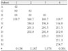
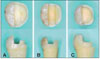 | Figure 1Different tooth preparations suggested for inlays: (A) Group I-conventional functional cusp capping/shoulder margin, (B) Group II-horizontal flat reduction of cups, (C) Group III-complete reduction of cusps/shoulder margin.
|
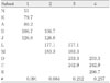
 | Figure 2Schematic drawings of preparations in Groups I, II and III, representing bucco-lingual cross sections. Reference points in Groups II and III were designated based on reference points in Group I.
|

References
1. Jedynakiewicz NM, Martin N. CEREC: science, research, and clinical application. Compend Contin Educ Dent. 2001. 22:6 Suppl. 7–13.
2. Mou SH, Chai T, Wang JS, Shiau YY. Influence of different convergence angles and tooth preparation heights on the internal adaptation of Cerec crowns. J Prosthet Dent. 2002. 87(3):248–255.

3. Nakamura T, Dei N, Kojima T, Wakabayashi K. Marginal and internal fit of Cerec 3 CAD/CAM all-ceramic crowns. Int J Prosthodont. 2003. 16(3):244–248.
4. Bindl A, Mormann WH. Clinical and SEM evaluation of all-ceramic chair-side CAD/CAM-generated partial crowns. Eur J Oral Sci. 2003. 111(2):163–169.
5. Jeon DK, Park SH. Single-visit appointment of Cerec inlay. J Korean Acad Conserv Dent. 2007. 32(3):298.
6. Kokubo Y, Ohkubo C, Tsumita M, Miyashita A, Vult von Steyern P, Fukushima S. Clinical marginal and internal gaps of Procera AllCeram crowns. J Oral Rehabil. 2005. 32(7):526–530.
7. Bindl A, Richter B, Mörmann WH. Survival of ceramic computer-aided design/manufacturing crowns bonded to preparations with reduced macroretention geometry. Int J Prosthodont. 2005. 18(3):219–224.
8. Akbar JH, Petrie CS, Walker MP, Williams K, Eick JD. Marginal adaptation of Cerec 3 CAD/CAM composite crowns using two different finish line preparation designs. J Prosthodont. 2006. 15(3):155–163.
9. Park MJ, Jin MU, Kim YK, Kim SK. Consideration of marginal adaptation in ceramic restoration using Cerec3. J Korean Acad Conserv Dent. 2007. 32(6):639.
10. Hickel R, Dasch W, Mehl A, Kremers L. CAD/CAM--fillings of the future? Int Dent J. 1997. 47(5):247–258.
11. Tsitrou EA, Northeast SE, van Noort R. Evaluation of the marginal fit of three margin designs of resin composite crowns using CAD/CAM. J Dent. 2007. 35(1):68–73.
12. Federlin M, Schmidt S, Hiller KA, Thonemann B, Schmalz G. Partial ceramic crowns: influence of preparation design and luting material on internal adaptation. Oper Dent. 2004. 29(5):560–570.
13. Bindl A, Mormann WH. Marginal and internal fit of all-ceramic CAD/CAM crown-copings on chamfer preparations. J Oral Rehabil. 2005. 32(6):441–447.
14. Federlin M, Sipos C, Hiller KA, Thonemann B, Schmalz G. Partial ceramic crowns. Influence of preparation design and luting material on margin integrity--a scanning electron microscopic study. Clin Oral Investig. 2005. 9(1):8–17.
15. Seo D, Yi Y, Roh B. The effect of preparation designs on the marginal and internal gaps in Cerec3 partial ceramic crowns. J Dent. 2009. 37(5):374–382.
16. Otto T, Schneider D. Long-term clinical results of chairside Cerec CAD/CAM inlays and onlays: a case series. Int J Prosthodont. 2008. 21(1):53–59.
17. Reiss B. Clinical results of Cerec inlays in a dental practice over a period of 18 years. Int J Comput Dent. 2006. 9(1):11–22.
18. Reiss B, Walther W. Clinical long-term results and 10-year Kaplan-Meier analysis of Cerec restorations. Int J Comput Dent. 2000. 3(1):9–23.
19. Otto T, De Nisco S. Computer-aided direct ceramic restorations: a 10-year prospective clinical study of Cerec CAD/CAM inlays and onlays. Int J Prosthodont. 2002. 15(2):122–128.
20. Sjögren G, Molin M, van Dijken JW. A 10-year prospective evaluation of CAD/CAM-manufactured (Cerec) ceramic inlays cemented with a chemically cured or dual-cured resin composite. Int J Prosthodont. 2004. 17(2):241–246.
21. Krämer N, Taschner M, Lohbauer U, Petschelt A, Frankenberger R. Totally bonded ceramic inlays and onlays after eight years. J Adhes Dent. 2008. 10(4):307–314.
22. Krämer N, Frankenberger R. Clinical performance of bonded leucite-reinforced glass ceramic inlays and onlays after eight years. Dent Mater. 2005. 21(3):262–271.
23. Frankenberger R, Kramer N, Lohbauer U, Nikolaenko SA, Reich SM. Marginal integrity: is the clinical performance of bonded restorations predictable in vitro? J Adhes Dent. 2007. 9:Suppl 1. 107–116.
 XML Download