

 ePub
ePub Citation
 Print
Print


| Korean J Sports Med. 2015 Dec;33(2):139-142. English. Published online December 07, 2015. https://doi.org/10.5763/kjsm.2015.33.2.139 | |
| Copyright © 2015 The Korean Society of Sports Medicine | |
|
Young Jun Kim,
Duke Whan Chung, | |
| Department of Orthopedic Surgery, Kyung Hee University School of Medicine, Seoul, Korea. | |
| Received October 07, 2015; Revised November 09, 2015; Accepted November 18, 2015. | |
|
This is an Open Access article distributed under the terms of the Creative Commons Attribution Non-Commercial License (http://creativecommons.org/licenses/by- | |
|
Abstract
| |
|
Ulnar tunnel syndrome (UTS) is a compressive neuropathy of the upper extremity that shows various clinical symptoms according to the anatomic region of the compression site. Numerous factors may cause UTS, and most publications are case reports describing various etiologies; thus, obtaining a correct diagnosis is often challenging. Giant cell tumor of the tendon sheath (GCTTS) is well described to be a common benign soft tissue tumor of the hand; however, it is rarely reported to cause UTS. We report a case of GCTTS in Guyon's canal causing UTS that was misdiagnosed as handlebar palsy. |
|
Keywords: Ulnar neuropathies; Ulnar nerve compression syndromes; Giant cell tumors |
|
|
Introduction
|
Giant cell tumor of the tendon sheath (GCTTS) is the second most common benign soft tissue tumor of the hand. GCTTS occurs particularly on the volar surface of the finger or hand, and tends to involve the radial three digits and distal interphalangeal joint region1). This tumor type has been known to impinge upon surrounding structures and may even erode the bony structure in a confined space. Numerous factors may cause ulnar tunnel syndrome (UTS). Any mass growing within Guyon's canal is a classical cause of UTS, and chronic repetitive trauma on the hypothenar eminence has been implicated as a common cause of UTS in cyclists2, 3).
Here, we report a case of UTS caused by GCTTS in Guyon's canal that was misdiagnosed as a sports-related neuropathy, handlebar palsy.
|
Case Report
|
A 53-year-old male patient presented with right-hand weakness and pain over the hypothenar eminence. He was a habitual cyclist who had experienced pain in the prior 7 months. He had been take a bike twice in a day, usually 1-1.5 hours of duration for one time, since 2 years ago. Also he was experienced aggravated numbness and pain when grip on mountain bicycle type handlebar. Weakness was noticeable by the patient 6 months before admission. There was marked atrophy of the first web space with a relatively spared hypothenar eminence, and he showed decreased grip strength compared with the contralateral side. Tinnel's sign was positive with no palpable mass over the ulnar side of the wrist. Egawa's sign was also positive. There was no sensory loss in the affected hand, and the patient complained of intermittent numbness over the ulnar two digits. Simple radiography showed no definitive bony lesion or alteration of the soft tissue density around the ulnar side of the wrist.
A nerve conduction study revealed that the first dorsal interossei muscles showed a small amplitude and delayed latency; additionally, sensory nerve conduction was normal. Electromyography revealed abnormal spontaneous activities and a decreased interference pattern of the first dorsal interossei muscles, suggesting incomplete ulnar neuropathy around the wrist level.
We considered that the cause of the ulnar neuropathy was repetitive stress injury related to sports activity, also known as handlebar palsy. Surgical exploration was performed without further imaging study, such as sonography or magnetic resonance imaging.
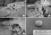
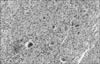
A skin incision was made directly over Guyon's canal. We performed ulnar nerve exploration from the proximal side of the canal. The mass was identified and was found to originate at the distal and deep portions of Guyon's canal (Fig. 1A). The deep branch of the ulnar nerve was obviously compressed, and tenting was shown by the mass. The mass was located directly under the deep branch of the ulnar nerve, distal to the bifurcation point of the nerve over the pisohamate ligament (Fig. 1B). After careful dissection, the mass was loosely associated with the tendon sheath of the flexor carpi ulnaris tendon (Fig. 1C). The canal was decompressed after mass excision. The mass was well capsulized to about in 1×1.5 cm in size and appeared yellowish and solid (Fig. 1D). The surgical finding was correlated with the electrodiagnosis of ulnar neuropathy which representing motor nerve pathology. Photomicrography revealed uniform distribution of mononuclear stromal cells with multinucleated giant cells characteristic of GCTTS (Fig. 2).
|
|
The patient had fully restored sensory function with improved motor function of the hand 6 months after the operation.
|
Discussion
|
Although the anatomic course of the ulnar nerve in the wrist has been well documented in standard texts, it is important to emphasize specific distinctions concerning the anatomy with respect to compression of the ulnar nerve. The ulnar tunnel originates at the proximal edge of the palmar carpal ligament and extends distally to the fibrous arch of the hypothenar muscles at the level of the hook of hamate4). During its course in the fibro-osseous tunnel, the ulnar nerve bifurcates into a superficial sensory and deep motor branch distal to the distal pole of the pisiform3). The motor branch to the hypothenar muscles separates from the main ulnar nerve just distal to the pisiform at the wrist. Thus, the hypothenar muscle was relatively unaffected by compression of the deep branch. The deep branch of the ulnar nerve courses around the hook of the hamate to innervate the other intrinsic muscles of the hand. This deep branch is susceptible to nerve compression from overlying tendinous bands in the hypothenar muscles and from its excursion as it courses around the hook of the hamate3). Therefore, it is important to inspect the course of the deep branch because it may require decompression and because satellite lesions may be present. The relationship between the three anatomical zones of ulnar nerve compression within Guyon's canal and symptoms can be classified into three categories4). Zone 1 refers to the region proximal or within Guyon's canal, before bifurcation of the ulnar nerve that manifests as motor weakness and sensory deficits, or pure sensory or pure motor deficits. Zone 2 surrounds the deep branch, and compression in this region results in paralysis of the intrinsic muscle and may involve or spare the hypothenar muscle. Zone 3 surrounds the superficial branch, and a lesion in this zone produces sensory symptoms. Clinical findings can appear as various aspects in the mass lesion of Guyon's canal depending on where the compression occurs.
UTS is caused by various etiologic factors. Common causes are acute or chronic trauma most likely related to an occupation, and ganglia5). Other causes include carpal bone fracture, ulnar artery or hypothenar muscle pathology, and concurrent case of idiopathic carpal tunnel syndrome. Chronic repetitive trauma over the hypothenar eminence has been implicated as a cause of UTS and is not uncommon among long-distance cyclists, classically described as handlebar palsy2, 6). Typical findings are an isolated lesion of the deep terminal branch, and an additional lesion of the superficial sensory branch may be present2). Handlerbar palsy is occurred mostly in long distance cycling or professional cyclist. Patterson et al.6) investigated the incidence of ulnar neuropathy in 25 cyclists. The author investigated cyclists, who underwent a 600 km tour over 4 days. 70% of the participants experienced some form of sensory or motor neurological symptoms by the end of the tour. There is a gap in the literature regarding evidence based information for the management of handlebar palsy. The most commonly recommended treatment is to prevent its occurrence, including avoid local pressure using some forms of gloves and/or modifications of handlebar grip. However, There are some treatment guidelines of UTS in European HANDGUIDE study in 20137). They concluded that main factor for treatment choice were severity of symptoms, duration of symptoms and previous treatment received. In the presented case, patient's first web space atrophy was severe, which suggesting chronic UTS. Surgery is an option to explore ulnar nerve compression in the canal.
GCTTS causing UTS located in Guyon's canal is rare. There have been few reports concerning GCTTS in the English-language literature, but it should be included in the possible pathologic structure of Guyon's canal and the differential diagnosis8, 9, 10).
This case demonstrates the possibility of a misdiagnosis of UTS. Clinical history and examination may be nonspecific or related to coexisting pathologies, making an accurate diagnosis confusing3). When external causes like handlebar palsy was suspected to cause compression of the ulnar nerve in Guyon's canal, appropriate imaging studies are advised in addition to electrodiagnostic studies. Careful exploration of the nerve down to the rest of the ulnar tunnel should be performed during surgical exploration because of the propensity for space-occupying lesions.
|
Notes
|
No potential conflict of interest relevant to this article was reported.
|
References
|
| 1. | Glowacki KA, Weiss AP. Giant cell tumors of tendon sheath. Hand Clinics 1995;11:245–253.
|
| 2. | Akuthota V, Plastaras C, Lindberg K, Tobey J, Press J, Garvan C. The effect of long-distance bicycling on ulnar and median nerves: an electrophysiologic evaluation of cyclist palsy. Am J Sports Med 2005;33:1224–1230.
|
| 3. | Chen SH, Tsai TM. Ulnar tunnel syndrome. J Hand Surg Am 2014;39:571–579.
|
| 4. | Gross MS, Gelberman RH. The anatomy of the distal ulnar tunnel. Clin Orthop Relat Res 1985;(196):238–247.
|
| 5. | Foucher G, Berard V, Snider G, Lenoble E, Constantinesco A. Distal ulnar nerve entrapment due to tumors of Guyon's canal: a series of ten cases. Handchir Mikrochir Plast Chir 1993;25:61–65.
|
| 6. | Patterson JM, Jaggars MM, Boyer MI. Ulnar and median nerve palsy in long-distance cyclists: a prospective study. Am J Sports Med 2003;31:585–589.
|
| 7. | Hoogvliet P, Coert JH, Friden J, Huisstede BM. European HANDGUIDE group. How to treat Guyon's canal syndrome? Results from the European HANDGUIDE study: a multidisciplinary treatment guideline. Br J Sports Med 2013;47:1063–1070.
|
| 8. | Budny PG, Regan PJ, Roberts AH. Localized nodular synovitis: a rare cause of ulnar nerve compression in Guyon's canal. J Hand Surg Am 1992;17:663–664.
|
| 9. | Francisco BS, Agarwal JP. Giant cell tumor of tendon sheath in Guyon's canal causing ulnar tunnel syndrome: a case report and review of the literature. Eplasty 2009;9:e8.
|
| 10. | Nucci F, Artico M, Antonini G, Millefiorini M, Bastianello S, Bozzao L. Compression of the ulnar nerve in Guyon's canal by a giant cell tumor. Zentralbl Neurochir 1989;50:196–198.
|