

 PDF
PDF ePub
ePub Citation
 Print
Print


Guyon's canal syndrome is a compression neuropathy of the ulnar nerve entrapment at the wrist or hand that can cause motor, sensory or combined motor and sensory loss depending on the localization of entrapment along the canal.12) The most common etiological factor is the compression of the ulnar nerve at the wrist by a ganglion. However, other conditions such as anomalous musculotendinous arches, lipomas, diseases of the ulnar artery, fractures of the hamate, direct trauma to the ulnar side of the hand.34) Schwannoma, also known as neurilemmoma of the hand is rare.5) We report a rare case of ulnar neuropathy caused by a schwannoma at the level of Guyon's canal.
CASE REPORT
A 57-year-old female presented the hypothena muscle atrophy and decreased little finger abduction power at the initial examination. She had no tingling sensation and burning pain in the little finger and ulnar side of the ring finger of right hand. There was marked atrophy of the dorsal and volar interossei, abductor digiti minimi, and adductor pollicis. Tinel's sign was negative at the level of Guyon's canal. Froment sign was positive.
We underwent electromyography (EMG) as involvement of a motor branch of an isolated ulnar nerve was seen on physical examination. The EMG result showed right ulnar nerve entrapment neuropathy at the wrist level before undergoing surgery. We did not perform EMG after surgery. The patient underwent magnetic resonance imaging (MRI) due to the possibility of a space occupying lesion.
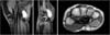
MRI showed a well-demarcated homogeneous low density 1.7×1.0 cm sized mass on spin-echo T1-weighted images, and high intensity on fast spin-echo T2-weighted images (Fig. 1).
Under brachial plexus block anesthesia, 5 cm incision was made directly over Guyon's canal. The mass was identified and was found to originate at the distal portion of Guyon's canal. The size of the mass was 1.7×1.0 cm. The mass was encapsulated inside the ulnar nerve motor branch sheath. Nerve sheath was incised longitudinally over the mass, and the mass was excised without ulnar nerve damage (Fig. 2).
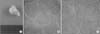
The mass was composed histologically of spindle cells with wavy appearing nuclei. S100 protein stain was positive in immonohistochemical stainning. The histological diagnosis was made as schwannoma (Fig. 3).
DISCUSSION
Guyon's canal syndrome is a compression neuropathy of the ulnar nerve at the wrist or hand, which presents the isolated motor, sensory or combined loss of motor and sensory function of the nerve.2) The 4.0 to 4.5 cm long tunnel known as Guyon's canal begins proximal to the hypothenar eminence and extends to the fibrous arch of the hypothenar muscles. The roof is composed of the palmar carpal ligament, palmaris brevis and fibrous tissue. The floor is composed of the tendons of the flexor digitorum profundus proximally, the transverse carpal ligament, and the pisohamate and pisometacarpal ligaments. The medial wall includes the flexor carpi ulnaris, the pisiform, and the abductor digiti minimi. The lateral wall is formed by the tendons of the finger flexors, transverse carpal ligament, and the hook of the hamate. 6)
Various lesions can cause ulnar nerve entrapment symptoms and signs at wrist and hand, such as ganglion, traumatic neuritis, ulnar artery disease and factures of carpal bones. While, other lesions such as lipomas, anomalous muscles, osteoarthritis and lesions of ulnar artery can cause ulnar nerve compression. Schwannoma, known as neurilemmoma of the hand are the commonest benign peripheral nerve sheath tumors though rare. 5) They arise from a proliferation of Schwann cells. Aetiology is unclear.5) Many schwannomas occur as solitary lesion and are often found incidentally.7) However, multiple schwannomas can occur mostly related to conditions comprising the neurofibromatosis (neurofibromatosis type II-NF2 and schwannomatosis). 8)
It was found that the risk of developing neurological deficits was more likely to be high in patients with larger tumors. Some authors reported that tumor size may be a risk factor for neurological deficits. 9) Park et al.10) observed that larger tumors tended to have more fascicles entering the tumor substance and were at greater risk of major neurological deficits after surgery.
MRI was found to be the useful diagnostic tool. Ganglion cysts and shwannomas may show similar findings on MRI. Although cysts show homogenous T2 high signal, cystc schwannomas can also show T2 high signal making differentiation difficult. In this case, contrast enhancement study may help as schwannomas should show internal enhancement.
In schwannoma of the ulnar nerve of the current case, only the pure motor component was involved. Even after mass excision, there was no sensory change on ulnar nerve territory. At 4 months after surgery, abduction power of the little finger recovered from grade IV to grade V. A rare case of symptomatic schwannoma in the Guyon's canal was reported together with the literature review.

 XML Download