

 PDF
PDF ePub
ePub Citation
 Print
Print


INTRODUCTION
For many years, Porcelain fused to metal (PFM) restorations have represented the most widely used restorative technique in fixed prosthodontics and this popularity may have attributed to the clinical longevity and accepted esthetics. However, these restorations are constantly exposed to challenges such as marginal leakage, which leads to the demise of the tooth pulp.1,2
When the crowns or bridges are going to be retained, there is very little information regarding the best way to restore the endodontic access opening. Clinicians routinely use either amalgam, composite resin or resin modified glass ionomer cement. Material of choice is critical, since coronal leakage of bacteria or their by products are an important factor in the failure of non-surgical root canal treatment (NSRCT).1
The newer generations of dentin bonding agents have better adhesion and marginal integrity in the laboratory but clinical data and long term results still need to be evaluated.3-5 The advantage of silane coupling agents appears to enhance the bond strength by promoting a chemical bond between the adhesive and the adherent.6 The dye leakage model has been used to determine if any of the dental materials in current clinical use has the ability to prevent coronal leakage in restored endodontic access openings in permanently fixed crowns following NSRCT.7 Exposing the samples to thermocycling speeds up the diffusion of water between the filling materials and the ceramic.8 This study was planned in order to evaluate the ability of filling materials such as amalgam, composite and compomer, to close access openings in PFM specimens.
MATERIALS AND METHODS
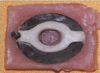
Ninety PFM specimens were constructed from green wax of 0.4 mm in thickness which were punched with 25 mm diameter ring and in the center with 6 mm diameter ring. Each of the 90 samples was invested, burned out and casted according to manufacturer's instructions. The metal samples were separated from their sprues, leaving 2 mm to facilitate handling during acrylic block constructions. All the samples were sandblasted with aluminum oxide particle to remove the excess oxide layer. Opaque and body porcelain were then applied according to manufacturer's instructions (The opaque layer around the perforation was 0.3 ± 0.1 mm while the body porcelain was 2.0 ± 0.3 mm in thickness). The PFM samples were filled with wax to prevent inward movement of the acrylic into the perforation during acrylic mold construction. The depths of the PFM samples were 2.0 ± 0.5 mm (Fig. 1).
The samples were divided into 3 equals groups according to the filling material.
Group A: The PFM perforations were filled with amalgam Septalloy NG-70 (Septodont, France) according to the manufacturer's instructions, regarding mixing and processing time. After 48 hours, the amalgam fillings were polished with green silicon polishing burs (Vivadent, Liechenstein) (with one bur for 10 samples).
Group B: The PFM perforations were filled with Tetric-Ceram composite (Vivadent, Liechenstein). The walls of the PFM perforations were treated with 37% phosphoric acid gel for 60 sec, washed with a copious amount of water for 15 sec, dried with oil-free air spray for 10 sec. The bonding agent, Excite (Vivadent, Leichtenstein), was applied to the treated walls of PFM samples using Vivadent applicator brush for 10 sec and finally, Tetric-Ceram was applied into the PFM perforation in one increment. After adapting resin with plastic instruments, the filling and perforation were covered with celluloid strip through which the light curing was done for 40 sec by holding the light emission window close to the strip and the fillings.
Group C: The PFM perforations were filled with Compoglass-F compomer (Vivadent, Liechenstein). The walls of the PFM perforations were treated with 37% phosphoric acid gel for 60 sec, rinsed and dried thoroughly and then the silane liquid Monobond-S (Vivadent, Liechenstein) was applied to the ceramic surface and left for 60 sec. The bonding agent, Syntac adhesive was applied with a brush, left for 2 sec, and dispersed with oil-free air spray until no movement of the liquid was visible. Then, with plastic instruments, sufficient amount of compomer was filled into the PFM perforations and light curing was done for 40 sec by holding the light emission window as close as possible to the celluloid strip and the filling. The fillings of group B and C were finished with gray silicon composite finishing burs and polished with green silicon polishing burs (one bur for 10 samples). The manual pressure during condensation of the amalgam and the adaptation of composite as well as the compomer was 5 ± 1 kg.
All the samples were incubated in normal physiological saline for one week at 37℃. Each group was further subdivided into three equal subgroups according to the thermocycling period (one week for 70 cycles, one month for 300 cycles and three months for 900 cycles). The dye used was Pelikan INK. The samples were put long enough in the dye solution to increase hydraulic pressure to enhance the dye penetration. Thermocycling was carried out to investigate the effect of temperature change on the adaptation of restorative materials to PFM interfaces and walls. The thermocycling device cycles the samples between two water bathes, one maintained at 5 ± 2℃ and the other at 55 ± 2℃. Each cycle was done for 30 sec in each bath. After completing the thermocycling, the samples were removed from the water bath and allowed to dry for 2 hrs at room temperature to facilitate the dye fixation.
All the samples were sectioned by a double-ended diamond wheel disk mounted on hand piece under water spray. The disk sectioned at the midline of the samples passing through the center of the filling materials (Fig. 1). The depth of dye penetration was measured from both sides for each PFM and filling materials interfaces using Co-ordinate Vernier Microscope (Griffin & George, UK) at × 20 magnification and scores calculated (Table 1). The data obtained was analyzed by SPSS, 2006 using one way ANOVA test and student t-test and significant difference level at (P<.01).
RESULTS
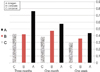
Two surfaces were obtained after sectioning of each sample (Fig. 1). The four readings gained from each sample were obtained together and then divided by four to get the mean penetration level. From Fig. 2, it is clear that the lowest level of the dye penetration occurred during the first week in all three groups including the subgroups. The value of dye leakage had significantly increased by time intervals in subgroups A and C. There was no significant difference in the time intervals between one month and three months in subgroups B and C.
DISCUSSION
The aim of this study was to determine if amalgam, composite or compomer has the ability to prevent coronal leakage in restored endodontic access opening in PFM specimen's in-vitro.
Septalloy NG-70 amalgam capsules (high copper alloy admixed type) were used in this study because amalgam has been widely used as a restorative material for more than a century in the field of dentistry.9 Tetric-Ceram has an advanced composite technology used for anterior and posterior fillings. It is a highly dispersed hybrid material that has continuous fluoride ion releasing effect and contains ceramic particles in its composition. 4,10 Compomer is a hybrid material composed of a single hydrophobic resin that is filled with acid-leachable glass particles bonded with functional primer and light cured, which results in its higher mechanical strength, improved marginal seal by hygroscopic expansion and improved bonding ability after storage in water.4,11 Therefore, when two dissimilar materials are to be attached, whether micromechanical or chemical is chosen, the use of silane coupling agents could increase attachment.12
An interpretation of the results indicated that all restorative materials showed leakage but the dye did not reach the metal part of the PFM, and this is due to low difference in linear coefficient of thermal expansion (LCTE) between Ni-Cr alloy and amalgam or composite, this is in agreement with the previous studies1,7,13,14
Group A (with highly significant difference) had the highest dye penetration compared to other groups and this may be due to the high difference between LCTE of porcelain and amalgam15 or due to the contact angel that formed between the PFM walls and the amalgam, which is considerably sufficient for the dye particles to penetrate.15
Our finding agreed with those1 who concluded that amalgam is not the material of choice for restoration of access opening through the PFM crowns, but did not agree with those13 who founded that the use of lathe-cut and admixed amalgam would reduce the marginal microleakage.
Dye penetration of group B (with significant difference) indicated that microleakage of Tetric-Ceram composite increased as the number of thermocycling increased (Fig. 2). This increase with aging may be related to disintegration of the bonding agent and gap formation at the composite filling and PFM interfaces.15 This could be explained by the initial polymerization shrinkage of Tetric-Ceram which resulted in a pulling back of the restoration and adhesive bonding toward the light source resulted in gap formation and increased microlekage.15 The incorporation of water into the composite may compensate the initial shrinkage of the Tetric-Ceram and resulted in secondary expansion, which could maintain the same level of leakage with time lapses.10,16 The use of Exite, 5th generation adhesive system which has both hydrophilic and hydrophobic groups, results in good bond with hydrophobic porcelain. Although our results did not agree with those7,13 who concluded that microleakage of Tetric-Ceram is very high during the first 48 hrs, it shows great reduction in leakage with aging due to water sorption.
Group C indicated a significant difference among samples thermocycled for week and samples thermocycled for one or three month. This is because compomer does not under-go dimensional changes by expansion and did not improve the marginal seal after storage in water for a week,11 but during one or three months absorption of water and hygroscopic expansion of compomer has resulted in improvement of bonding ability after storage in water. On the other hand, there is no significant difference between samples thermocycled for one and three months and this may be due to the effect of silane bonding agent, which minimizes the gap formation at compomer and PFM interfaces. Also, compomer is a single component hydrophobic resin, composed of combination of both composite and GI, so the LCTE of it is in the range of GI and anterior or posteriors composite, which is similar to that of hydrophobic porcelain.3,15 Some authors8,11 have reported that Compoglass-F produced smoother surfaces than conventional GI cements, which lead to the reduction of susceptibility to bacterial adhesion that comes as a consequence of gap formation.
It is obvious from Fig. 2 that increasing the number of thermocycling resulted in more dye penetration in all subgroups, which may be due to excessive contraction of amalgam during the first days and the polymerization shrinkage of Tetric-Ceram and Compoglass-F and this corresponds with Grobler et al.17 Septalloy NG-70 amalgam subgroups showed the highest level of dye penetration because the time provided is not long enough and the high-copper alloys did not readily produced corrosion product. Our results did not agree with those7,14 who said that the dual-cured composite and glass ionomer had the greatest level of dye penetration in PFM crowns comparing with that of amalgam.
There is no significant difference between one and three months in subgroup B and C; this may be due to polymerization shrinkage of Tetric-Ceram and Compoglass-F during the first days, and water absorption and secondary expansion afterwards. In subgroups C, etching and silane application prior to placement of dentin bonding was significantly effective as it pulled resin-based composite toward walls of the PFM resulting in reduction of microleakage and better marginal integrity in vitro.4,5,18 Several authors3,17 have concluded that compomer is alleged to produce less shrinkage and compensate for internal stress by its elasticity and through water resorption therefore, it has the potential to minimize the initial shrinkage due to polymerization.
This in vitro and short time study only simulates the actual used condition and it should not be used alone to predict the clinical performance of these materials, and therefore, more long time studies are necessary in the future.
 XML Download