

 PDF
PDF ePub
ePub Citation
 Print
Print


Introduction
The World Health Organization (WHO) has declared obesity a disease in need of treatment, and one of the most serious social problems for the people's health [1]. According to the 2010 Korean National Health and nutrition Survey (KNHNS), 30.8% of Koreans over 19 are obese, a significant increase from 26% of 1998 [2]. Obesity is a state in which adipose tissues are excessively accumulated, representing hyperplasia and hypertrophy of adipocytes. As preadipocytes increase cells differentiate, leading to an accumulation of lipid droplets within the adipocytes, thus differentiating into a mature adipocyte [3]. In the preliminary stages of differentiation transcription factors such as CCAAT/enhancer-binding protein (C/EBP)β is activated, followed by an activation of peroxisomal proliferator activated receptor (PPAR)γ and C/EBPα in its middle stages; in the latter stages, fatty acid binding protein (FABP)4 induces the differentiation into mature adipocytes [4,5]. Furthermore, the proliferation and differentiation of adipose tissues are facilitated by angiogenesis which provides oxygen and nutrients within adipose tissues [6,7].
Resveratrol is a type of polyphenol usually found in grapes, wines and peanuts, a natural antibiotic created naturally to fight against infections from bacteria or germs [8]. The resveratrol in grapes exists generally in its skin resulting in a 1-5 µg/mL of resveratrol, while white wine contains 0.1-0.5 µg/mL [9]. Furthermore, resveratrol does not dissolve in water but dissolves well in alcohol, and so red wine contains greater resveratrol than grape juice [10].
Numerous studies of resveratrol have reported on its physiological functions, including anti~inflammatory [11], antioxidant [12], prevention of heart and vascular diseases [10,13], and life expension [14]. Furthermore, resveratrol suppressed lipogenesis by activating the 5' adenosine monophosphate-activated protein kinase (AMPK) pathway and thus stimulating the oxidation of fatty acid [15,16]. However, there has been limited research regarding the effects of resveratrol on the protein expression of transcription factors regarding adipocyte differentiation and activity of angiogenesis-related matrix metalloproteins (MMPs). This research tried to determine the effects of resveratrol on protein expression of transcription factors that lead to proliferation and differentiation of adipocytes, as well as its effects on the activities of MMPs.
Materials and Methods
Resveratrol (Sigma, St. Louis, MO, USA) was dissolved in dimethyl sulphoxide (DMSO) (Sigma, St. Louis, MO, USA) to make a 40 mmol/L stock. This stock solution was stored at -20℃ and diluted in Dulbecco's modified Eagle's medium (DMEM) (WelGENE, Daegu, Korea) for each experiment. Unless otherwise indicated, all other reagents were purchased from Sigma.
Cell culture
3T3-L1 mouse fibroblast preadipocytes were obtained from the American Type Culture Collection (Rockville, MD, USA). Cells were grown to confluence in DMEM (WelGENE, Daegu, Korea) and were supplemented with 10% bovine calf serum (BCS) (WelGENE, Daegu, Korea), 100 units/mL penicillin, and 100 µg/mL streptomycin (WelGENE, Daegu, Korea) to make a regular medium (RM) [17]. The medium was replaced every 2 days and incubated in 5% CO2 atmosphere at 37℃ until the medium reached confluence state. When cells reached 70 - 80% confluence, cells were rinsed twice with phosphate buffered saline solution (PBS) and treated with trypsin-EDTA (WelGENE, Daegu, Korea). The separated cells were then sub-incubated with the medium being replaced every 2 days.
Adipocyte differentiation induction
To differentiate 3T3-L1 preadipocytes into adipocytes, 10% fetal bovine serum (FBS) (WelGENE, Daegu, Korea), 0.5 mmol/L isobuthylmethylxanthine (IBMX), 10 µg/mL insulin, 1 µmol/L dexamethasone (Dex), 100 units/mL penicillin, and 100 µg/mL streptomycin was added to DMEM to make Differential Media (DM), which was used to induce differentiation [18]. Two days after differentiation, 10% FBS, 10 µg/mL insulin, 100 units/mL penicillin, and 100 µg/mL streptomycin was added to DMEM to make post differentiation media (PM). Adipocytes were incubated in PM, which maintains the adipocyte state.
Adipocyte proliferation
MTT assay was conducted to determine the effects of resveratrol on adipocyte proliferation [19]. 3T3-L1 preadipocytes were incubated with DM to induce differentiation, and then PM, treated with 0, 10, 20, and 40 µmol/L of resveratrol, was added to mature adipocytes. Cell viability was estimated by adding 1mL of MTT solution (1 mg/mL 3-(4,5-dimethylthiazol-2-yl)-2,5-diphenyltetrazolium bromide) and by then incubating 37℃, 5% CO2 atmosphere for 3 h. After incubation of 0, 2, 4, and 6 days, each MTT solution was diluted with 0.5 mL of isopropyl alcohol and absorbance rate was measured at 490 nm with spectrophotometer (Tecan, Austria GmbH, USA).
Oil-Red-O-Staining
After induction of differentiation, cells were stained with oil-red O solution using adipogenesis assay kit (Chemicon International Inc., Temecula, USA) [20]. Cells were washed twice with 100 µL PBS, and then 50 µL of oil-red O solution was added and they were incubated at room temperature for 15 min. After incubation, the stained oil droplets were dissolved in isopropanol and were measured for absorbance at 450 nm using a spectrophotometer (Tecan, Austria GmbH, USA).
Intracellular triglyceride concentration
Intracellular triglyceride accumulation was measured with a commercially available triglyceride assay kit (Wako chemicals GmbH, Neuss, Germany) [21]. Cells were treated with 0, 10, 20, and 40 µmol/L of resveratrol and were rinsed twice with PBS. Cells were resuspended in 137 mmol/L NaCl, 20 mmol/L Tris-Cl, 1% triton X-100, 10% glycerol, 1 mM/L sodium orthovanadate, 1 mmol/L PMSF, 20 µg/mL aprotinin, 10 µg/mL antipain, 10 µg/mL leupeptin, and 80 µg/mL benzamidine HCl. Cells were then lysed by sonification at 4℃ and centrifuged at 12,800 rpm for 5 min. After centrifugation, the aliquot was mixed with an equal volume of chloroform and was then centrifuged again. The chloroform layer was carefully dried and resuspended in a 1% Triton X-100. The Sample was treated with 300 µL of color reagent and was incubated at 37℃ in a 5% CO2 incubator for 5 min. Absorbance was then measured at 600 nm using spectrophotometer (Tecan, Austria GmbH, USA).
Activity of Glycerol-3-phosphate dehydrogenase (GPDH)
GPDH activity was measured with GPDH activity kit (Takara Bio Inc., Japan) [22]. Cells were plated at a density of 1 × 10⁴ cells/mL in 96 well plate. Cells were treated with 0, 10, 20, and 40 µmol/L of resveratrol every two days. At six days after differentiation induction, cells were rinsed with PBS and enzyme extraction buffer was added to the cells. Cells were then diluted with a solution containing 0.1 mol/L 2-mercaptoethanol. The change in absorbance density was measured at 340 nm, 30℃ every minute for 10 min using a spectrophotometer (Tecan, Austria GmbH, USA). GPDH activity was calculated as the amount of enzyme used to consume 1 µmol/L of substrate in one min at 30℃, converting this unit to mg protein/mL. Protein in the supernatant was quantitatively measured with a protein assay kit (BioRad, Richmond, CA, USA).
Western blotting analysis
Cell lysates were performed before western blotting analysis [22]. The harvested cells were transferred to 50 mL tubes on ice, centrifuged for 3 min, and the supernatant was discarded. Cells were then rinsed with ice-cold PBS buffer. The protein of the sample were separated using gel electrophoresis employed with polyacrylamide gels and with buffers loaded with 4-20% gradient sodium dodecyl sulfate (SDS) (SDS-PAGE) [23]. It was then transferred overnight to immobilon TMp membrane (Millipore, Bedford, MA, USA) at 4℃. Blocking of non-specific binding was achieved by placing the membrane in 10 g/L Bovine serum albumin (BSA) or 50 g/L milk in TBST (Tris-buffered saline (20 mmol/L Tris-Cl, and 150 mmol/L of NaCl), and 1 g/L Tween 20, pH7.5). After blocking, proteins in membrane were then immunoblotted with antibodies to PPARγ (Abcam, Cambridge, England), C/EBPα, C/EBPβ (Santa Cruz Biotechnology, Santacruz, USA), and FABP4 (Abcam, Cambridge, England) [24-28]. After rinsing off the membrane with TBST, anti-mouse 1 g horseradish peroxidase/TBST or anti-rabbit 1 g horseradish peroxidase/TBST (Amersham, Buckinghamshire, England) were incubated with the membrane. After a final wash with TBST, the western blot analysis was performed by using the enhanced Chemiluminescence method, and the Super Signal West Dura Extended Duration Substrate (Piece, Rockford, IL). The density of each band image was analyzed by Image J Launcher (Provided by NCBI), which evaluates the relative amount of protein staining and quantifies the results in terms of optical density. Protein expression level of the control group was set as 100% and the results to each resveratrol treatment groups were compared with the control group.
Activity of matrix metalloproteinases (MMPs)
MMPs activity was determined as previously described [29]. After cells were incubated with various concentrations of resveratrol, 0, 10, 20, and 40 µmol/L, for 3 days and centrifuged for 10 min at 3,000 rpm, the supernatants were collected and MMP activity was determined by electrophoresis on a polyacrylamide gel containing 1% gelatin. The gel was shaken twice for 30 min under renaturation buffer (Triton X-100 2.5% in water (v/v)) and also under developing buffer (0.5 mmol/L Tris, Tris-HCl, NaCl, CaCl2, Brij), and incubated for 17 h at 37℃. The gel was stained for 30 min with 0.25% Commassie blue and then the activated parts of each band under the dyed gel was scanned with digitization by image program Image J Launcher (provide by NCBI). The MMPs were detected as clear bands against a blue background of undegraded substrate.
Results
Effects of resveratrol on adipocyte proliferation
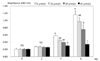
According to MTT assay at 0, 2, 4, and 6 days, resveratrol did not show significance on cell proliferation at 0 and 2 days after incubation. However, cell proliferation was found to be decreased significantly in a dose-dependent manner of resveratrol treatments after 4 days of incubation; When the non-treatment group of resveratrol was said to express as standard 100%, cell proliferation was decreased by 77.2%, 68.3%, and 52.1%, at the 4th day of incubation, and by 84.2%, 65.3%, and 29.6% at the 6th days of incubation, with increasing resveratrol concentrations of 10, 20, and 40 µmol/L (P < 0.05) (Fig. 1).
Effects of resveratrol on adipocyte differentiation
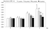
In the oil-red O dye staining, two days after resveratrol treatments of 10, 20, and 40 µmol/L, lipid accumulation in adipocyte was significantly inhibited by 93.8%, 92.4%, and 91.5% respectively, compared to the control (P < 0.05) (Fig. 2). At four days of incubation, adipocyte differentiation was determined to be 90.6% with 10 µmol/L of resveratrol and 61.4% with 40 µmol/L resveratrol, respectively. The resveratrol treatments at day 6 caused significant differences in adipocyte differentiation, with 90.6% differentiation for 10 µmol/L resveratrol treatments and 59.7% for 40 µmol/L treatments.
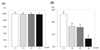
Triglyceride concentrations were very slightly, but significantly decreased by 99%, 98.8%, and 97.7% according to resveratrol treatments with 10, 20, and 40 µmol/L, respectively, compared to control (100%) that contained no resveratrol (P < 0.05) (Fig. 3A).
When GPDH activity in 3T3-L1 treated with resveratrol at 10, 20, and 40 µmol/L were measured, incubation at day 6 caused significantly decreases of GPDH activity in a dose-dependent manner by 63%, 59.9%, and 25.1% with resveratrol treatments of 10, 20, and 40 µmol/L respectively (P < 0.05) (Fig. 3B).
Effects of resveratrol on protein expression of transcription factor related adipocyte differenciation
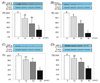
The protein expression of C/EBPβ, which induces initial stages of differentiation, decreased significantly in a dose-dependent manner to 78% 56%, and 30% compared to the control after resveratrol treatments of 10, 20, and 40 µmol/L respectively, (P < 0.05) (Fig. 4A). The protein expression of PPARγ, which induces the middle stages of differentiation, was significantly reduced by 57% and 15% with resveratrol treatments of 20 and 40 µmol/L, respectively (P < 0.05) (Fig. 4B) while the protein expression of C/EBPα was also significantly decreased by 74% and 38% with resveratrol treatment of 20 and 40 µmol/L (P < 0.05) (Fig. 4C). The protein expression of FABP4, which induces final stages of differentiation, was decreased significantly by 88%, 72%, and 44% with the aforementioned increased resveratrol concentrations (P < 0.05) (Fig. 4D).
Effects of Resveratrol on MMP activity
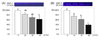
The activity of MMP-2 was decreased significantly 84%, 70%, and 63% after different resveratrol treatment concentrations of 10, 20, and 40 µmol/L, respectively (P < 0.05) (Fig. 5A). MMP-9 activity was also decreased to 74%, 62%, and 39% that of the control, in a dose-dependent manner (P < 0.05) (Fig. 5B).
Discussion
Resveratrol is a type of stilbene phytoalexin that is naturally produced by plants [10]. Although several studies determined the effect of resveratrol on adipocyte differentiation and various protein transcription factors [16,17,26,27,30-32], only limited information is available about inhibitory action of resveratrol on MMPs activity in adipocyte differentiation [33,34]. Thus, in the present study, the effects of resveratrol on MMP-2 and MMP-9 activity were investigated to provide a mechanistic basis for the protective effect of resveratrol on adipocyte differentiation.
This study showed a significant drop in cell proliferation and differentiation when 3T3-L1 adipocytes were incubated in concentrations of 0, 10, 20, and 40 µmol/L. Even with 2 days of incubation, significant inhibition effects with 40 µmol/L resveratrol treatments were observed, and these findings suggest that resveratrol inhibits hormonal mixture induced 3T3-L1 adipocyte proliferation. Rayalam et al. [27] reported that resveratrol decreased proliferation and induced significant apoptosis showing when mature adipocytes were treated with 100 and 200 µmol/L resveratrol, Other studies also supported the inhibitory effect of resveratrol on adipocyte diffentiation and proliferation [24-27,30-32].
Glycerol-3-phosphate in adipose tissues is produced reversibly from dihydroxyacetone phosphate by glycerol-3-phosphate dehydrogenase (GPDH), combining with fatty acyl-CoA to produce triglyceride [35]. In this study, GDPH activity was decreased, greatly depending on increased concentration of resveratrol, and this result was consistent with Chen's study [15] which showed that resveratrol-amplified grape skin extracts with 200 ug/mL and 400 ug/mL led to a significant decrease of GDPH activity does-independently.
Many transcription factors are needed for the differentiation of adipocytes. In the early stages, transcription factors such as C/EBPβ are active, while in the metaphase (middle stages) PPARγ and C/EBPα are active, and in the final stages, factors such as FABP4 are active [28]. Several studies have demonstrated that resveratrol treatment inhibited PPAR and C/EBPs expression [30-32], which are consistent with this study suggesting that resveratrol inhibits adipocyte differentiation and proliferation by modulating the expression of transcription factors, such as C/EBPβ, PPARγ, C/EBPα, and FABP4.
One of the most important factors of obesity is angiogenesis; the oxygen and nutrients that adipocytes received are used to form new blood vessels, which are further used to facilitate the differentiation of adipocytes [36]. Remodeling of the extracellular matrix (ECM) appears during obesity-related fat mass development, and this mechanism is thought to be related with regulated production of MMPs [37].
This study, by treating resveratrol with adipocytes, showed that there was a significant decrease in the activities of MMP-9 and MMP-2, with MMP-9 being more significantly affected than MMP-2. Since MMP-2 and MMP-9 are key enzymes for the first step of angiogenic process [37], it may be suggested that inhibiting actions of resveratrol on MMP's activities are related to suppress proliferation and differentiation of adipocytes. Two other studies supported our study results; Gregoire et al. [38] reported that the concentration of MMP-9 in obese children was a significantly higher compared to that of normal children. Alexander et al. [39] reported that as 3T3-L1 preadipocytes undergo differentiation, MMP-2 and MMP-9 are produced, and MMP inhibitors play an important role in inhibiting adipocyte differentiation.
In conclusion, resveratrol treatment of 3T3-L1 adipocytes significantly inhibits adipocyte proliferation in a dose dependent manner. Resveratrol inhibits adipocyte differentiation by modulating the protein expression of transcription factors such as C/EBPβ, PPARγ, C/EBPα, and FABP4, and by decreasing MMP-9 activity.
 XML Download