

 PDF
PDF ePub
ePub Citation
 Print
Print


Introduction
Folate deficiency is the most common sign of malnutrition in patients with chronic alcoholism [1,2]. The frequent finding of folate deficiency in patients with chronic alcoholism suggests a role for folate in the pathogenesis of alcoholic liver disease (ALD). Folate plays an integral role in methionine metabolism. 5-Methyltetrahydrofolate and homocysteine (Hcy) are substrates for methionine synthase to produce endogenous methionine which is a precursor of S-adeonosylmethionine (SAM). Because folate helps maintain normal concentrations of Hcy, methionine, and SAM, folate deficiency can disturb methionine metabolism, leading to hyperhomocysteinemia and SAM depletion. These metabolic changes are important features of ALD [3].
According to extensive studies in animal models, chronic ethanol consumption increases hepatic oxidative stress, and ethanol-induced liver injury is associated with free radical formation [4]. Folate deficiency accelerates ethanol-induced changes in hepatic methionine metabolism while producing enhanced oxidative liver injury [5]. Chronic alcoholism is associated with abnormal Hcy metabolism in humans and animal models.
Increasing evidence suggests that Hcy is involved in the pathogenesis of ALD [6]. Abnormal Hcy metabolism increases fat accumulation, inflammation, and injury to hepatocytes from rats fed alcohol [7,8]. The injury induced by Hcy involves oxidative damage [9-12] and the elevated Hcy plays an important role in various pathologies by increasing H2O2 production [13-15], affecting antioxidant defense systems [16], and promoting DNA damage [17]. Increased DNA damage triggers a form of programmed cell death called apoptosis. Thus, the oxidative insults induced by folate depletion and elevated Hcy may play a major role in various pathogenic states.
Previous studies have demonstrated that peroxidative damage increases with ethanol intake [18,19]. The mechanisms for ALD include activation of CYP2E1 [20-22] and NADPH oxidase [23]. These mechanisms involve production of reactive oxygen species (ROS), and oxidative liver injury is the net result of enhanced generation of ROS and depletion of antioxidants such as glutathione [20-23].
Although oxidative stress is one of the important mechanisms contributing to Hcy-induced tissue injury, it has not been examined whether exogenous administration of folic acid attenuates the oxidative stress and hepatic toxicity induced by ethanol. This study was undertaken to establish whether the disturbances in some antioxidant activities and hepatic methionine metabolism induced by chronic ethanol consumption are a mechanism linking chronic ethanol consumption to hepatic injury. We also investigated the in vivo effect of folic acid supplementation on oxidative stress and hepatic toxicity in ethanol-fed rats.
Materials and Methods
Materials
Lactobacillus rhamnosus (ATCC 7469) was obtained from the American Type Culture Collection (Manassas, VA, USA). Folic acid depleted casein medium was obtained from Difco Laboratories (Detroit, MI, USA) and 7-fluoro-benzo-2-oxa-1, 3-diazole-4-sulfonate (SBDF) was obtained from Wako Chemicals (Osaka, Japan), respectively. L-Homocystine, tri-n-butylphosphine, DL-methionine, met-myoglobin, 2,2'-azino-bis(3-ethylbenzothiazoline-6-sulfonic acid) diammonium salt (ABTS), heparin, and cyclohexane were purchased from Sigma (St. Louis, MO, USA). All chemicals were of the highest purity commercially available.
Animal model
The Lieber-DeCarli liquid diet [24], for administering alcohol orally, has provided an excellent means to reproduce the early lesions of ALD, which include steatosis and oxidative stress. Thirty-two male Wistar rats (initial weight, 170-180 g) were obtained from Orient Bio (Seoul, Korea). After a 7 day acclimation, animals were randomly assigned to one of four groups of eight rats each: (i) Control group (C); rats were a fed liquid diet that was essentially the same as the diet described by Lieber and DeCarli [24] with the exception of a reduction in total lipid content from 39.6 g/L to 23 g/L and a dextrin-maltose supplement to account for the energy deficit. The control liquid diet consisted of the following components (g/L): carbohydrate, 153 (dextran-maltose); protein, 41.4 (casein, vitamin free > 85%); lipids, 23 (soybean oil); cellulose, 10; minerals plus vitamins (AIN 76); choline bitartrate 0.53; and, xanthan gum 3. Ethanol was gradually introduced into the diets over 5 days. (ii) Low ethanol group (LE); ethanol was added to the liquid diet at 12% of total calories instead of dextrin-maltose. (iii) High ethanol group (HE); ethanol was added to the liquid diet at 36% of total calories. (iv) Ethanol plus folic acid group (FE); ethanol was added to the liquid diet at 36% of total calories with 10 mg/L folic acid supplementation.
Animals in pair-fed groups (C, LE, FE) were fed the same amount of HE diet as that consumed by the HE group over the preceding 24 hrs. The amount of the diets consumed was monitored daily, and body weight was measured once per week. The diets were fed for 5 weeks.
Rats were housed individually in plastic cages in a temperature (23 ± 1℃) and humidity controlled (50 ± 5%) room with a daily light cycle from 06:00 to 20:00 hr. Animal experiments followed protocols established by the NIH Guide for the Care and Use of Laboratory Animals, and this study was approved by the institutional animal care and use committee.
Sample collection
At the end of the 5-wk feeding period, rats were fasted overnight and then anesthetized with carbon dioxide. Blood samples were collected by cardiac puncture into heparinized syringes. Blood was immediately centrifuged for 15 min at 1,500 × g and 4℃ to collect plasma. Liver tissues were removed, washed in ice-cold saline, and rapidly frozen in liquid nitrogen. Samples were stored at -70℃ until analysis.
Plasma alanine transaminase and aspartate transaminase
Plasma alanine transaminase (ALT) and aspartate transaminase (AST) activities were measured using a photometric autoanalyzer (ERBA Chem Pro, Transasia Bio-Medicals, Mumbai, India).
Plasma and liver triglycerides
Liver tissues were homogenized, and hepatic total lipids were extracted and resolved in 5% Triton-X 100 in water. Hepatic triglyceride (TG) content was determined by an enzymatic colorimetric method using commercially available kits (Sigma). Plasma TG content was determined by an enzymatic method using a photometric autoanalyzer.
Plasma homocysteine
Total plasma Hcy levels were measured by high pressure liquid chromatography (HPLC) with fluorometric detection (excitation at 385 nm and emission at 515 nm) according to Araki and Sako [25]. Hcy was separated with a Hypersil ODS analytical column (250 × 4.6 mm I.D., 5 µm particle size) (Thermo, Runcorn, UK).
Plasma and liver folate
Folate was analysed by a microplate assay method using L. rhamnosus (ATCC 7469) according to Tamura [26]. Portions of liver were homogenized and autolysed for hydrolysis of γ-glutamyl residues in the presence of sodium ascorbate at 37℃. The supernatants of liver homogenates and plasma samples were used for the folate assay.
S-Adenosyl methionine and S-adenosylhomocysteine
Portions of frozen liver were homogenized with 0.4 M HClO4. Samples were centrifuged at 12,000 × g for 4℃ for 30 min. Each supernatant was filtered through a 0.45-µm filter. SAM and SAH were measured on a Shimadzu LC-10 HPLC system equipped with 250 × 4.6 mm Ultrasphere 5-µm ODS Betasil analytical column (Thermo) according to Wagner et al. [27].
Plasma total radical-trapping antioxidant potential
Total radical-trapping antioxidant potential (TRAP) was analyzed using an inhibition assay according to Rice-Evans and Miller [28]. Plasma samples were incubated with a peroxidase (met-myoglobin) and H2O2 to produce the cation ABTS+. A relatively stable blue-green color occurred and was measured at 30℃ at 740 nm. Antioxidants in the added plasma sample cause suppression of this color production to a degree that is proportional to their concentration. The standard procedure consisted of determination of the Trolox equivalent antioxidant capacity (mmol/L).
Conjugated diene formation of LDL
Oxidation of LDL was identified by measuring the formation of conjugated dienes (CD), which is an early lipid peroxidation event. LDL was isolated with buffered heparin as described previously [29,30]. To estimate LDL oxidation, lipids were extracted from LDL samples by chloroform-methanol (2:1) and dried under nitrogen, then redissolved in cyclohexane. The quantity of CDs in LDL was assessed by monitoring the change in absorbance at A234 at the indicated time points [31]. The results are expressed as µmol/L.
Statistical analysis
Results are expressed as mean ± standard error (SE). Data were analyzed by one-way analysis of variance (ANOVA) followed by Duncan's multiple range test. Data were considered significantly different at P < 0.05. Pearson's correlation procedure was used for the correlation analysis. All statistical analyses were performed using SPSS 18.0 for Window (SPSS, Inc., Chicago, IL, USA).
Results
We evaluated the effects of folic acid supplementation to correct the ethanol-induced perturbation in liver methionine metabolism, oxidative stress parameters, and experimental liver injury. As shown in Table 1, the 36% ethanol diet increased both final body weight (P < 0.001) and weight gain (P < 0.001) compared with those in rats fed the control diet. Folic acid supplementation of the ethanol diet resulted in a significant decrease in body weight compared with rats fed the HE diet. However, the LE diet had no apparent effect on body weight. When liver weight was expressed as a percent of body weight, a significant increase was found in all ethanol-fed rats (LE, HE, and FE) compared with rats fed the control diet (P < 0.05).
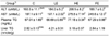
As shown in Table 2, liver folate concentrations remained unchanged in rats fed the LE or HE diets, and increased slightly in rats fed the FE diet compared with those in the control group. However, the HE diet tended to decrease plasma folate concentration slightly as compared to rats fed the control or LE diets. Plasma folate increased significantly in rats fed the FE diet compared with rats fed the HE diet (P < 0.01). Both plasma Hcy (P < 0.001) and cysteine (P < 0.05) increased significantly in rats fed the HE diet compared to rats fed the control diet (12.07 nmol/mL vs. 8.29 nmol/mL, 329.4 nmol/mL vs. 268.5 nmol/mL, respectively) (Table 2). Folic acid supplementation decreased plasma Hcy slightly in ethanol-fed rats. However, liver SAM, SAH, and the SAM/SAH ratio remained unchanged by feeding either the HE diet or by folic acid supplementation.
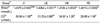
Plasma AST and AST activity was measured to assess whether folic acid supplementation influenced the development of ethanol-induced liver injury (Table 3). The HE diet significantly elevated plasma ALT (P < 0.05) and AST (P < 0.05) activities indicating hepatic toxicity. Supplementation of ethanol-fed rats (FE) with folic acid reduced ALT and AST activity significantly compared to that in rats fed the HE ethanol diets, which demonstrated that ethanol-induced liver injury can be alleviated by folic acid supplementation.
The high ethanol diet caused significant increase in plasma TG levels compared to that in controls, but the LE diet had no apparent effect on plasma TG (Table 3). Supplementation of ethanol-fed rats with folic acid resulted in decreased plasma TG to control levels (P < 0.05). However, liver TG remained unchanged either by ethanol administration or by folic acid plus ethanol administration.
TRAP, which represents non-enzymatic plasma antioxidant activity, decreased significantly in plasma of rats fed the LE or HE diets (P < 0.01) (Table 4). Folic acid supplementation elevated plasma TRAP in the ethanol-fed animal models. Levels of CDs in isolated LDL were analyzed as an oxidative stress parameter in plasma (Table 4). CD levels in rats administered the HE diet tended to be significantly higher than those of the control group, and the FE diet decreased CD to the lowest levels in this study (P < 0.05). CD levels remained unchanged in the plasma of the LE group compared to that in the control group.
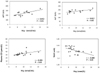
We performed a correlation analysis between plasma Hcy and plasma aminotransferase activities and oxidative parameters to investigate whether Hcy levels were associated with the parameters related to ethanol-induced hepatic toxicity and oxidative stress (Fig. 1). We found a significant correlation between Hcy and AST (r = 0.652, P < 0.01), Hcy and ALT (r = 0.612, P < 0.05), Hcy and CD levels (r = 0.495, P < 0.05), and Hcy and TRAP levels (r = -0.486, P < 0.05). These results indicate that Hcy may cause hepatic toxicity by elevating oxidative stress in rats fed a chronic ethanol diet.
Discussion
Numerous reports have demonstrated that oxidative stress plays an important role in the development of alcoholic liver disease [32-34]. Both alcoholic patients and rats fed ethanol have elevated Hcy and decreased folate levels [35-37]. Although folate deficiency and hyperhomocysteinemia seem to be involved in the pathogenesis of alcoholic liver disease, practically nothing has been reported on the effects of exogenous administration of folic acid on oxidative stress and hepatic toxicity in ethanol-fed rats. The present study is the first to provide data on the ability of folic acid supplementation to correct ethanol-induced perturbations in methionine metabolism and experimental liver injury in ethanol-fed rats, an animal model of alcoholic liver disease.
Previous studies have shown that ethanol-induced Hcy elevation triggers the endoplasmic reticulum (ER) stress response that mediates oxidative and inflammatory processes in the development of both vascular injury and hepatic steatonecrosis [6,38]. Ethanol and its metabolites, such as acetaldehyde and fatty acids, could generate a large amount of ROS during metabolism by the microsomal enzyme CYP2E1. ROS can damage liver cells directly or by enhancing the sensitivity of hepatocytes to lipid peroxidation [39,40]. Pathogenesis of alcohol-induced hepatic steatosis and steatohepatitis is thought to be dependent on oxidative stress, whereby lipid peroxidation products facilitate the inflammatory response. Oral folic acid supplementation improves cardiovascular functions in patients with mild homocysteinemia [41].
We observed that chronic ethanol feeding (HE) significantly elevated plasma Hcy, AST, ALT, and TG. In contrast, folic acid supplementation partially reduced plasma Hcy, ALT, AST, and TG in ethanol-fed rats. These results suggest that folic acid supplementation offers a hepatoprotective effect in ethanol-induced liver injury by decreasing Hcy. It has become apparent that abnormal hepatic methionine metabolism is integral to hepatic toxicity, and that hyperhomocysteinemia resulting from a disturbance in methionine metabolism plays an important role in alcohol-induced liver injury [42]. Because ethanol-induced hyperhomocysteinemia is toxic to liver cells, we also examined the correlation between plasma Hcy and aminotransferase levels (Fig. 1). We found that plasma Hcy concentration was positively correlated with plasma ALT and AST levels in rats fed the ethanol diets.
Total antioxidant defense, represented by the TRAP measurement, decreased significantly in plasma of rats fed the HE diet but was partially restored by folic acid supplementation (P < 0.001). Elevated TRAP following folic acid supplementation may imply improved antioxidant capacity to prevent damage associated with free radicals. Hyperhomocysteinemia is associated with ER stress, leading to the activation of ER-dependent apoptosis and upregulation of lipid synthesis in hepatocytes [6]. We also observed strong correlations between plasma Hcy and oxidative stress markers in ethanol-fed rats (Fig. 1). CD levels in the isolated LDL samples from the HE group were significantly higher than those in the control group (P < 0.05) (Table 3), suggesting that a high concentration ethanol diet induced production of oxygen free radicals leading to lipid peroxidation. We observed that a decreased CD level following folic acid supplementation in ethanol-fed rats. These results suggest that decreasing Hcy to a certain level by folic acid supplementation might partially prevent oxidative stress.
In this study, oxidative status markers were modified by folic acid supplementation in the sense of lower oxidative stress. However, recent studies have suggested that folate may have protective effects independent of Hcy-lowering effects through free-radical scavenging activity [43,44]. The protective effects of folic acid supplementation on endothelial dysfunction may be due to a direct effect of folate on free-radical oxidation of LDL lipids. The plausible rationale is free radical scavenging activity of folate [44]. Because liver injury by free radicals is a potential participating mechanism in alcoholic liver disease, and oral folic acid supplementation may be a safe and inexpensive way to reducing oxidative damage, further studies are needed to elucidate the possible mechanism underlying these protective effects.
In summary, the results show that folic acid supplementation has potential implications for alleviating hepatic toxicity by preventing homocysteinemia in ethanol-fed rats. Considering the oxidative stress elicited by Hcy, folic acid supplementation could contribute to correct the observed decrease in antioxidant potential and the increase in oxidative stress induced by chronic ethanol treatment.


 XML Download