

 PDF
PDF ePub
ePub Citation
 Print
Print


Introduction
Plants generate a board variety of biologically active substances, some of which are associated with special flavors or tastes, and others that evidence antioxidant and/or antimicrobial activities. Antioxidants reduce damage to cells and biomolecules attributable to reactive oxygen/nitrogen species (ROS/RNS) [1]. In the past, both ROS and RNS have been implicated in the oxidative destruction of food products as well as in the pathogenesis of several human diseases including atherosclerosis, diabetes mellitus, chronic inflammation, neurodegenerative disorders including Alzheimer's disease, and certain types of cancer [2,3]. Among the dietary antioxidants, phenolic compounds and secondary metabolites from plants are the most abundant [1].
A variety of natural products with biological properties that enhance the host defense system have been previously applied as cancer immunotherapies [4-7], where they are becoming increasingly viewed as alternatives to cytotoxic drugs [8]. This is relevant, considering that innate immune system activation may perform a critical role in defense against foreign antigens including tumors [9].
Mosidae (Adenophora remotiflora) is a perennial, wild plant of the Campanulaceae family, which inhabits the Korean peninsula. Mosidae has been traditionally utilized in Korea as a Complementary and Alternative Medicine (CAM) for the treatment of certain diseases including expectoration, chills, fever, poisoning, and phlegm discharge [10]. The principal constituents of Mosidae leaves (ML) are the organic acids, saponin and inulin, and unlike individual constituent crude drugs its efficacy and quality can be altered. Additionally Mosidae has also been reported to exert antioxidant effects [11] in a few in vitro and in vivo studies [12]. However, the anti-metastatic effects of Mosidae have yet to be clearly elucidated.
In this study, we evaluated the antioxidant activity of Mosidae leaves ethanol extract (MLE) which was extracted with 70% ethanol solution in water bath at 30℃ for 1 hour, by assessing its reducing power, total phenolic compounds, DPPH radical scavenging activity, and hydroxyl radical scavenging activity. We also evaluated the anti-metastatic activity of MLE through the prophylactic inhibition of lung metastasis of colon26-M3.1 carcinoma cells. The cytotoxic effects of MLE were evaluated using an experimental metastasis model featuring murine splenocyte and tumor cells (B16BL6 and colon26-M3.1).
Materials and Methods
Materials
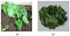
Two-month-old fresh (a) and vacuum freeze-dried (b) Mosidae (Adenophora remotiflora) leaves were donated by Eco-Sprout Co, Ltd. (Gyeonggido, Korea), an environmentally-friendly agricultural company (Fig. 1). The material was filtered through a 0.2 um micro filter and stored at 4℃ until use.
The proximal compositions of fresh Mosidae leaves (ML), moisture, protein, fat and ash (%), were determined by following AOAC [13] procedures.
Antioxidant activities
Total phenolic compound content
Total phenolic content of the MLE was measured using Folin-Ciocalteau reagent [14,15]. Folin-Ciocalteau reagent was diluted by 2 times using deionized water. The 2.0 mL of MLE solution (25 mg/mL) was mixed with diluted reagent (0.4 mL) and 2.0 mL of 2% sodium carbonate solution. Then mixed solution was held at room temperature for 30 min. After the absorbance of the solution was determined at 750 nm by an UV/visible spectrophotometer (UV-1601, Shimadzu, Tokyo, Japan). Standard curve was drawn using a tannic acid and calculated from it.
DPPH free radical scavenging activity
The free radical scavenging activities of MLE were evaluated according to the method described by Huang et al. [16], albeit with slight modification. Briefly, 0.2 mL of MLE was mixed with 1.0 mL of 0.2 mM DPPH ethanol solution. After 30 min at room temperature, absorbance values were measured by a UV-visible spectrophotometer set at 517 nm. The percent inhibition of DPPH radical by the sample extract was calculated by the following formula: DPPH free radical scavenging activity (%) = (1- As/Ab)×100, where Ab is the blank absorbance and As is the absorbance of the extract sample [16]. α-Tocopherol and BHT were used as positive standards.
Reducing power
The reducing power was determined according to the method of Oyaizu [14]. The 1.0 mL of sample solution was mixed with 0.5 mL of 50 mM sodium phosphate buffer (pH 6.5) and 0.7 mL of 1% potassium ferricyanide. The mixture was incubated at 50℃ for 20 min. After 1.0 mL of 10% trichloroacetic acid and 1.0 mL of distilled were added, the mixture was centrifuged at 3,000 rpm for 10 min. The 1.0 mL of upper layer was mixed with 0.1 mL of 0.1% of ferric chloride, and the absorbance was measured at 700 nm. The assays were carried out in triplicate and the results were expressed as mean values standard deviations. BHT and α-tocopherol were used as standards.
Hydroxyl radical scavenging effect
The effect of hydroxyl radical was assayed by using the 2-deoxyribose oxidation method [17]. The reaction mixture contained 1.0 mL of 0.1 M sodium phosphate buffer (pH 7.4), 0.20 mL of 10 mM 2-deoxyribose, 0.20 mL of 10 mM FeSO4-EDTA, 0.20 mL of 10 mM hydrogen peroxide and 0.20 mL of extract solution in a tube. After incubation at 37℃ for 1 hour, the reaction was stopped by adding 1.0 mL of 1.0% trichloroacetic acid and 1.0 mL of 1.0% of thiobarbituric acid. The mixture was boiled for 10 min, cooled in ice and then measured at 532 nm. Hydroxyl radical-scavenging effect was evaluated as the inhibition rate of 2-deoxyribose oxidation by hydroxyl radical.
Mice and cell cultures
Specific pathogen-free female BALB/c mice (6 weeks old) were purchased from NARA Biotech. (Seoul, Korea) and housed in a specific pathogen free environment. All experiments were conducted in accordance with the guidelines established by the Animal Care and Use Committee of Yuhan University (2009E-001). Water and dietary pellets were supplied ad libitum. Highly lung metastatic sublines of colon 26 carcinoma (colon26-M3.1) and murine B16-BL6 melanoma cells were maintained as monolayer cultures in Eagle's minimal essential medium (EMEM) supplemented with 7.5% fetal bovine serum (FBS), sodium pyruvate, nonessential amino acids and L-glutamine, all of which were purchased from Gibco BRL (Gibco: Carlsbad, CA, USA). Splenocytes from normal mice were cultured in RPMI-1640 (Gibco: Carlsbad, CA, USA) supplemented with 7.5% fetal bovine serum and glutamine [18].
Experimental lung metastasis
Experimental lung metastasis was induced via i.v. inoculation of colon26-M3.1 carcinoma cells (3×10⁴) into BALB/c mice [19]. To assess the anti-metastatic activity of MLE, groups of five mice each were i.v. treated with MLE (50 ug- 10 mg/kg mice) 2 days prior to tumor cell inoculation. The mice were sacrificed 14 days later and their lungs were fixed in Bouin's solution. Lung tumor cell colonies were counted under a microscope [18].
Cytotoxicity assay
For the cytotoxcity assay, various doses of MLE (0.1-500 ug/mL) in culture medium were added to each well of a 96-well plate containing either murine tumor cells (B16-BL6 cell and colon26-M3.1) (1×10⁵/well) or splenocytes (2×10⁵/well) from normal mice. After incubation for 72 hours, cytotoxcity was assayed by WST-1 based colorimetric assay (Wako Pure Chemicals Industrials, Ltd., Osaka, Japan) [19]. The absorbance of each well was monitored at 450 nm using a micro-plate reader (Molecular Devices, Sunnyvale, CA, USA).
Results
Proximate compositions
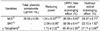
The proximate compositions of fresh ML are shown in Table 1. The fresh ML consisted of moisture (91.92%), protein (1.63%), fat (0.04%) and ash (0.6%).
Antioxidant activities
In this study, the antioxidant activity of MLE was evaluated by measuring reducing power, total phenolic compounds, DPPH radical scavenging, and hydroxyl radical scavenging. As shown in Table 2, the concentration of total phenolic compounds in MLE was 39 mg/100 mL. MLE evidenced a high reducing capacity (1.24), similar to that of α-Tocopherol (1.72). The DPPH scavenging activity of MLE was 88.89%, a value much lower than that of BHT (99.06%) and α-Tocopherol (95.45%). Nonetheless, we noted no significant differences between MLE and α-Tocopherol. The hydroxyl radical scavenging activity of MLE (22.10%) was significantly lower than that of BHT (29.37%), but did not differ significantly from that of α-Tocopherol (27.71%).
Inhibitory effect of MLE on lung metastasis
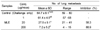
In order to determine whether MLE has any inhibitory effect on lung tumor metastasis, we assessed the effects of MLE on the experimental lung tumor metastasis of colon26-M3.1 cells. As shown in Table 3, prophylactic i.v. MLE treatment significantly inhibited lung metastasis in a dose-dependent manner. Specifically, a dose of 200 ug MLE per mouse strongly inhibited lung metastasis by 88.90%. Thus, MLE treatment within a certain range exerted no apparent side effects, such as a reduction in body weight (data not shown).
Cytotoxicity assay
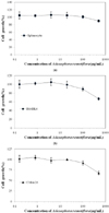
The cytotoxic effects of MLE on murine splenocyte and tumor cells (B16BL6 and colon26-M3.1) were examined in vitro as shown in Fig 2. MLE doses up to 100 ug/mL had no effect on the growth of normal murine splenocytes. Additionally, the survival of normal murine splenocytes was not shown to be influenced at MLE doses below 500 ug/mL. However, MLE doses up to 500 ug/mL resulted in decreased tumor cell growth in the B16BL6 (67% alive) and colon26-M3.1 (62% alive) cells. These results demonstrate that MLE had no effect on normal murine splenocytes, but demonstrated its cytotoxic effects against tumor cells.
Discussion
Mosidae (Adenophora remotiflora) has been traditionally used as an alternative medicine in Korea. However, there are currently no reports concerning its biological activity on the basis of the scientific evidence. Our study focused on the antioxidant activities of Mosidae leaf ethanol extract (MLE) by measuring reducing power, total phenolic compounds, DPPH radical scavenging, and hydroxyl radical scavenging. We also evaluated the anti-metastatic activity of MLE with regard specifically to its prophylactic lung metastasis inhibitory activity in colon26-M3.1 carcinoma cells. The cytotoxic effects of MLE were examined in an experimental metastasis model of murine splenocyte and tumor cells (B16BL6 and colon26-M3.1).
The proximate compositions of fresh ML are provided in Table 1. The moisture content of the fresh ML was 91.92%, the protein content of fresh ML was 1.63%, the fat content was 0.04%, and the ash content of fresh ML was 0.6%. An increasing amount of evidence suggests that the production of reactive oxygen species underlies a variety of disorders, and is also responsible for cellular damage [20]. The reducing capacity of cellular damage may be a significant indicator of any potential antioxidant activity [21]. Accordingly, MLE might contain a sizable amount of reductant, which may react with free radicals to stabilize and terminate free radical chain reactions. In this study, the reducing capacity of MLE was similar to that of vitamin E, but less profound than that of BHT. Phenolic compounds are thought to perform important functions as dietary antioxidants for the prevention of oxidative damage in living systems. Cho [22] reported that tannins extracted from fresh ML harbored high levels of total phenolic compounds as compared with spinach and aster, and moreover exhibited high DPPH radical scavenging activities. In this study, the total phenolic content of MLE was 39 mg/100 mL. The DPPH assay is known to provide reliable information concerning the antioxidant potential of a particular compound [23]. The results in Table 3 show that the DPPH radical scavenging activity of MLE is similar compared with α-Tocopherol and BHT. Kim et al. [10] did report the DPPH radical scavenging activity of fresh MLE (55.63%), which was lower than the value we reported (88.89%). However, such a difference could have been caused by the different cultivation conditions in our experiments. For instance, the samples utilized in this study were cultivated by the environmentally-friendly agricultural company, Eco-Sprout Co, Ltd.
The hydroxyl radical is considered a quick initiator of lipid peroxidation through the abstraction of hydrogen atoms from unsaturated fatty acids. Hence, the hydroxyl radical scavenging activity of MLE was evaluated and the results are shown in Table 2. In this study, the hydroxyl radical scavenging activity of MLE was 29.37%, a level similar to that of BHT and α-Tocopherol. Therefore, MLE appears likely to be an effective radical scavenger. Furthermore, the results of this study show that MLE is a good scavenger of DPPH and hydroxyl radical.
In an experimental tumor metastasis model, natural products commonly used in Oriental medicine were deemed useful in suppressing tumor growth and inhibiting tumor metastasis [18]. Unlike conventional chemotherapeutic agents, natural products have been shown to be relatively nontoxic to normal cells and to enhance host defense by stimulating immune-related cells [24]. In fact, many previous reports have demonstrated that some Oriental medicines inhibit the proliferation of macrophages, as well as metastasis in experimental tumor models [7,18,25]. In this study, prophylactic i.v. treatment with MLE inhibited the lung metastasis of colon26-M3.1 carcinoma cells in a dose-dependent manner. Specifically, a 200 ug dose of MLE per mouse strongly inhibited lung metastasis by 88.90%. Therefore, MLE treatment in this range exerted no apparent side effects, such as reduction in body weight or piloerection (data not shown).
Since the inception of the National Center for CAM, there has been an enormous increase in the number of basic science studies and therapy-based clinical trials designed to explore Complementary and Alternative Medicine (CAM). This subspecialty of immunology represents a particularly productive area with a large number of therapies shown to affect the immune system. Some recent studies have uncovered a biochemical mechanism that may potentially be involved in the immune modulatory pathways exploited by a variety of herbs [5,7,8]. Clinically, hundreds of trials have examined the effects of CAM on asthma, allergic rhinitis, and atopic dermatitis [26-28]. In our cytotoxicity assay, MLE did not affect the growth or survival of normal murine splenocytes. However, MLE doses up to 500 ug/mL reduced the percentage of tumor cell growth in B16BL6 (67% alive) and colon26-M3.1 (62% alive) cells. These results demonstrate that MLE has no effect on normal cell, but does demonstrate its cytotoxic effects against tumor cells.
Pine pollen extract showed strong antioxidant and anti-inflammatory activity [29], and the results of this study also demonstrated that MLE has strong antioxidant activity by measuring reducing power, DPPH radical scavenging, and hydroxyl radical scavenging. MLE may prove to be useful in preventing lung tumor metastasis, because it appears to exert no additional side effects. Therefore, MLE may be useful in the development of new plant materials as CAMs against diseases whose principal pathology is oxidative stress.

 XML Download