

 PDF
PDF ePub
ePub Citation
 Print
Print


Abbreviations
NAFLD
Non-alcoholic fatty liver disease
EAF
Ethanol extract of Allium fistulosum
IR
Insulin resistance
TG
Triglyceride
FFAs
free fatty acids
VLDL
very low density lipoproteins
SREBP1c
Sterol regulatory element-binding protein 1
FASN
fatty acid synthase
ACLY
ATP citrate lyase
WD
Western diet
FER
Food efficiency ratio
AST
Aspartate aminotransferase
ALT
alanine aminotransferase
ALP
alkaline phosphatase
INTRODUCTION
Non-alcoholic fatty liver disease (NAFLD) is one of the more prevalent metabolic disorders around the world and emerges as a simple form of steatosis before progressing to steatohepatitis and, ultimately, cirrhosis of the liver [123]. Chronic lipid accumulation is thought to be the underlying cause of NAFLD, due to its effects on dysregulated lipid metabolism in the liver. The disease is often associated with the most common clinical features of metabolic syndrome including obesity, dyslipidemia, hypertension, insulin resistance (IR), diabetes, and certain types of cancer [456]. Although NAFLD is a serious medical problem, its pathogenesis is not yet completely described. It is currently hypothesized that four pathogenic mechanisms are responsible for the accumulation of triglyceride (TG)-based lipid droplets, namely (i) an increased uptake of free fatty acid (FFAs) from high-fat foods by adipocytes in body fat, (ii) an increased synthesis of FFAs in the liver from glucose or acetate by IR, (iii) decreased mitochondrial β-oxidation of FFAs caused by a multitude of drugs, and (iv) decreased secretion of TGs in very low density lipoproteins (VLDL) by the liver [789].
Currently, there are no U. S. Food and Drug Administration (FDA)-approved pharmacologic agents or FDA guidelines for the treatment of NAFLD, despite it being the most common liver disease in the USA [10]. Because NAFLD is strongly associated with obesity, IR, and dyslipidemia, pharmacologic therapy directed at weight loss, IR, and/or dyslipidemia have been considered potential therapeutic approaches. However, due to the potentially hazardous side effects of anti-obesity drugs, including orlistat and sibutramine, a number of natural phytochemical compounds to treat NAFLD have been explored. Interestingly, epigenetic and environmental factors such as exercise and diet are known to interact in the definition of the NAFLD phenotype and to determine its progression [1112]. For this reason, lifestyle modifications, similar to those recommended for obesity, remain the currently recommended therapeutic option [13].
Allium fistulosum, a perennial herb in the genus Allium of the Alliaceae family, is used widely as an ingredient in Chinese, Japanese, and Korean cuisine [14]. In addition, A. fistulosum has been traditionally applied to treat common colds, headache, abdominal pain, and cardiovascular disease [15]. Previously, researchers have reported that A. fistulosum also exhibits antiplatelet, anti-oxidative, anti-hypertensive, and anti-hyperlipidemic effects [161718], and other members of the Allium family have been reported to have inhibitory activity against pathogenic bacteria, fungi, mycotoxins, and putrefactive bacteria [19]. A recent study described the anti-obesity effect of an A. fistulosum extract [14].
In the present study, by assessing its effect on various parameters relevant to NAFLD in vivo and in vitro, we investigated the potential of an ethanol extract of A. fistulosum (EAF) as a candidate compound for suppression of NAFLD development.
MATERIALS AND METHODS
Cell culture
Human hepatocellular carcinoma (HepG2) cells were purchased from the American Type Culture Collection (Mannassas, VA, USA) and cultured in a humidified atmosphere of 5% CO2 at 37℃ with high-glucose Dulbecco's modified Eagle's medium (DMEM) supplemented with 10% fetal bovine serum and antibiotics (Welgene, Daegu, Republic of Korea). Cells were incubated with 1% BSA low glucose DMEM (ND), 0.5 mM oleic acid in 1% BSA low glucose DMEM (OA), or with 0.5 mM oleic acid DMEM supplemented with 100 or 200 µg/mL EAF for 24 h.
Preparation of Allium fistulosum extract
The A. fistulosum was purchased from a local market (Sungnam-si, Republic of Korea) and identified by Prof. Sang-In Shim in the Department of Agronomy, Gyeongsang National University, Republic of Korea. A voucher specimen was deposited in the Korea Food Research Institute (KFRI). The A. fistulosum samples were cleaned and extracted in a 10-fold volume of 70% ethanol by shaking for 24 h at 25℃, and the precipitate was removed by centrifugation at 8,000 × g for 30 min. The supernatant was lyophilized in a freeze-drier (II Shin, Dongdochum-Si, Korea).
Cell toxicity
HepG2 cells (5 × 10⁴) were seeded in 24-well plates, and after reaching approximately 70% confluence, cells were treated in the presence or absence of OA, alone or in combination with EAF at 100 or 200 µg/mL. After incubation for 24 h, the cells were treated with 10 µL of WST-1 solution (Enzo Life Sciences, Farmingdale, NY, USA) for 3 h. Subsequently, 100 µL of supernatant was transferred to a 96-well plate, and absorbance was measured at 450 nm (Molecular Devices, Sunnyvale, CA, USA).
Oil red O staining
HepG2 cells (5 × 10⁴) were seeded in 24-well plates and, after reaching approximately 70% confluence, were treated in the presence or absence of OA, alone or in combination with EAF at 100 or 200 µg/mL. After incubation for 24 h, the cells were washed with 200 µL of PBS and fixed with 200 µL of 4% paraformaldehyde for 15 min at room temperature. The cells were then washed three times with PBS and incubated with 200 µL of 60% isopropanol for 5 min, followed by staining with 200 µL of 0.1% oil red O staining solution (Sigma-Aldrich, St. Louis, MO, USA) for 1 h. After additional washing with water (1 mL), images were captured under a light microscope (Olympus IX51; Olympus, Central Valley, PA, USA). For lipid quantification, isopropanol was added to each well to dissolve the lipid-stained red dye. After 10 min, the absorbance was measured at 510 nm (Molecular Devices).
Quantitative real-time PCR
HepG2 cells (5 × 10⁴) were seeded in 24-well plates and, after reaching approximately 70% confluence, were treated in the presence or absence of OA, alone or in combination with EAF at 100 or 200 µg/mL. After incubation for 18 h, total RNA was isolated by using TRIzol reagent (Invitrogen, Carlsbad, CA, USA). Real-time RT-PCR was performed with an I Cycler iQ (Bio-Rad, Hercules, CA, USA) using SYBR Green PCR master mix (Thermo Fisher Scientific, Waltham, MA, USA). PCR amplification was carried out with the following primers: for sterol regulatory element-binding protein 1 (SREBP-1c), forward 5′-AAACTCAAGCAGGAGAACCTAAGTCT-3′, reverse 5′-GTCAGTG TGTCCTCCACCTCAGT-3′; for ATP citrate lyase (ACLY), forward 5′-TACCACCTCAGCCATCCAGA-3′, reverse 5′-GACCCCAACGAGACCAAGTT-3′; for fatty acid synthase (FASN), forward 5′-AACCGGCTCTCCTTCTTCTTCGACTT-3′, reverse 5′-TCCGAGCGGCAGTACCCATTC-3′; and for glyceraldehyde 3-phosphate dehydrogenase (GAPDH) forward 5′-ATGTTCGTCATGGGTGTGAAC-3′, reverse 5′-GCATGGACTGTGGTCATGAGT-3′.
All mRNA expression was normalized by using GAPDH. Reactions were performed in triplicate, and relative expression levels and standard deviation (SD) values were calculated by applying the comparative method.
Animal experiments
Animal experiments were conducted according to the Guide for the Care and Use of Wonkwang University (WKU16-21). Twenty-one male C57/BL6 mice (7 weeks old) were divided into three groups of 7 and housed in a temperature- and humidity-controlled room with a 12 h light-dark cycle. After 1 week of acclimation, the groups of mice fed on a Western diet (high-fat high-sucrose diet; WD, n = 7) or a WD containing 1% EAF (wt/wt; WD+ 1% EAF, n = 7). Mice fed on a normal chow diet (ND, n = 7) in the absence of EAF were used as controls. The WD was purchased from Research Diets (#D12079B, Research Diets, New Brunswick, NJ, USA). Mouse body weight was measured at the beginning of the experiment and at 1-week intervals for 12 weeks. The extent of food consumption by each group was recorded every week for 12 weeks. At the end of the experiment, the mice were sacrificed to collect serum and tissue samples.
Hematoxylin and eosin staining
Mouse liver specimens were fixed in 4% buffered formalin, embedded in paraffin, and cut into 4–5 µm-thick sections. The sections were stained with hematoxylin and eosin (H&E). Liver morphology was examined and tissue images were captured with a microscope (Nikon ECLIPSE 80i; Nikon Instruments, Melville, NY, USA).
Measurement of aminotransferase enzymes
The levels of aspartate aminotransferase (AST), alanine aminotransferase (ALT), and alkaline phosphatase (ALP) in serum were measured by applying an enzymatic approach using a commercially available kit (Asan Pharm, Seoul, Republic of Korea).
Statistical analysis
The results from the in vitro experiment were analyzed by using one-way analysis of variance (one-way ANOVA). Data are expressed as mean ± standard deviation (SD) values. For the in vivo data, values are expressed as mean ± standard error (SE) values, and the results were analyzed by using one-way ANOVA with Newman Keul's post-hoc test. The statistical analyses were conducted by using SPSS software (Ver. 20; SPSS, Chicago, IL, USA). A value of P < 0.05 was considered to indicate statistical significance.
RESULTS
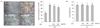
EAF reduces lipid accumulation in HepG2 cells
During screening of a natural substance library for anti-lipid accumulation, we observed that EAF reduced lipid accumulation in HepG2 cells. To evaluate whether EAF effectively blocks lipid accumulation, its anti-lipogenic properties were examined by Oil Red O staning (Fig.1A, left panel). Lipid accumulation was observed to be significantly increased in the OA-treated cells compared to that in the control group. Importantly, EAF treatment appeared to attenuate the OA-induced lipid accumulation in a dose-dependent manner. For quantitative analysis, Oil Red O dye was dissolved in isopropanol, and absorbance was measured at 510 nm (Fig. 1A, right panel). Colorimetric analysis indicated that OA treatment significantly increased lipid accumulation compared to that in the control group. In OA-treated cells, EAF attenuated lipid accumulation in a dose-dependent manner. We next examined whether the effect of EAF on the suppression of lipid accumulation occurred as a byproduct of cytotoxicity. HepG2 cells were incubated in the presence or absence of OA, alone or in combination with EAF. As shown in Fig. 1B, EAF did not affect the viability of HepG2 cells. Taken together, EFA efficiently blocked OA-induced lipid accumulation in HepG2 cells without inducing cytotoxicity.
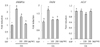
EAF inhibits transcriptional activity of SREBP-1c and FASN genes, but not the ACLY gene in HepG2 cells
To examine whether the reduction of lipid accumulation by EAF was related to the transcriptional regulation of lipogenesis-associated genes, we examined changes in the expressions of SREBP-1c, FASN, and ACLY by using qRT-PCR. It was observed that OA treatment significantly increased the mRNA expression levels of both the SREBP-1c and FASN genes, and both of which were dramatically decreased by EAF treatment in a dose-dependent manner (Fig. 2). Of particular note, the increase in transcriptional activity of SREBP-1c and FASN by OA was almost normalized to the control group following treatment of 200 µg/mL and 100 µg/mL EAF, respectively. By contrast, there were no differences in ACLY expression between the groups.
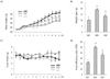
Dietary supplementation with 1% EAF significantly attenuates body weight in WD-fed mice without influencing food intake
To further examine whether EAF attenuates weight gain and fat accumulation in vivo, mice were fed with either ND or WD in the presence or absence of 1% EAF supplementation. At the 5-week mark, the WD group had a significantly higher average body weight than that in the ND group (P < 0.05), while 1% EAF supplementation with WD significantly blocked weight gain from 8 weeks after the beginning of supplementation (Fig. 3A). The total weight gain at 12 weeks in the WD group was approximately 20 g, an almost two-fold increase over that in the ND group (≒ 10 g). Relative to the WD group, significantly less weight gain was observed in the 1% EAF supplementation group fed the WD (Fig. 3B). Mice fed only on WD ate less than their ND counterparts, but there were no significant differences in daily food intake between the WD and 1% EAF supplementation groups and the WD group (Fig. 3C). The food efficiency ratio was higher in the WD group than in the ND group, which was significantly decreased after 12 weeks of EAF supplementation (Fig. 3D).
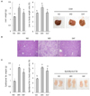
Dietary supplementation with 1% EAF reduces hepatic lipid accumulation, as well as both liver and epididymal fat weight elevated by WD in mice
The liver is the primary site of dietary fat metabolism and regulates fat levels in the blood. To investigate further whether liver weight was responsible for the increases in weight gain, we assessed liver weight and liver to body weight ratio between the groups. In comparison to the ND controls, the WD group had significantly higher liver weight and liver to body weight ratio at 12 weeks (P < 0.001), and the increases were relatively lower in the 1% EAF supplementation group (Fig. 4A, left panel). Hepatomegaly was observed in mice fed on WD when compared to ND-fed mice. Liver weights were significantly lower in the 1% EAF supplementation group; although there were no visual differences in liver size between the WD and EAF supplementation groups (Fig. 4A, right panel). To examine whether the reduction in liver weight was attributable to decreased hepatic lipid accumulation, liver samples after 12 weeks of supplementation were fixed, sectioned, and stained. Histological staining (H&E stain) revealed that the WD group exhibited greater lipid accumulation than that in the ND group, and the WD-induced fat accumulation was markedly attenuated by 1% EAF supplementation (Fig. 4B). Moreover, EAF supplementation significantly reduced the WD-mediated increases in epididymal fat weight (Fig. 4C, left panel) and effectively blocked hypertrophy of epididymal fat; similar to the effects observed in the ND controls (Fig. 4C, right panel). However, there was no statistically significant difference in epididymal fat to body weight ratios between the WD and 1% EAF-fed groups, indicating the increase or decrease of epididymal fat weight induced by either WD or EAF supplementation was associated with the changes in body weight. Altogether, the results indicate that EAF supplementation reduced both liver weight and size, which were increased by WD, thereby preventing hepatic lipid accumulation in vivo.
Dietary supplementation with 1% EAF blocks increases in plasma indicators for liver abnormalities
Plasma ALT levels rise dramatically with acute liver-specific damage, and the plasma level of AST is used as an indicator of hepatic and extrahepatic tissue damage. The mice in the WD group had significantly higher plasma levels of both ALT (P < 0.000) and AST (P < 0.016) than those in the ND group (Fig. 5A, B). The group fed 1% EAF with WD had a lower ALT level (P < 0.002) than that in the WD group (Fig. 5A). The plasma levels of AST were also lower in the mice fed the 1% EAF supplement, but there was no statistical significance to the difference (P < 0.066, Fig. 5B). However, as shown in Fig. 5C, we observed that 1% EAF supplementation normalized the AST:ALT ratio (P < 0.000), which was dramatically decreased in the WD-fed group (P < 0.004). An increased ALP level is another indication of liver abnormalities, including obesity. The increased plasma levels of ALP induced by WD were also significantly reduced by 1% EAF supplementation (Fig. 5D).
DISCUSSION
NAFLD is the most common liver disease and is a hepatic manifestation of obesity and metabolic syndrome [20]. Due to serious side effects associated with drugs used to treat the symptoms of NAFLD, dietary intervention using natural phytochemicals has gained interest as a potentially important concept in therapeutic intervention and public health. During ongoing library screening to identify natural phytochemicals for the treatment of NAFLD, we observed that EAF attenuates OA-induced lipid accumulation without cytotoxicity (Fig. 1). To validate whether EAF could reduce the development of NAFLD, we investigated OA-induced NAFLD in HepG2 cells in vitro [2122] and an WD-induced NAFLD model in vivo. Anti-lipogenic mechanisms in the liver are generally associated with the expression of lipogenic enzymes, cholesterol biosynthesis, TG biosynthesis, and fatty acid β-oxidation in HepG2 cells [23]. In the present study, we first demonstrated that EAF significantly prevents NAFLD by reducing the transcriptional activities of both SREBP-1c and FASN genes, which are representative lipogenesis-related genes in the liver (Fig. 2). Lipid accumulation in the liver is known to be the result of enhanced de novo lipogenesis, the activation of lipid uptake, and the lowering of its catabolism. These mechanisms are associated with the expression of SREBP-1c, which is the transcription factor responsible for fatty acid and triglyceride synthesis in the liver [24252627]. FASN encodes a key enzyme critical for de novo fatty acid and TG synthesis and catalyzes the final step in fatty acid biosynthesis; thus, FASN is believed to be a major determinant of the maximal hepatic capacity to generate fatty acids by de novo lipogenesis. The effect of A. fistulosum on the transcriptional activity of lipogenesis-related genes has yet to be investigated. However, there is a study showing the anti-inflammatory effect of A. fistulosum through inhibition of tumor necrosis factor-a and attenuation of excessive nitric oxide and prostaglandin generation [282930], which are considered as pro-inflammatory mediators, induces inflammation [3132]. It has also been reported that hyper inflammation is a major factor contributing to the development of NAFLD [3334]. Lipogenic targets such as PPAR-γ, SREBP-1c, and FASN were overexpressed in the liver of the patients with abnormally increased inflammation from hepatitis C virus infection with hepatic steatosis [35]. Based on the results in previous studies, the antiinflammatory property of A. fistulosum is probably a strong predictable mechanism that mediates prevention of NAFLD by reducing the transcriptional activity of lipogenesis-related genes in the liver. However, further studies are required to elucidate the exact molecular mechanisms involved.
Classic Western diets are high in both saturated fat and sugar, and the WD was originally developed as a model for NAFLD progression in mice [363738]. It also appears to model obese humans with mild non-alcoholic steatohepatitis, as recently reported in a thorough analysis of liver pathophysiology phenotypes [39]. On that basis, the WD consumption model has been used in studies related to chronic conditions associated with obesity. In this study, we adopted the WD-fed mouse model to examine whether EAF could prevent the development of NAFLD. The EAF supplementation group showed a significant decrease in body weight gain, liver to body weight ratio, and epididymal white fat to body weight ratio compared to the WD group, and the EAF had no cytotoxic effect (Fig. 5); moreover, there was no difference in daily food intake among the groups (Fig. 3, 4). Hepatic lipid accumulation in the WD group was higher than that in the ND group (Fig. 4B). The data for the WD-fed mice are consistent with the previously reported results for a phenotype associated with HFD-induced obesity [4041]. EAF significantly suppressed increases in various risk factors, including gain in body weight, liver to body weight ratio, and the degree of hepatic lipid accumulation, all of which are known to be involved in the progression of NAFLD (Fig. 3, 4).
There have been many reports on the hypolipidemic effects of Allium species. Yamamoto (2010) reported that welsh onion attenuates hyperlipidemia in rats fed on a high-fat high-sucrose diet [18]. Also, extracts of garlic, which is another Allium crop, have also been reported to lower plasma lipids in rats fed on a diet with or without cholesterol [424344]. However, there were no significant changes in lipid profiles in our data (data not shown). These results are consistent with those reported by Aoyama et al. [32], thus supporting our observation that the anti-lipidemic effect of A. fistulosum in liver was caused by attenuating the transcriptional activity of lipogenic-related genes, not through its hypolipidemic property.
Safe and effective extract dosing is necessary, regardless of the purpose of the supplementation. In animal experiments using extract administration, conversion methods based on the common perception of scaling of dose based on the body weight (mg/kg) have been used [45]. However, the animal experiments in our study adopted the method of food supplementation through diet; thus, it was not reasonable to apply dose scaling based on body weight. Therefore, we simply used a method that calculated the amount of food intake. The EAF supplemented diet contained 1% EAF, and the average of daily food intake of the study mice was 2.9 g over the 12-week experimental period, indicating that the average mouse ate 29 mg of EAF through the diet. Based on 24-hour dietary recall data obtained from 7,042 subjects in the 2016 Korea National Health and Nutrition Examination Survey, on average, Korean subjects ate 9,828 mg of A. fistulosum (data not shown). Considering the body weights of both mouse and human, we used a relatively high dosage of EAF in this study. Fortunately, there was no hepatic toxicity detected (Fig. 5).
Taken together, our results demonstrate that an ethanol extract of A. fistulosum attenuates the development of NAFLD, with EAF eliciting anti-lipogenic activity in liver. Therefore, EAF is a promising candidate for the development of novel therapeutic drugs or drug combinations for the prevention and treatment of NAFLD. Although functioning of the representative phytochemicals within A. fistulosum has been shown in hepatic lipogenesis at the phenotype level, there are no previous reports showing its anti-lipogenic effect via attenuation of lipogenic-related genes in the liver, even if it was limited in vitro. Regardless, further studies should be conducted to elucidate the exact molecular mechanism involved in the capacity of A. fistulosum to regulate lipogenesis in liver. In addition, to accurately determine the amount of Allium fistulosum required to protect against the development of NAFLD, an in-depth study is needed.

 XML Download