

 PDF
PDF ePub
ePub Citation
 Print
Print


INTRODUCTION
Diabetes mellitus is a chronic metabolic disease affecting 4% of the population worldwide [1]. The disease is divided into two main forms, insulin-dependent diabetes mellitus (IDDM) and non-insulin-dependent diabetes mellitus (NIDDM), and approximately 90% of patients are NIDDM with increased concentrations of glucose in the blood [2]. Epidemiological studies and clinical trials strongly support the notion that hyperglycemia is the principal cause of diabetic complications affecting the eyes, kidneys, nerves, and arteries [3]. Thus, effective blood glucose control is the key to preventing or reversing diabetic complications and improving quality of life in NIDDM patients.
α-Glucosidases are a group of key intestinal enzymes involved in the digestion of carbohydrates [4]. Therapeutic measures for the treatment of NIDDM include the use of α-glycosidase inhibitors, such as acarbose, miglitol, and voglibose, to delay the absorption of carbohydrates from the small intestine and thus lower postprandial blood glucose [5]. However, these drugs have certain gastrointestinal adverse effects, which may limit long-term compliance to therapy [6]. Currently, there is growing interest in plant foods due to their less frequent adverse effects compared with therapeutic agents for the treatment of diabetes mellitus [78].
Jerusalem artichoke (Helianthus tuberosus Linne) is native to North America and is in the daisy family; its tubers are available in the produce section of many grocery stores worldwide, including Korea, and are a commercial source of fructan [9]. Several researchers have reported that Jerusalem artichoke decreases fasting serum glucose levels, and this effect may be related to its fructan [101112]. Generally, fructan with a degree of polymerization from 2 to greater than 60 is labeled inulin, which is the major form in the Jerusalem artichoke. Fructooligosaccharides, which are produced by the partial enzymatic hydrolysis of inulin, are defined by a degree of polymerization of less than 10 [13].
In this study, the α-glucosidase inhibitory effects of purple Jerusalem artichoke (PJA) hydrolyzed by Lactobacillus plantarum, Bacillus subtilis, and Bacillus subtilis (produced by S&D Co.Ltd, Chuncheon, Korea) were investigated and compared in vitro. Furthermore, we studied the effect of hydrolyzed L. plantarum-fermented PJA (LJA) on blood glucose and α-glucosidase activity in a type 2 diabetic animal model (C57BL/ksJ, db/db mouse).
MATERIALS AND METHODS
Materials
α-Glucosidase, acarbose, aniline, diphenylamine, phosphoric acid, sulfuric acid, bovine serum albumin, sodium azide, p-nitrophenyl-α-d-glucopyranoside, sodium chloride, potassium chloride, edetic acid, sucrose, maltose, and lactose were purchased from Sigma-Aldrich Chemical Co. (St. Louis, MO, USA). Methanol was purchased from Avantor Performance Materials, Inc. (Center Valley, PA, USA). All other chemicals and solvents, unless otherwise specified, were guaranteed reagent grade and purchased from Sigma-Aldrich Chemical Co. (St, Louis, MO, USA).
Preparation of extract samples and fermented samples
The PJA was purchased from a local market in Chuncheon, Korea. The specimens were authenticated by Emeritus Prof. H.J. Chi, Seoul National University, Korea. The dried PJA powder (5 g) was extracted by water (500 ml) with sonication at 45℃ for 1 h. The PJA extract was fermented by L. plantarum, B. subtilis, and B. subtilis (produced by S&D Co.Ltd, Chuncheon, Korea) at 37℃ for 36 h. After centrifuging the samples at 5,741 × g for 15 min, the supernatants were freeze dried.
Measurement of fructan concentration
The total fructan concentration of LJA was measured according to the AOAC method 999.03 using the Megazyme Fructan Assay Kit (Megazyme International Ireland Ltd., Wicklow, Ireland). Fructan flour was used as a control. The fructan concentration was calculated as follows: where ΔA is the difference in absorbance between the sample and the blank, V is the extract volume, W is the weight of the extracted sample, F is the conversion factor from absorbance to the weight of fructose.
α-Glucosidase assay
The α-glucosidase assay was performed as previously described, with slight modifications [14]. Briefly, distilled water (500 µl), sample solution (50 µl, 50 mg/ml in distilled water), and p-nitrophenyl-α-d-glucopyranoside solution (150 µl, 2 mM in 0.4 mg/ml phosphate-buffered saline (PBS) containing 0.4 mg/ml bovine serum albumin and 0.04 mg/ml NaN3, pH 7) were mixed in a tube. Then, 300 µl of enzyme solution (0.5 units/ml in 0.4 mg/ml PBS buffer containing 0.4 mg/ml bovine serum albumin and 0.04 mg/ml NaN3, pH 7) was added to each mixture, and absorbance was measured at 405 nm using a spectrophotometer (Secoman, Alès, France). The inhibitory activity was calculated as follows: where A1 is the absorbance of the test samples with the enzyme, A2 is the absorbance of the test samples without the enzyme, A3 is the absorbance of the solvent with the enzyme, and A4 is the absorbance of the solvent without the enzyme.
Animals
Six-week-old C57BL/KsJ db/db mice and C57BL/KsJ db/+ mice were purchased from Central Lab, Animal Inc. (Seoul, Korea). All animals were acclimatized to the laboratory environment for 1 week before the experiment. Mice were allowed free access to drinking water and food under a constant room temperature (22 ± 1℃) and humidity (60 ± 5%) and an automatic 12-h light/12-h dark cycle. The mice were cared for and treated in accordance with the guidelines of the Committee on Care and Use of Laboratory Animal Resources, National Research Council, USA. The mice were randomly divided into 4 groups: lean mice were db/+ mice fed the standard diet only as a control; con were db/db mice fed the standard diet as diabetic control; LJA were db/db mice fed the standard diet with LJA (1.5 g/kg of diet); AB were db/db mice fed the standard diet with acarbose (0.5 g/kg of diet) as a positive control. During the 7-week experiment, food intake, body weight, and fasting blood glucose were measured once every week. All animal experiment procedures were conducted in accordance with the guidelines and approval of the Institutional Animal Care and Use Committees (IACUC) of Hallym University (Hallym-2012-37).
Oral glucose tolerance test
The mice were fasted for 10 h prior to the experiment, and then the soluble starch (1 g/kg of body weight) was orally administered with or without additives. LJA 200 was given LJA 200 mg/kg of body weight; LJA 400 was given LJA 400 mg/kg of body weight; AB 60 was given acarbose 60 mg/kg of body weight. The blood was withdrawn from the tail vein at 0 min, 30 min, and 120 min after glucose administration. Blood glucose levels were determined by the glucose oxidase method.
Blood biomarkers
Blood samples were drawn from an orbit vein. Serum was separated immediately by centrifugation (1,722 × g at 4℃ for 15 min) and stored at -70℃. Serum concentrations of triglycerides, total cholesterol, and non-esterified fatty acids were determined using Blood Chemistry Kornelab 20XT (Thermo, Vantaa, Finland). Serum HDL-cholesterol levels were measured using a commercial kit (Asan Pharmaceutical Corp., Seoul, Korea). Blood glycated hemoglobin (HbA1c) was measured on an HbA1c Analyzer (HLCr-723GHb G7, Tosoh, Aarhus, Denmark). Serum insulin and adiponectin were analyzed using a mouse insulin ELISA (enzyme-linked immunosorbent assay) kit and a mouse/rat high-molecular-weight adiponectin ELISA kit (both from Shibayagi Co. Ltd., Shibukawa, Japan).
Measurement of intestinal α-glycosidase activity
The small intestine was removed and washed with ice-cold saline. The intestine was cut into three equal segments (proximal, middle, and distal). Each section was homogenized with buffer (0.5 M NaCl, 0.5 M KCl, 5 mM EDTA, pH 7.0) and centrifuged at 8,037 × g for 30 min. The pellet was washed by saline and centrifuged again. The pellet was homogenized in saline and centrifuged at 200 × g for 10 min and the supernatant was used as the enzyme solution. The intestinal glycosidase was measured. Briefly, the enzyme solution (100 µl), disaccharides (100 µl, 2 mM maltose, 40 mM lactose, or 10 mM sucrose), and phosphate PBS (200 µl, 0.1 M, pH 7.0) were incubated at 37℃ for 30 min. Then, the protein was denatured by heat at 100℃ for 5 min and removed by centrifugation. The glucose concentrations were determined by Trinder's method [15] and protein concentrations by the Lowry method [16].
RESULTS
Fermentation of purple Jerusalem artichoke extract
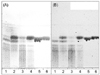
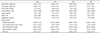
The PJA extract was fermented by L. plantarum, B. subtilis, and B. subtilis (produced by S&D Co.Ltd, Chuncheon, Korea). The composition, yield, and fructan concentration of fermented and original samples were analyzed and the results are shown in Fig. 1 and Table 1. The yields of fermented and original samples were similar, but the composition of PJA changed after fermentation, which resulted in the formation of glucose and fructose. Moreover, the compositions of PJA fermented by different microbes were different. L. plantarum and B. subtilis (produced by S&D Co.Ltd, Chuncheon, Korea) had higher PJA fermentation activities than B. subtilis owing to their lower fructan concentrations.
Evaluation of fermented PJA α-glucosidase inhibitory activity in vitro
The α-glucosidase inhibitory activities of fermented and original PJA were compared in vitro. As shown in Table 1, the α-glucosidase inhibitory activities of original PJA, LJA, B. subtilis-fermented PJA, and B. subtilis (produced by S&D Co.Ltd, Chuncheon, Korea)-fermented PJA were 12.01%, 49.34%, 12.45%, and 21.18%, respectively. LJA showed the best inhibitory effect on α-glucosidase and was used for further in vivo study.
Anti-diabetes effect of LJA in vivo
Effect of LJA on oral glucose tolerance
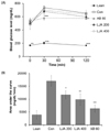
Oral glucose tolerance tests were performed to determine the effect of a single oral dose of LJA on glucose tolerance using db/db mice. The results are summarized in Fig. 2. Compared with the lean group, the glucose level of the control group was substantially higher. However, the LJA-treated groups had suppressed blood glucose levels at 30 and 120 min after starch load (Fig. 2A). When the area under the curve was compared between groups, the LJA 200 and LJA 400 groups showed 31.34% and 42.25% reductions, respectively, compared to the control group (Fig. 2B).
Effect of LJA on blood glucose levels
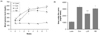
We studied the effects on blood glucose after the administration of LJA at 1, 3, 5, and 7 weeks in db/db mice. The results showed the presence of a high blood glucose level in the control group, but lower levels in the LJA-treated group (Fig. 3A). However, their differences were not significant, except at first week, and the decline in blood glucose levels reached a maximum after 1 week. When the AUC was compared between groups, the LJA groups showed 30.46% reduction than the control group (Fig. 3B).
Effect of LJA on body weight, food intake, and FER
We studied the effects in body weight, food intake, and feed efficiency ratio (FER) after LJA administration for 7 weeds in db/db mice. The differences in body weight and food intake between the mice treated with LJA and controls were not significant (Table 2). Changes in body weight and FER did not differ between the groups.
Effect of LJA on intestinal glycosidase activity
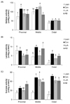
The activities of intestinal glycosidases (sucrose, maltase, and lactase) in the small intestine after LJA administration are shown in Fig. 4. LJA showed no significant inhibitory effect on maltase activity. However, it inhibited sucrose activity in the distal segment of the intestine. Moreover, lactase activity in the proximal segment was inhibited significantly by treatment with a low dose of LJA. Small intestinal glycosidase activity can be partially inhibited by treatment with LJA in db/db mice.
Effect of LJA on serum metabolic parameters
To determine the mechanism underlying the effect of LJA, we studied the effect of LJA on serum metabolic parameters, including insulin concentration, HbA1c percentage, adiponectin concentration, and serum lipid concentration (Table 2). The serum insulin levels in the LJA-treated mice were increased significantly, by 1.3-fold, compared to the control group. Furthermore, the HbA1c levels were significantly lower (10.32% lower) in LJA-treated mice than control mice. However, adiponectin concentrations did not differ between mice treated with LJA and the control group.
Triglyceride levels were 65.27% lower in the LJA group than the control group. The non-esterified fatty acid concentration of LJA-treated mice was also significantly lower than that of the control group. The total cholesterol level in the LJA-treated mice showed no significant differences, but the HDL-cholesterol levels in the LJA group were significantly higher (by 38.89%) than those in the control group. Finally, a significant increase (13.52%) in HDL-total cholesterol ratio (HTR) in the LJA group was observed compared with the control group.
DISCUSSION
Sustained reductions in hyperglycemia are associated with a decreased risk of developing complications in NIDDM patients [17]. However, it is difficult to reverse the dietary and lifestyle trends of NIDDM patients. Thus, the dietary supplements are used for regulation of blood glucose and the α-glucosidase inhibitors are used as a dietary intervention.
It has been reported that the tubers of Jerusalem artichoke contained sugars, fructans, coumarins and lectins as its main components [181920]. Jerusalem artichoke showed α-glucosidase inhibitory activity without adverse effects and may increase insulin sensitivity, which may be related to its inulin [1321]. Also, Jerusalem artichoke decrease fasting serum glucose levels perhaps due to it is rich in fructan and coumarins [1322]. Jerusalem artichoke had anti-diabetic effects due to high in fructoligossacharides that may decrease insulin resistance by mechanisms that remain unknown [23]. Therefore, the degree of polymerization of fructan is considered as a factor that influences hyperglycemia amelioration and α-glucosidase inhibitory activity.
The aim of this study was to investigate the α-glucosidase inhibitory effect and hyperglycemia amelioration of fermented PJA for NIDDM treatment. Therefore, the α-glucosidase inhibitory activity of the PJA extract and its fermented products were compared in vitro. Our results demonstrated that the inhibitory effect of PJA against α-glucosidase depends not only on the degree of polymerization of fructan, but also on the change in its composition by fermentation with different microbes. LJA showed the highest α-glucosidase inhibition activity and was used to investigate its effects on hyperglycemia using NIDDM animal models.
LJA does not influence body weight, food intake, or FER. However, we observed that LJA significantly reduces blood glucose and HbA1c levels in db/db mice, indicating that there is a significantly higher rate of glucose disposal after LJA treatment and the effect of LJA on glucose metabolism can last for a long period. There was also a significant increase in the levels of serum insulin after LJA administration to db/db mice. Accordingly, LJA may exert hypoglycemic action in diabetic mice by potentiating the effect of insulin in serum or by increasing either the pancreatic secretion of insulin from existing beta cells or its release from the bound form. Our OGTT data in db/db mice indicated that, after LJA treatment, glucose disposal increased significantly, suggesting an improvement in glucose tolerance via LJA, presumably due to an increase in insulin sensitivity.
The main features of insulin resistance include dyslipidemia, which is characterized by high triglycerides, high non-esterified fatty acids, low high-density lipoproteins, and low adiponectin in the blood. Even though there were no changes in adiponectin after LJA treatment, there were significant decreases in triglycerides and non-esterified fatty in the LJA-treated groups. Moreover, although a slight increase in total cholesterol was observed after LJA treatment, HDL-cholesterol and HTR increased significantly. The inclusion of fructan in the diet of saturated fat-fed rats significantly reduced the triglyceride content in the blood and liver. Delzenne and Kok [23] suggested that the triacylglycerol-lowering effect of oligofructose occurs via a reduction in low-density lipoprotein-triacylglycerol secretion from the liver as a result of the reduction in the activity of lipogenic enzymes, and in the case of fatty acid synthase, via modification of lipogenic gene expression.
Small intestinal α-glucosidase activity was found to be partially inhibited by LJA. In particular, LJA significantly inhibited sucrose in the distal segment and lactase in the whole intestine. For this reason, carbohydrate digestion can be delayed. Therefore, another possible mechanism underlying the anti-hyperglycemic effect of LJA may be the decreased activity of small intestinal α-glucosidase, which converts carbohydrates into glucose, preventing the rapid increase in postprandial blood glucose levels and slow absorption of carbohydrates in the small intestine.
We observed that the inhibitory effect of PJA on α-glucosidase activity could be improved dramatically by L. plantarum fermentation. LJA has a significant anti-hyperglycemia effect in db/db mice by increasing the level of insulin, decreasing insulin resistance, and delaying the absorption of carbohydrates. LJA has potential as a food supplement for the treatment of NIDDM.

 XML Download