

 PDF
PDF ePub
ePub Citation
 Print
Print


INTRODUCTION
Glioma is acknowledged as an incurable brain tumor, which has high propensity for invasion and cell migration [12]. Cancer migration occurs in response to stimuli such as growth factors and cytokines [34]. In particular, stromal cell-derived growth factor 1 (SDF-1) controls the enhanced migration and proliferation of glioma [5], and its receptor, chemokine receptor type 4 (CXCR4), is highly expressed in various cancer cells including melanoma, colon cancer, breast cancer, ovarian cancer, and glioma [56789]. Interaction between CXCR4 and SDF-1 is crucial to tumor growth and metastasis [1011].
SDF-1 and CXCR4 propagate a down-stream signal, activating phospholipase C (PLC) followed by activation of protein kinase C (PKC), which plays a key role in malignant cancer progression. PKC activation is associated with various cellular functions, including proliferation, adhesion, malignant transformation, and migration [1213]. On the other hand, extracellular signal-regulated kinase (ERK), a down-stream target of SDF-1/CXCR4, is also involved in cell migration and proliferation [14]. Cancer motility is promoted by the activated matrix metalloproteinases (MMPs) under the influence of phosphorylated kinases.
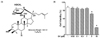
Panax ginseng C.A. Meyer has been in use as an oriental herbal medicine for more than 2000 years [15]. The major functional component, saponin, otherwise known as ginsenoside, was reported to exhibit pharmacological properties including antiinflammation, anti-diabetic, and anti-tumor [161718]. While more than 30 ginseng saponins have been identified thus far, their chemical structure renders their absorption difficult, when taken orally [1920]. CK, 20-O-β-(d-glucopyranosyl)-20(S)-protopanaxadiol (Fig. 1A) is a metabolite of ginseng saponin, produced by intestinal bacteria after oral administration of ginseng. It was reported to have therapeutic potential in cancer therapy [21222324]. Hence, the purpose of this work was to determine the anti-migration property of CK and define the mechanism of its action through the suppression of wound-healing in SDF-1-stimulated C6 glioma cells. However, the effect of CK on SDF-1-induced migration of glioma cells remains unclear. Our study is the first to report evidence for CK-mediated regulation of C6 glioma cell migration through down-regulation of CXCR4 via the PKC/ERK-mediated pathway.
MATERIALS AND METHODS
Chemicals
CK was purchased from BioMechatronic (Seoul, Korea). Cell culture materials and EZ-cytox cell viability assay kit were purchased from Gibco BRL (Gaithersburg, MD, USA) and Daeil Lab service (Seoul, Korea), respectively. SDF-1 was obtained from PeproTech Inc. (Rocky Hill, NJ, USA). Diff Quick stain was purchased from Sigma (St. Luis, MO, USA). PKCα and phospho-PKCα antibodies were obtained from Cell Signaling Technology Inc. (Beverly, MA, USA), ERK1/2 and phospho-ERK1/2 antibodies were obtained from Santa Cruz (Santa Cruz, MA, USA). All other chemicals were of analytical grade.
Cell culture and cell viability assay
Rat C6 glioma cells (Korean Cell Line Bank, Seoul, Korea) were cultured in DMEM medium (containing 10% fetal bovine serum (FBS) and 1% penicillin-streptomycin) at 37℃, humidified with 5% atmospheric CO2. Cell viability analyses were performed as previously reported [25]. Briefly, C6 glioma cells (5 × 10³ cells/well) were seeded and incubated in a 96-well micro-plate for 24 h. The cells were replaced with serum-free medium and incubated for the next 24 h. Subsequently, the cells were treated with different concentrations of CK (0.03 µM to 10 µM dissolved in 0.05% DMSO and diluted in serum-free medium), followed by re-incubation for another 24 h. Cell viability was measured using an EZ-cytox cell viability assay kit. Absorbance was determined using ELISA at a wavelength of 450 nm. Each assay was performed in triplicate.
Scratch wound healing assay
Scratch wound healing analyses were performed as previously reported [25]. Briefly, C6 glioma cells were plated in 6-well plates (2 × 10⁵ cells) and incubated in medium containing 10% FBS. After the cells were grown to 70% confluence, the medium was replaced with serum-free medium and incubated for 24 h. A wound scratch was made on the monolayer of cultured cells across the center of each well using a 200 µL pipette tip. The cells were washed twice with phosphate-buffered saline for removal of non-adherent cells and subsequently treated with or without CK or SDF-1 (100 ng/ml) for 24 h. An image of the cells that had migrated into the cell-free wound scratch area was observed under an inverted microscope (Nikon, Tokyo, Japan) and photographed. The migration distance was measured using Image J software.
Boyden chamber assay
Boyden chamber assay was performed using a 48-well chemotaxis chamber (Neuro-Probe, Gaithersburg, MD, USA) to confirm the inhibitory effect of CK on migration of C6 glioma cells. Scratch wound healing analyses were performed as previously reported [25]. Briefly, C6 glioma cells were suspended in serum-free medium at a density of 1 × 10⁶ cells/ml, and 50 µl of the cell suspension was loaded in the upper chamber wells. Treatment with SDF-1 with or without CK at different concentrations was performed in the lower chamber with incubation at 37℃ and 5% CO2 atmosphere for 90 min. Cells on the lower surface of the membrane were fixed and stained using Diff Quick solutions. After staining, the migrated cells were photographed and counted under a microscope (Nikon, Tokyo, Japan). The migrated cells were measured using Image J software.
Western blot analysis
Protein levels were analyzed by western blotting as previously reported [25]. Briefly, the proteins (20 µg) were loaded, separated, and transferred to polyvinylidene difluoride membranes in transfer buffer. After blocking overnight at 4℃ in 5% skim milk (dissolved in Tris-buffered saline buffer), the membrane was treated with Tris-buffered saline containing 0.1% Tween 20 (TBS-T) and washed three times. The membrane was incubated with phospho-ERK1/2 and phospho-PKCα, ERK1/2 and PKCα antibodies at 1:1000 dilutions. The membranes were washed with TBS-T, followed by incubation with a 1:1000 dilution of secondary antibody conjugated to horseradish peroxidase. The proteins were analyzed by a chemiluminescent reaction (ECL plus kit, Amersham Pharmacia Biotech, Buckinghamshire, UK); the bands were detected using Hyperfilm ECL (Amersham Pharmacia Biotech, Buckinghamshire, UK). Image J Software was used for visualization and quantification of proteins.
Gelatin-zymography
Gelatin-zymography analyses were performed as previously reported [26]. Briefly, the supernatant medium obtained from SDF-1 with or without CK treated C6 cells was re-suspended in a sample buffer and loaded without boiling in 10% polyacrylamide gel containing 0.25% (w/v) gelatin. The supernatants were loaded and separated on running-gel. After electrophoresis, the gel was soaked in 2.5% Triton X-100 at room temperature and then washed in ddH2O. The gel was incubated at 37℃ for 24 h and placed in a tank containing incubation buffer (50 mM Tris HCl, 0.15 M NaCl, and 10 mM CaCl2, pH 7.8).To determine activity of bands, clear bands were visualized using destaining solution.
Statistical analysis
The results were expressed as the mean ± standard error (SE) from at least three independent experiments. Statistical significance was analyzed using Student's t-test for each paired experiment using GraphPad Prism 4.0 software (version 4.00 for Windows, San Diego, CA, USA) with significance defined at 0.05 (P < 0.05).
RESULTS
CK is non-cytotoxic to C6 Glioma cells at a minimal dose
Cytotoxicity of CK in C6 glioma cells was determined by quantitative assessment of cell viability using the EZ-cytox cell viability assay kit. As shown in Fig. 1B, CK had no cytotoxic effect until the dose of 1 µM. However, CK exhibited cytotoxicity at higher concentrations (3 µM and 10µM) (Fig. 1B).
CK reduced scratch wound-healing in SDF-1-stimulated C6 cells
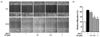
The scratch wound-healing assay was performed to assess the inhibitory effect of CK on SDF-1-induced migration of C6 glioma cells. Scratched C6 glioma cells were incubated in CK (0.1 µM to 1 µM) in serum-free medium for 1 h, followed by stimulation with SDF-1 (100 ng/ml) for the next 24 h. As shown in Fig. 2, compared to control condition, SDF-1 (SDF-1 alone) induced cell migration to approximately 215%. SDF-1-induced migration of C6 glioma cells was inhibited by 0.3 µM and 1 µM CK to 76% and 88% of SDF-1 alone control (Fig. 2).
CK inhibits migration of SDF-1-stimulated C6 cells
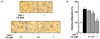
Next, the inhibitory effect of CK on SDF-1-induced migration of C6 cells was tested using the Boyden chamber assay. As shown in Fig. 3, SDF-1 significantly induced the cell migration to 226.3% compared to the untreated group. In contrast, CK treatment attenuated the migration of SDF-1-stimulated C6 cells at a minimum dosage of 0.3 µM and the maximum inhibitory effect (95.3% of SDF-1 alone control) was shown at 1 µM. The results also indicated that CK inhibits SDF-1-stimulated migration of C6 cells without inducing cytotoxicity.
CK down-regulated the phosphorylation of PKCα and ERK
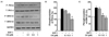
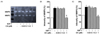
The downstream-signaling pathway of SDF-1 and CXCR4 activates PKCα and ERK1/2 in various cells [1314]. Western blot with phospho-specific antibodies of PKCα and ERK1/2 was performed to determine inhibitory effects of CK. C6 glioma cells were treated with SDF-1 (100 ng/ml) and CK (0.1 µM to 1 µM). As shown in Fig. 4, SDF-1 increased PKCα phosphorylation (194.0%) and ERK1/2 phosphorylation (163.96%) compared to the control group. CK (1 µM) demonstrated the maximal inhibitory effect on phosphorylation of PKCα (94.67% of SDF-1 alone control) and ERK1/2 (94.67% of SDF-1 alone control) (Fig. 4).
DISCUSSION
In the current study, we examined the anti-migratory effect of CK. Scratch wound-healing assay, Boyden chamber assay, western blot, and gelatin-zymography assay were performed in SDF-1-stimulated C6 glioma cells. Because gliomas have a tendency of rapid migration and invasion of the surrounding tissues, surgery and radio-irradiation methods are not viable options [2728]. The severity of cancer is often aggravated by metastasis [2]. Therefore, targeting cancer migration is an anti-cancer strategy for glioma [25]. Our study demonstrated that SDF-1-induced migration of glioma was suppressed by CK.
In oriental medicine, ginseng, derived from the root of P. ginseng Meyer, was used for centuries to cure both acute and malignant diseases. Ginsenoside, commonly known as saponin, is the major active component of this herbal medicine [1920]. Although several reports have demonstrated that ginseng has a therapeutic effect on cancer, oral administration of this herb renders it difficult to absorb by the body [20]. Therefore, development of functional food is an alternative pharmacological approach [25]. Alternatively, a particular ginsenoside metabolite of ginseng, compound K (CK), was reported to be easily absorbed and metabolized by the human body [29]. The metabolite of saponin produced during fermentation might be used in development of functional foods. Hence, based on its inhibitory effect of glioma migration, we suggest CK as a potential candidate for use in development of functional foods.
Numerous studies have demonstrated the interaction of chemokine SDF-1 to its receptor CXCR4 [30]. SDF-1 acts as the critical signaling factor, activating various cellular responses in tumor cells including angiogenesis, invasion, and migration [313233]. Rapid transmission of these cellular signals in the brain is a key trait of glioma, potentiating malignant progression of brain tumor [12627]. CK was reported to suppress cell growth in several types of cancer cells [23]. However, its pharmacological activity in the receptor signaling pathway is still unclear. In the current study, CK (1 µM) strongly inhibited SDF-1-induced C6 glioma migration in wound healing assay and Boyden chamber assay. Our data demonstrate that CK is an effective inhibitor of C6 glioma cell migration and has the potential for treatment of early stage or prevention of malignant progression of glioma.
Cancer cell migration and proliferation were mediated by activation of SDF-1/CXCR4 downstream signaling molecules, PKC and mitogen-activated protein kinase (MAPK) [33]. Stimulation with SDF-1 results in activation of PLC and formation of diacylglycerol (DAG); these pathways are known to activate PKC signaling [31]. Seven different isotypes of PKC, α, β1, β2, γ, δ, ɛ, ζ, η, θ, λ, and µ are present in cells and were implicated in diverse cellular functions [34]. Among members of the PKC superfamily, phosphorylation of PKCα regulates robust cell migration and malignant progression of glioma [35]. CXCR4 and SDF-1 exhibit a signal transduction mechanism, not only via PKCα phosphorylation but also via ERK1/2 phosphorylation [35]. ERK activity was reported to regulate migration and proliferation of diverse types of cancer cells, including glioma [3236]. Our results indicate that 1 µM CK inhibits glioma cell migration through reduced phosphorylation of PKCα and ERK1/2. These results suggest that CK acts as a CXCR4 antagonist and inhibits SDF-1 induced C6 glioma cell migration by inhibiting CXCR4 receptor-mediated activation of PKCα and ERK1/2.
Increased expression of extracellular endopeptidases such as MMPs was observed in various cancers [25]. MMP 2 and 9 are involved in cell migration and proliferation, particularly in cancer [37]. Interaction between chemokine and MMPs induces the development of cancer. Thus, the design of this study involved down-regulation of both cell migration and MMPs as a therapeutic strategy for glioma. Our results indicate that CK (1 µM) significantly inhibited the expression of MMP2 and MMP9 in SDF-1-stimulated C6 glioma cells.
In conclusion, CK regulates SDF-1-stimulated C6 glioma migration. CK (1 µM) inhibited the migration of SDF-1-stimulated C6 glioma cells as demonstrated by the scratch wound-healing assay and Boyden chamber assay. CK treatment resulted in down-regulated phosphorylation of PCKα and ERK1/2, and expression of MMP2 and MMP9 in SDF-1-stimulated C6 glioma cells. These data suggest that CK inhibits SDF-1-stimulated C6 glioma cell migration by down-regulating the activation of PKCα and ERK1/2. The crucial observation of our study is that CK can alter the downstream signaling of the CXCR4/CXCR12 pathway in C6 glioma cells. Further studies are required to determine the CK-derived mechanism to the blood brain barrier as their structure means transcellular diffusion will be limited. The cellular mechanisms of CK derived to brain via blood brain barrier remain to be elucidated.
 XML Download