

 PDF
PDF ePub
ePub Citation
 Print
Print


INTRODUCTION
Nevus sebaceous is a congenital tumor, usually located on the scalp or face, and presents as an alopecic patch or a slightly elevated yellowish plaque1-3. Various tumors can arise from the nevus sebaceous, including trichoblastoma4. Trichoblastoma is primarily located on the head and neck area and usually presents as a solitary, non-ulcerated, skin-colored papule or nodule5. Histologically, trichoblastoma is usually classified into several forms according to its cellular arrangement6. Cases of non-pigmented trichoblastoma from the nevus sebaceous have been reported in Korea7,8, but not for pigmented trichoblastoma. We herein report a case of pigmented trichoblastoma presenting as a nodule on the forehead.
CASE REPORT
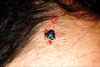
A 28-year-old healthy man presented with a dome-shaped, dark-pigmented nodule within a yellowish hairless plaque on the right side of forehead. The plaque had existed since birth. The central, pigmented nodule began to appear three years ago and enlarged gradually. The patient had no personal or family history of cutaneous or internal malignancies. Physical examination of the forehead revealed a 2.0×1.5 cm yellowish, verrucous, hairless plaque. A distinct 0.6×0.5 cm dark pigmented nodule appeared within the yellowish plaque (Fig. 1). A biopsy specimen was taken from the central pigmented nodule.
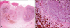
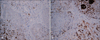
Histopathologic findings of the central pigmented lesion showed a well-circumscribed tumor island located in the dermis without connection to the overlying epidermis. Tumor cells were arranged in nodular nests of palisading basaloid cells affecting the dermis and fibrocellular stroma around the basaloid island, but showed lack of high-grade cytologic atypia and no clefting artifact between the basaloid tumor islands and stroma. The lesion showed heavy melanin deposits within and around the tumor nests (Fig. 2). None of tumor cells were immunolabeled with bcl-2 (Fig. 3A), and stromal cells around the tumor nests were weakly positive for CD10 (Fig. 3B). The patient was referred to the plastic surgery department, where he received a total excision. The excised lesion around the pigmented tumor revealed typical histopathologic findings of nevus sebaceous. Therefore, the pathologic diagnosis was pigmented trichoblastoma arising from the nevus sebaceous.
DISCUSSION
Nevus sebaceous commonly appears as a yellowish patch or plaque at birth or in early childhood. Several benign and malignant tumors can develop from nevus sebaceous1-3, with trichoblastoma and syringocystadenoma papilliferum4 the most common benign forms. Trichoblastoma, occurring primarily or secondarily as dermal neoplasm, appear as a solitary, small, non-ulcerated nodule on the scalp or face5. Trichoblastoma was described by Headington in his reviews of hair follicle tumors, including trichoblastoma, trichoblasitic fibroma, trichogenic trichoblastoma, and trichogenic myxoma9. However, Ackerman's classification is widely accepted now, with trichoblastoma subdivided into five histopathologic patterns: large nodular (including pigmented), small nodular, cribriform, racemiform, and retiform6. Thereafter, several less common forms such as adamantinoid, columnar, and rippled-pattern, subcutaneous, and superficial forms were added as variants10-13. Pigmented variants of trichoblastoma are very rare and only a few cases have been reported in the literature14-17. The non-pigmented form of trichoblastoma has been reported in Korea, but not the pigmented form.
The essential histologic findings of trichoblastoma are basaloid proliferation in which the tumor cells are arranged in cords, sheets, or discrete clusters surrounded by fibrous stroma6. Pigmented trichoblastoma contains melanin depositions but no significant melanocyte hyperplasia18. Nodular basal cell carcinoma (BCC) and trichoepithelioma must be differentiated histologically. The present case revealed a fibrocellular stroma around the basaloid tumor cells rather than myxoid stroma and stromal retraction or clefting around basaloid islands, two characteristic histologic features of BCC. This case did not show well-formed horn cysts, a characteristic feature of trichoepithelioma. Here, we report the first Korean case of pigmented trichoblastoma arising from the nevus sebaceous.
 XML Download