

 PDF
PDF ePub
ePub Citation
 Print
Print


Abstract
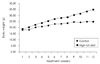
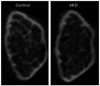
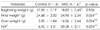
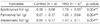
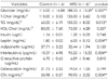
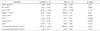
Obesity not only reduces bone mineral density but also increases inflammatory markers. Therefore, we examined the change in inflammatory markers and morphological microstructure of the bones using a mouse model fed a high-fat diet. C57BL/6J 4-week-old male mice were divided into a control group (n = 6) and a experimental group (n = 6); the control group was provided with 10% Kcal fat diet, and the high-fat diet group was provided with 45% Kcal fat diet for 12 weeks using the free provision method. Blood was analyzed for inflammatory markers, and micro-computed tomography was used to measure the morphological microstructure of the femoral bone. The weight increases in the control group and high-fat diet group were 5.85 ± 1.84 g and 16.06 ± 5.64 g, respectively (p < 0.01), glucose was 115.00 ± 16.88 mg/dL and 188.33 ± 13.29 mg/dL (p < 0.01), and triglycerides were 65.00 ± 6.19 mg/dL and 103.33 ± 8.02 mg/dL (p < 0.05) respectively. Leptin and interleukin (IL)-6 were significantly higher in the high-fat diet group than that in the control group (p < 0.01). As a result of a biochemical index analysis of bone metabolism, osteocalcin tended to be lower in the high-fat diet group, whereas CTx was significantly higher in the high-fat diet group compared to that in the control group (p < 0.01). The thickness of the bony trabecula was significantly narrower in the high-fat diet group than that in the control group (p < 0.05), and the gap in the bony trabecula was significantly wider in the high-fat diet group than that in the control group (p < 0.05). IL-6 and the gap in the bone trabecula, which was a morphological microstructure of the bones, showed a positive correlation (p < 0.05). Taken together, inducing obesity through a high-fat diet in mice during the growth phase caused a change in bone microstructure and was correlated with the inflammation index. Accordingly, restriction of excessive fat intake may be needed to suppress the inflammatory reactions and promote normal bone formation.
Figures and Tables

References
1. Ministry of Health and Welfare. Korea national health ans nutrition examination survey report (KNHANES IV). 2009.
2. Kim SH, Kim JY, Ryu KA, Sohn CM. Evaluation of the dietary diversity and nutrient intakes in obese adults. Korean J Community Nutr. 2007. 12(5):583–591.
3. Recker RR, Heaney RP. Peak bone mineral density in young women. JAMA. 1993. 270(24):2926–2927.

4. Goulding A, Jones IE, Taylor RW, Manning PJ, Williams SM. More broken bones: a 4-year double cohort study of young girls with and without distal forearm fractures. J Bone Miner Res. 2000. 15(10):2011–2018.
5. Oh YK, Sohn C. Comparative study on nutrients intake, physical activities and bone mineral density of specialized game high school students according to obesity level. Korean J Community Nutr. 2010. 15(3):393–402.
6. El Hage R, Moussa E, Jacob C. Bone mineral content and density in obese, overweight, and normal-weighted sedentary adolescent girls. J Adolesc Health. 2010. 47(6):591–595.
7. Ducher G, Bass SL, Naughton GA, Eser P, Telford RD, Daly RM. Overweight children have a greater proportion of fat mass relative to muscle mass in the upper limbs than in the lower limbs: implications for bone strength at the distal forearm. Am J Clin Nutr. 2009. 90(4):1104–1111.
8. Leonard MB, Shults J, Wilson BA, Tershakovec AM, Zemel BS. Obesity during childhood and adolescence augments bone mass and bone dimensions. Am J Clin Nutr. 2004. 80(2):514–523.
9. Ahima RS, Flier JS. Adipose tissue as an endocrine organ. Trends Endocrinol Metab. 2000. 11(8):327–332.
10. Berner HS, Lyngstadaas SP, Spahr A, Monjo M, Thommesen L, Drevon CA, Syversen U, Reseland JE. Adiponectin and its receptors are expressed in bone-forming cells. Bone. 2004. 35(4):842–849.
11. Zoico E, Zamboni M, Di Francesco V, Mazzali G, Fantin F, De Pergola G, Zivelonghi A, Adami S, Bosello O. Relation between adiponectin and bone mineral density in elderly post-menopausal women: role of body composition, leptin, insulin resistance, and dehydroepiandrosterone sulfate. J Endocrinol Invest. 2008. 31(4):297–302.
12. Ouchi N, Kihara S, Funahashi T, Nakamura T, Nishida M, Kumada M, Okamoto Y, Ohashi K, Nagaretani H, Kishida K, Nishizawa H, Maeda N, Kobayashi H, Hiraoka H, Matsuzawa Y. Reciprocal association of C-reactive protein with adiponectin in blood stream and adipose tissue. Circulation. 2003. 107(5):671–674.
13. Kim M, Kim H, Sohn C. Relationship between vitamin K status, bone mineral density, and hs-CRP in young Korean women. Nutr Res Pract. 2010. 4(6):507–514.
14. Lee Y, Kim M, Choi K, Kim J, Bae W, Kim S, Sohn C. Relationship between inflammation biomarkers, antioxidant vitamins, and bone mineral density in patients with metabolic syndrome. Nutr Res Pract. 2011. 5(2):150–156.
15. Yasojima K, Schwab C, McGeer EG, McGeer PL. Generation of C-reactive protein and complement components in atherosclerotic plaques. Am J Pathol. 2001. 158(3):1039–1051.
16. Manolagas SC, Jilka RL. Bone marrow, cytokines, and bone remodeling. Emerging insights into the pathophysiology of osteoporosis. N Engl J Med. 1995. 332(5):305–311.
17. Russell M, Mendes N, Miller KK, Rosen CJ, Lee H, Klibanski A, Misra M. Visceral fat is a negative predictor of bone density measures in obese adolescent girls. J Clin Endocrinol Metab. 2010. 95(3):1247–1255.
18. Ciarelli MJ, Goldstein SA, Kuhn JL, Cody DD, Brown MB. Evaluation of orthogonal mechanical properties and density of human trabecular bone from the major metaphyseal regions with materials testing and computed tomography. J Orthop Res. 1991. 9(5):674–682.
19. Kumasaka S, Kawamata R, Okada T, Miyake M, Kashima I. Relationship between bone mineral density and bone stiffness in bone fracture. Oral Radiol. 2005. 21(1):38–40.
20. Thomsen JS, Ebbesen EN, Mosekilde L. Relationships between static histomorphometry and bone strength measurements in human iliac crest bone biopsies. Bone. 1998. 22(2):153–163.
21. Lee CJ, Lee W, Lee BD. Morphometric analysis of bone in the ovariectomized rat using in vivo micro-CT. Korean J Oral Maxillofac Radiol. 2008. 38(1):29–37.
22. Song YH, Lee W, Lee CJ, Ji JH, Lee BD. Study of bony trabecular characteristics using bone morphometry and micro-CT. Korean J Oral Maxillofac Radiol. 2007. 37(1):27–33.
23. Kim A, Park T. Diet-induced obesity regulates the galanin-mediated signaling cascade in the adipose tissue of mice. Mol Nutr Food Res. 2010. 54(9):1361–1370.
24. Patsch JM, Kiefer FW, Varga P, Pail P, Rauner M, Stupphann D, Resch H, Moser D, Zysset PK, Stulnig TM, Pietschmann P. Increased bone resorption and impaired bone microarchitecture in short-term and extended high-fat diet-induced obesity. Metabolism. 2011. 60(2):243–249.
25. Shih CC, Lin CH, Lin WL. Effects of Momordica charantia on insulin resistance and visceral obesity in mice on high-fat diet. Diabetes Res Clin Pract. 2008. 81(2):134–143.
26. Lee JH, Son CW, Kim MY, Kim MH, Kim HR, Kwak ES, Kim S, Kim MR. Red beet (Beta vulgaris L.) leaf supplementation improves antioxidant status in C57BL/6J mice fed high fat high cholesterol diet. Nutr Res Pract. 2009. 3(2):114–121.
27. Kim MA, Jeong YS, Chun GT, Cha YS. Antihyperlipidemic and glycemic control effects of mycelia of inonotus obliquus including protein-bound polysaccharides extract in C57BL/6J mice. J Korean Soc Food Sci Nutr. 2009. 38(6):667–673.
28. Bugianesi E, McCullough AJ, Marchesini G. Insulin resistance: a metabolic pathway to chronic liver disease. Hepatology. 2005. 42(5):987–1000.
29. Díez JJ, Iglesias P. The role of the novel adipocyte-derived hormone adiponectin in human disease. Eur J Endocrinol. 2003. 148(3):293–300.
30. Asayama K, Hayashibe H, Dobashi K, Uchida N, Nakane T, Kodera K, Shirahata A, Taniyama M. Decrease in serum adiponectin level due to obesity and visceral fat accumulation in children. Obes Res. 2003. 11(9):1072–1079.
31. Bacha F, Saad R, Gungor N, Arslanian SA. Adiponectin in youth: relationship to visceral adiposity, insulin sensitivity, and beta-cell function. Diabetes Care. 2004. 27(2):547–552.
32. Fasshauer M, Paschke R, Stumvoll M. Adiponectin, obesity, and cardiovascular disease. Biochimie. 2004. 86(11):779–784.
33. Yamamoto Y, Hirose H, Saito I, Tomita M, Taniyama M, Matsubara K, Okazaki Y, Ishii T, Nishikai K, Saruta T. Correlation of the adipocyte-derived protein adiponectin with insulin resistance index and serum high-density lipoprotein-cholesterol, independent of body mass index, in the Japanese population. Clin Sci (Lond). 2002. 103(2):137–142.
34. Weiss R, Dufour S, Groszmann A, Petersen K, Dziura J, Taksali SE, Shulman G, Caprio S. Low adiponectin levels in adolescent obesity: a marker of increased intramyocellular lipid accumulation. J Clin Endocrinol Metab. 2003. 88(5):2014–2018.
35. Feingold KR, Grunfeld C. Role of cytokines in inducing hyperlipidemia. Diabetes. 1992. 41:Suppl 2. 97–101.
36. Trayhurn P, Wood IS. Adipokines: inflammation and the pleiotropic role of white adipose tissue. Br J Nutr. 2004. 92(3):347–355.
37. Ahn KY, Choi YS, Lee DW, Kim KY, Kim JH, Sung HN, Choi KM, Kim SM, Lee CB. Relationship between C-reactive protein and obesity in children. Korean J Obes. 2009. 18(4):131–137.
38. Bistrian BR, Khaodhiar L. Chronic systemic inflammation in overweight and obese adults. JAMA. 2000. 283(17):2235–2236.
39. Rosen HN, Moses AC, Garber J, Iloputaife ID, Ross DS, Lee SL, Greenspan SL. Serum CTX: a new marker of bone resorption that shows treatment effect more often than other markers because of low coefficient of variability and large changes with bisphosphonate therapy. Calcif Tissue Int. 2000. 66(2):100–103.
40. Brown JP, Delmas PD, Malaval L, Edouard C, Chapuy MC, Meunier PJ. Serum bone Gla-protein: a specific marker for bone formation in postmenopausal osteoporosis. Lancet. 1984. 1(8386):1091–1093.
41. Ducy P, Desbois C, Boyce B, Pinero G, Story B, Dunstan C, Smith E, Bonadio J, Goldstein S, Gundberg C, Bradley A, Karsenty G. Increased bone formation in osteocalcin-deficient mice. Nature. 1996. 382(6590):448–452.
42. Kim HS. Biochemical marker of bone metabolism. J Korean Soc Pediatr Endocrinol. 2006. 11(2):131–137.
43. Ma H, Torvinen S, Silvennoinen M, Rinnankoski-Tuikka R, Kainulainen H, Morko J, Peng Z, Kujala UM, Rahkila P, Suominen H. Effects of diet-induced obesity and voluntary wheel running on bone properties in young male C57BL/6J mice. Calcif Tissue Int. 2010. 86(5):411–419.
44. Hsu YH, Venners SA, Terwedow HA, Feng Y, Niu T, Li Z, Laird N, Brain JD, Cummings SR, Bouxsein ML, Rosen CJ, Xu X. Relation of body composition, fat mass, and serum lipids to osteoporotic fractures and bone mineral density in Chinese men and women. Am J Clin Nutr. 2006. 83(1):146–154.
45. Park SJ, Ahn Y, Min HS, Oh KS, Park C, Cho NH, Kimm K. Osteoporosis prevalence of radius and tibia and related factors using multiple bone sites quantitative ultrasound measurement of the Korean health and genome study cohort women. Korean J Community Nutr. 2005. 10(4):536–545.
46. Parhami F, Tintut Y, Beamer WG, Gharavi N, Goodman W, Demer LL. Atherogenic high-fat diet reduces bone mineralization in mice. J Bone Miner Res. 2001. 16(1):182–188.
47. Cao JJ, Gregoire BR, Gao H. High-fat diet decreases cancellous bone mass but has no effect on cortical bone mass in the tibia in mice. Bone. 2009. 44(6):1097–1104.
48. Ali AA, Weinstein RS, Stewart SA, Parfitt AM, Manolagas SC, Jilka RL. Rosiglitazone causes bone loss in mice by suppressing osteoblast differentiation and bone formation. Endocrinology. 2005. 146(3):1226–1235.
49. Lorentzon M, Lorentzon R, Nordström P. Interleukin-6 gene polymorphism is related to bone mineral density during and after puberty in healthy white males: a cross-sectional and longitudinal study. J Bone Miner Res. 2000. 15(10):1944–1949.
 XML Download