

 PDF
PDF ePub
ePub Citation
 Print
Print


REFERENCES
1.Tashiro K., Kikuchi S., Itoyama Y., Tokumaru Y., Sobue G., Akiguchi I, et al. Nationwide survey of juvenile muscular atrophy of distal upper extremity (Hirayama disease) in Japan. Amyotroph Lateral Scler. 2006. 7:38–45.

2.Zhou B., Chen L., Fan D., Zhou D. Clinical features of Hirayama disease in mainland China. Amyotroph Lateral Scler. 2010. 11:133–139.
3.Sonwalkar HA., Shah RS., Khan FK., Gupta AK., Bodhey NK., Vottath S, et al. Imaging features in Hirayama disease. Neurol India. 2008. 56:22–26.
4.Hassan KM., Sahni H., Jha A. Clinical and radiological profile of hirayama disease: a flexion myelopathy due to tight cervical dural canal amenable to collar therapy. Ann Indian Acad Neurol. 2012. 15:106–112.
5.Yin B., Liu L., Geng DY. Features of Hirayama disease on fully flexed position cervical MRI. J Int Med Res. 2011. 39:222–228.
6.Finsterer J., Loscher W., Wanschitz J., Baumann M., Quasthoff S., Grisold W. Hirayama disease in Austria. Joint Bone Spine. 2013. 80:503–507.
7.Wang XN., Cui LY., Liu MS., Guan YZ., Li BH., DU H. A Clinical neurophysiology study of Hirayama disease. Chin Med J (Engl). 2012. 125:1115–1120.
Go to : 
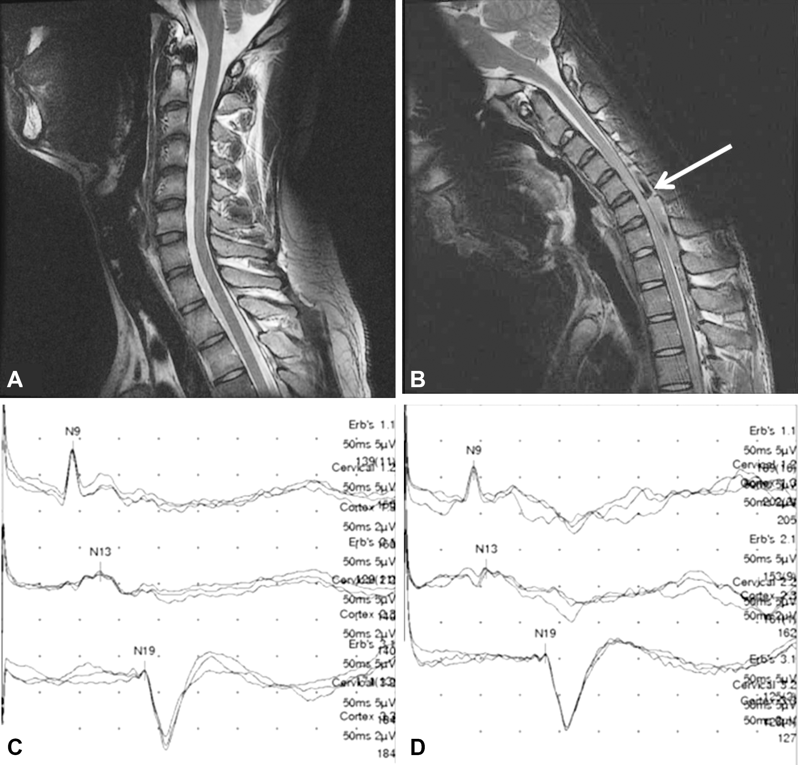 | Fig. 1.Cervical magnetic resonance imaging (MRI) and right median somatosensory evoked potentials (SEP) of the patient. The T2 weighted MRI with neck in neutral position was unremarkable while T2 weighted MRI with neck in flexion shows anterior displacement of the posterior dural sac (A, B). The reversible inter-peak latency between N13 and N19 on supine (5.5 ms) (C) and flexed (7.45 ms) (D) somatosensory evoked potentials (SEP) in the symptomatic side also support the diagnosis of Hirayama disease. (Normal SEP interpeak latency < 6.59 ms). |
 XML Download