In a distal-extension removable partial denture (RPD), there is a discrepancy in displacement between the tooth and the mucosa covering the edentulous area on application of an occlusal load. This causes the RPD to rotate around the fulcrum line and may harm the surrounding oral tissues if stress distribution is not well distributed over the teeth and the edentulous area.1, 2 To avoid these harmful effects, first, the abutment teeth should be strong enough to transfer the load to the periodontal ligament apparatus. Second, the denture base should be properly extended for stress distribution and functional form, i.e., maximal base extension without overextension (snowshoe effect) should be achieved, and a functional impression of the edentulous ridge should be made. Especially, in case of long span Kennedy Class I edentulous ridge, functional edentulous form is more important for denture support and stability. Therefore, functional impression techniques have been used to record the functional form of the edentulous ridge. McLean's overimpression technique, the altered cast impression technique, and Applegate's fluid wax impression technique are examples of functional impression techniques.3, 4, 5, 6
Among them, McLean's overimpression technique employs a custom tray for the edentulous area. When functional impression is completed using the appropriate impression material, this impression is repositioned in the mouth. Then, an alginate overimpression is made while applying pressure to the previously completed functional impression, resulting in the functional form of the edentulous area and the anatomic form of the remaining teeth.7
The overextension of denture border in Class I and II RPDs displace the denture away from the tissues by causing it to rotate around the primary fulcrum line during movement of the surrounding oral structures such as the tongue, frenum, and cheeks. This results in harmful lateral stress on the abutment teeth and continuous patient discomfort.7 Therefore, this is a good impression method which not overextends the border and can record functional form of edentulous ridge in distal-extension base RPD. This overextension can be attributed to the viscous nature, thermal plasticity, and short working time of modeling plastic.
Another issue to be solved during impression making for RPDs is accurate seating of individual trays in the oral cavity, despite the preparation of tray stops on the remaining teeth. The purpose of this case report is to introduce an alternative technique for functional impression with maximal denture base extension without overextension and proper seating of tray.
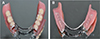
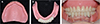
A 67-year-old female presented with the desire to have a new maxillary complete denture (CD) and a mandibular class I RPD. The patient exhibited severe alveolar bone resorption in the mandibular molar region. There was a crown fracture on the right mandibular second premolar, crown detachment on right mandibular first premolar and fracture of denture teeth on the left maxillary central incisor, lateral incisor, canine and left mandibular second premolar (Fig. 1). Interim prostheses (maxillary CD and mandibular Class I RPD) and provisional fixed restorations were fabricated in advance and delivered on the same day of the mandibular crown removal, therefore, the patient could use the dentures immediately (Fig. 2).
|
|

|
Fig. 1
Initial photo. (A) Right, (B) Frontal, (C) Left. Severe alveolar bone resorption in the mandibular molar region was observed. Extracoronal attachment was used on the left mandibular canine.
|
|
 Click for larger image Click for larger image |
|
|
|
|
Fig. 2
Interim denture. (A) Maxillary interim denture (B) Provisional fixed restorations and mandibular interim denture (C) Interim denture delivery. Provisional fixed restorations and interim dentures were fabricated.
|
|
Click for larger image |
|
All sore spots were eliminated, and the overextended borders were adjusted to relieve patient discomfort. Once the patient was comfortable with her provisional dentures after adjustments in follow-up visits, a functional impression was made using the mandibular provisional denture with a thin-mix pink colored irreversible hydrocolloid (SELECTION-J, Youdent Corporation, Chiba, Japan) (Fig. 3A). An overimpression was made with orange colored irreversible hydrocolloid (ALGINoplast, Heraeus, Hanau, Germany) while maintaining the provisional denture with the functional impression surface in its intended position. This was similar to McLean's physiologic impression method, with the difference being that the method was used for individual tray fabrication, not for final RPD impression, in this case (Fig. 3B). The cast was then fabricated using Type IV dental stone (Hi-Koseton, Maruishi Gypsum Corporation, Osaka, Japan), multiple layers of baseplate wax (Modeling wax, Kim's International Inc., Seoul, Korea) were adapted on the abutment teeth to maintain space for abutments impression or the pick-up impression of surveyed crowns, and minimal block-out was performed on edentulous undercut areas. Tin-foil was applied to easily remove the individual tray from wax (Fig. 3C).
|
|

|
Fig. 3
Functional impression. (A) Functional impression using provisional denture with irreversible hydrocolloid (pink color), (B) Over-impression with irreversible hydrocolloid (orange color). (C) Minimal relief was done on distal extension base undercut area and sufficient space was prepared for impression material on abutment teeth with baseplate wax, which covered tinfoil.
|
|
Click for larger image |
|
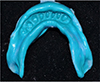
The individual tray (Ostron 100, GC Corporation, Tokyo, Japan) was fabricated on the cast to the full length of the border and trimmed with a denture bur to make the tray border 0.5 mm shorter for impression material. The borders were smoothened (Fig. 4). This tray was used for two procedures (impression for abutments and RPD impression). First, an impression for the abutment teeth prepared to receive metal-ceramic surveyed crowns (Fig. 5) and surveyed metal-ceramic crowns were fabricated in harmony with the maxillary wax denture (Fig. 6). Subsequently, a pick-up impression for the RPD was made without modeling compound only using polyvinyl siloxane impression material (Examixfine regular type, GC Corporation, Tokyo, Japan). Thus, a functional impression of the ridge with well-formed borders was obtained (Fig. 7) and master cast was fabricated (Fig. 8).
|
|

|
Fig. 4
Individual tray. (A) Right, (B) Occlusal, (C) Left. Border is extended approximately 0.5 mm shorter than full length of cast border.
|
|
Click for larger image |
|
|
|
|
Fig. 5
Impression of the mandibular abutments. This impression covers retromolar pad which is useful for occlusal plane determination.
|
|
Click for larger image |
|
|
|

|
Fig. 6
Maxillary wax denture and mandibular surveyed crowns. (A) Left, (B) Frontal, (C) Right. Mandibular abutment teeth were fabricated with reference to retromolar pad and maxillary wax denture.
|
|
Click for larger image |
|
The mandibular metal framework was fabricated (lingual bar and RPI (rest, proximal plate, I-bar) system), followed by adjustment, bite registration (vertical dimension and centric relation), mandibular wax denture fabrication, and denture curing (Fig. 9). Clinical remounting was done and bilateral balanced occlusion was acquired. The maxillary CD and mandibular class I RPD were delivered to the patient (Fig. 10).
It is generally believed that border molding with modeling compound is a prerequisite for achieving retention and stability of conventional RPDs. However, obtaining precisely extended border is very time-consuming, particularly in patients with severe alveolar bone loss. In the technique described here, modeling compound was not used and a final impression using an individual tray with borders similar to those of the final denture was obtained. Accurate replication of details in the individual tray is accomplished by provisional denture relining using irreversible hydrocolloid, which also provides a functional impression similar to that obtained using Mclean's overimpression method. The difference between our method and Mclean's method is that the latter uses a custom tray for functional impression and makes an overimpression with irreversible hydrocolloid for RPD master cast. On the other hand, our technique applied irreversible hydrocolloid (pink color) on interim dentures to take functional impression and then made an overimpression with irreversible hydrocolloid (orange color) for individual tray fabrication. This individual tray can be used for making impressions of abutment teeth prepared to receive surveyed crowns and final impressions of functional RPDs without modeling compound. This method is not technique-sensitive in terms of providing a perfect border, using only polyvinyl siloxane impression materials for the final impression. Consequently, this method decreases the chair time by eliminating the border molding procedure with modeling compound and accelerating the impression-making procedure without overextension.
With regard to the technique of maximal base extension without overextension in implant overdenture impressions, Lee et al.8 reported the use of a tissue-conditioned provisional denture for fabricating an individual tray, which was the used for making a pick-up impression without modeling compound border molding. An overextended border is often experienced because of improper heating and tempering of modeling compound by clinician, and patient's insufficient movement of oral structure. But Tan et al.9 found that the borders molded using modeling plastic tended to be thicker and shorter than those molded using polyether material.
In the above method, functional impression was taken by using interim denture as a “custom tray”, therefore, there was no overextension of the border and border molding with modeling compound was also not required. Only a polyvinylsiloxane impression material was used in the final impression for fabricating a class I RPD. This allows the individual tray to seat in the correct position by using a long edentulous area for the tray stop. This method is particularly useful for patients with the long edentulous area, in whom correct positioning of the individual tray is difficult because of very few remaining teeth.
Another advantage of this method is that it is easy to determine the occlusal plane during the fabrication of surveyed crowns, because impression of mandibular abutments also covered limiting anatomical structures such as the retromolar pad (half to two-third the height of the retromolar pad is the posterior reference point for the mandibular occlusal plane).
An alternative impression technique based on McLean's functional impression method was proved to be useful because of the following advantages: no need for border molding with modeling compound, easy to determine the occlusal plane during surveyed crown fabrication and decreased chair time for impression-making while preventing overextension of border.


 ePub
ePub Citation
 Print
Print