

 PDF
PDF ePub
ePub Citation
 Print
Print


INTRODUCTION
Various technical and practitioner factors influence the precision of constructing the impression. Most of these factors can be controlled by careful manipulation. However, various factors related to the patient are essentially out of the dentist' s control, including the gag reflex, microstomia, limited mouth opening, difficulties in saliva control, and bleeding. Especially in the case of a full arch impression for multiple prepared teeth in full mouth rehabilitation, the inherent limited working time of the impression material coupled with patient factors makes the prosthetic procedure challenging. Some modifications have been made to techniques of impression-tray fabrication in order to make a complete impression for the cases of the limited mouth opening, the full mouth rehabilitation and so on.1-11
The purpose of this article is to design and fabricate a segmental tray system and introduce an effective way in making an accurate impression of multiple teeth in a fixed partial denture in a difficult case of saliva control and mouth opening.
TECHNIQUE
Tray fabrication procedure
The impression tray was fabricated using the following procedure:
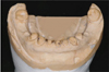
1. The diagnostic cast was constructed (Fig. 1).
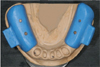
2. The arch for the impression was divided into two or three segments, each of which usually included two or three prepared teeth for ease of management, and each segment was marked with a pencil.
3. The individual segmental tray was constructed (Fig. 2). One sheet of baseplate wax was covered for relief and then the resin mixture in the dough stage was extended 1 - 2 mm over the cervical margins of the prepared teeth. This extension of the segmental tray acted as a tray stop. A small wing was attached to the buccal or labial side of the segmental tray to allow the simultaneous removal of both segmental trays and an overlay tray.
Impression procedure
The impression was made using the following procedures:
1. After a conventional gingival displacement procedure, an appropriate adhesive was applied to the internal surface of all trays, and particularly the external surfaces of the segmental trays.
2. After removing of retraction cords, impression material (Pentamix, 3M ESPE, Germany) was syringed around the prepared teeth involved in a segment, and a segmental tray containing the impression material was positioned. Excessive material was removed from around the tray to ensure the precise vertical positioning of the overlay tray.
3. After the impression material of a segmental tray was set, the above procedure was repeated for the next segment without removing the pre-taken impression segmental tray.
4. To reduce the number of segmental trays, an overlay tray can be used to take an impression of the remaining prepared teeth.
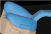
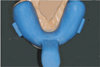
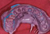
5. After the impression material in the overlay tray hardened, all trays should be removed together by holding both wings of the segmental trays and a handle or margin of an overlay tray to avoid dimensional change and flexing deformation of the impression material despite the presence of segmentally different paths (Fig. 6).
DISCUSSION
Full arch impressions are the most difficult to manage in prosthetic dental treatment, but they have been made frequently in many cases. These impressions pose many problems to dentists, including dimensional change of impression material and dental stone, along with the limited working time of the impression material. A total working and setting time of 4 minutes with a snap set is generally regarded as adequate for most procedures.12 These difficulties are likely to make it necessary to repeat impression procedures in a patient.
Gardener proposed an intraoral coping technique for making impressions of multiple preparations.6
Vasilakis proposed a cast impression coping technique that removed the need for a retraction technique, which provided a better impression environment.7 However, in these techniques, the individual cast coping had no stops, making it questionable whether the material will have a uniform thickness and whether the stock tray will trap all the cast copings undermining the subgingival area upon removal of a stock tray. In addition, the flexing deformation still remains as a problem in both techniques.
The above-mentioned problems can be solved by ensuring the union of segmental trays and an overlay tray during their removal from the mouth and also by attaching a structure such as a handle to each coping or individual tray for the stable removal of segmental impression without flexing deformation.13 In this technique, a handle was attached to each segmental tray that was strong enough to sustain the force required for removal. Its thickness was 1.5 mm and its length was determined by the impression area. An overlay tray had an indent around the handle of each segmental tray and covered all segmental trays sufficiently for precise positioning, which increased the dimensional stability of the overall impression.
One of the advantages of this segmental technique is that it allows the clinician to focus on syringing around no more than two or three teeth, which will improve the accuracy of the margin and a narrow zone of unprepared tooth apical to the finishing line,2,12 even in difficult cases such as compromised gingival health, the use of chemical agents for bleeding control,12,13 and an uncooperative patient, and allowing for the limited working time of the materials.
In conclusion, the segmental impression technique applied in this study has several advantages, including higher impression quality, fewer impressions, fewer remakes, and being more comfortable for the patient and less stressful for the clinician. Although the clinical results have been satisfactory, scanning electron microscopy should be used to characterize the dimensional stability of the master cast and the accuracy of the margin comparing to the conventional impression method.
 XML Download