Journal List > Immune Netw > v.11(6) > 1033320

Park, Im, Kim, and Lee: Immunomodulatory Effects of Hypocrellin A on MHC-restricted Antigen Processing

Abstract

Hypocrellin A has gained much attention in recent years due to its light-induced antitumor, antifungal and antiviral activities. Here we report that hypocrellin A exerts immunomodulatory effects on MHC-restricted presentation of antigen. Hypocrellin A inhibited class II-MHC restricted presentation of exogenous antigen, but not class I MHC-restricted presentation of exogenous antigen, in dendritic cells. Hypocrellin A also inhibited the cytosolic pathway of endogenous antigen presentation. However, hypocrellin A did not inhibit the expression of class I and class II MHC molecules on dendritic cells (DCs), the phagocytic activity of DCs, or the H-2Kb-restricted presentation of a synthetic peptide, SIINFEKL. These results show that hypocrellin A differentially modulates the MHC-restricted antigen presentation pathways.

Hypocrellin A is a naturally occurring perylenequinone compound isolated from the stromata of Hypocrella bambusae and Shiraia bambusae , which are parasitic fungi of bamboo (1). Hypocrellin A has long been known as an excellent photosensitizer and has gained much attention in recent years because of its light-induced antitumor, antifungal and antiviral activities (2-7). Hypocrellin A appears to exert photodynamic anticancer and antiviral activities through its abilities to generate reactive oxygen species and to inhibit protein kinase C activity (8-10). Hypocrellin A has also been shown to exert antimicrobial and antileishmanial activities in vitro (11).
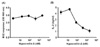
The present study reports that hypocrellin A differentially modulates MHC-restricted antigen presentation pathways. To examine the effects of hypocrellin A on the MHC-restricted antigen presentation, DC2.4 cells, a dendritic cell (DC) cell line, were incubated with hypocrellin A for 2 h, and then biodegradable microspheres containing ovalbumin (OVA, 50µg/ml as OVA) were added to the cultures for 2 h. The process used to fabricate OVA-containing biodegradable microspheres was previously described in detail (12). The cells were then fixed with paraformaldehyde and washed thoroughly with PBS. The amounts of OVA peptides presented via class I MHC molecules were assessed in B3Z cells (H-2b), which express β-galactosidase upon recognition of H-2Kb-OVA peptide complexes, as described previously (13). The effects of hypocrellin A on the class II MHC-restricted presentation of exogenous OVA were examined in bone marrow-derived DCs (BM-DCs), which were generated from BM cells of BALB/c mice (H-2d) by culturing 6 days in the presence of 200 units/ml of GM-CSF. After culturing BM-DCs with biodegradable microspheres containing OVA (50µg/ml as OVA) for 2 h, the BM-DCs were fixed with paraformaldehyde, washed, and then co-cultured with DOBW cells, which express IL-2 upon recognition of I-Ad-OVA peptide complexes, as described previously (14). As shown in Fig. 1A, hypocrellin A did not inhibit class I MHC-restricted presentation of exogenous OVA. However, hypocrellin A dose-dependently inhibited class II MHC-restricted presentation of exogenous OVA (Fig. 1B). The IC50 was approximately 80 nM. These results show that hypocrellin A preferentially inhibits the class II MHC-restricted presentation pathway of exogenous antigen.
The effects of hypocrellin A on the cytosolic pathway of endogenous antigen presentation were also examined in DCs. In this experiment, DC2.4 cells were incubated with hypocrellin A for 2 h, washed, and then soluble OVA was loaded into the cytosol by osmotic shock as described previously (13). After 2-h incubation, the amounts of H-2Kb-OVA peptide complexes were assessed with B3Z cells. As shown in Fig. 2A, hypocrellin A profoundly inhibited class I MHC-restricted presentation of endogenous OVA. To confirm the inhibitory effects of hypocrellin A on the endogenous antigen presentation pathway, recombinant Vaccinia virus-encoded OVA (rVV-OVA) was utilized as another source of cytosolic antigen as described previously (14). Briefly, DC 2.4 cells were infected with rVV-OVA at MOI of 10 for 20 min and then incubated with different concentrations of hypocrellin A for 6 h. The amounts of OVA peptides presented on class I MHC molecules were then assessed using B3Z cells. As shown in Fig. 2B, hypocrellin A dose-dependently inhibited class I MHC-restricted presentation of endogenous OVA. The IC50 was approximately 200 nM.
To prove that hypocrellin A inhibits an intracellular event in the antigen processing pathways, we examined whether hypocrellin A inhibited the expression of MHC molecules on DCs. DC2.4 cells were incubated with hypocrellin A (2,000 nM) for 2 h, and then the expression levels of class I and class II MHC molecules were determined using anti-H-2Kb and anti-I-Ad monoclonal antibodies. As shown in Fig. 3A and B, the expression levels of H-2Kb and I-Ad molecules were not decreased by hypocrellin A to a discernable degree. We then examined whether hypocrellin A inhibited the phagocytic activity of DCs using microspheres containing both OVA and FITC; we found that it did not inhibit the phagocytic activity of DCs (data not shown). In addition, we examined the effects of hypocrellin A on the class I MHC-restricted presentation of the H-2Kb-binding OVA peptide, SIINFEKL. As shown in Fig. 3C, hypocrellin A did not inhibit presentation of exogenously added synthetic peptide, SIINFEKL.
Two distinct pathways have been identified for the MHC-restricted presentation of exogenous antigens (15). In the classical paradigm of exogenous antigen presentation by professional APCs, antigens internalized by phagocytosis are processed and loaded on class II MHC molecules in a post-Golgi compartment. Professional APCs, however, process exogenous antigens for presentation on class I MHC molecules. This process, termed cross-presentation, is currently recognized as an obligatory mechanism for the generation of CTL responses to antigens that are expressed only in nonprofessional antigen-presenting cells (APCs) (16,17). Various mechanisms have been proposed to explain crosspresentation such as the cytosolic alternate pathway, the vacuolar alternate MHC-I pathway, and the fusion of the endoplasmic reticulum (ER) with the endosome/phagosome (18,19). It was recently shown that phagocytosed OVA-microspheres are cross-presented via an alternate MHC-I processing pathway in DCs (12). It appears that hypocrellin A does not inhibit the alternative pathway of exogenous antigen presentation, which involves transfer of exogenous antigenic proteins from the vacuolar compartment to the cytosol, where they are partially digested by the proteosome, transferred into ER by transporter associated with antigen presentation (TAP), and then loaded on class I MHC molecules (20). However, hypocrellin A does inhibit the classical cytosolic pathway of endogenous antigen presentation, as shown in Fig. 2. Thus, it appears that hypocrellin A inhibits a post-Golgi compartment in the exogenous antigen processing pathway, probably inhibiting intraphagosomal processing of the antigen and export of the antigen from the phagosome to the cytosol. Although the precise mechanism underlying the inhibitory activity of hypocrellin A has not been elucidated, it is tempting to speculate that the mechanism for the inhibition of the class II MHC-restricted antigen processing pathway by hypocrellin A is different from that of chloroquine or ammonium chloride, which inhibit the class II MHC-restricted presentation of exogenous antigens by reducing the acidification of the endocytic compartment (21), because hypocrellin A is a hydrophobic peryloquinone derivative (22).

Figures and Tables

Figure 1
Effects of hypocrellin A on the MHC-restricted presentation of exogenous OVA. (A) DC2.4 cells were incubated with the indicated amounts of hypocrellin A for 2 h, and then biodegradable microspheres containing OVA (50µg/ml as OVA) were added to the cultures for 2 h. The cells were then fixed with paraformaldehyde, and the amounts of OVA peptides presented on MHC class I molecules were assessed by a LacZ T cell activation assay using OVA-specific CD8 T cell hybridoma B3Z cells. (B) BM-DCs generated from bone marrow cells of BALB/c mice were incubated with the indicated amounts of hypocrellin A for 2 h, and then biodegradable microspheres containing OVA (50µg/ml as OVA) were added to the cultures for 2 h. The cells were then washed, fixed with paraformaldehyde, and the amounts of OVA peptides presented on MHC class II molecules were assessed using OVA-specific CD4 T cell hybridoma DOBW cells.
in-11-412-g001
Figure 2
Effects of hypocrellin A on the cytosolic pathway of endogenous antigen presentation. (A) DC2.4 cells were incubated with the indicated amounts of hypocrellin A for 2 h, washed, and then soluble OVA was loaded into cytosol by osmotic shock as described previously (13). After 2-h incubation, the amounts of H-2Kb-OVA peptide complexes were assessed using OVA-specific CD8 T cell hybridoma B3Z cells. (B) The indicated amounts of hypocrellin A were added to cultures of DC2.4 cells that had been infected with rVV-OVA (MOI 10). After 6 h, the DCs were washed, fixed with paraformaldehyde, and then the capacity to activate OVA-specific CD8 T cells was assessed using OVA-specific CD8 T cell hybridoma B3Z cells.
in-11-412-g002
Figure 3
Hypocrellin A does not inhibit the expression levels of class I and class II MHC molecules or the presentation of a synthetic OVA peptide SIINFEKL. Hypocrellin A (2,000 nM) was added to cultures of DC2.4 cells for 2 h. The cells were harvested, washed, and then stained with anti-mouse H-2Kb monoclonal antibody (A) or anti-I-Ab monoclonal antibody (B). The shaded histograms represent the expression levels of class I and class II MHC molecules in the presence of hypocrellin A, and the thick lines represent the expression levels of class I and class II MHC molecules in the absence of hypocrellin A. The dotted lines represent isotype controls. (C) Indicated amounts of hypocrellin A were added to cultures of DC2.4 cells together with the antigenic peptide of OVA, SIINFEKL. The cells were harvested, washed, and then the amount of SIINFEKL-H-2Kb complex was measured by a LacZ T cell activation assay using OVA-specific CD8 T cell hybridoma B3Z cells.
in-11-412-g003

ACKNOWLEDGEMENTS

This work was supported by the research grant of the Chungbuk National University in 2010.

Notes

The authors declare no financial or commercial conflicts of interest.

References

1. He YY, Jiang LJ. Synthesis and EPR investigations of new aminated hypocrellin derivatives. Free Radic Biol Med. 2000. 28:1642–1651.
crossref
2. Wan XY, Chen YT. Hypocrellin A, a new drug for photochemotherapy. Chinese Sci Bull. 1981. 26:1040–1041.
3. Ma JS, Yan F, Wang CQ, An JY. Hypocrellin-A sensitized photooxidation of bilirubin. Photochem Photobiol. 1989. 50:827–830.
crossref
4. Schinazi RF, Chu CK, Babu JR, Oswald BJ, Saalmann V, Cannon DL, Eriksson BF, Nasr M. Anthraquinones as a new class of antiviral agents against human immunodeficiency virus. Antiviral Res. 1990. 13:265–272.
crossref
5. Daub ME, Ehrenshaft M. The photoactivated cercospora toxin cercosporin: Contributions to Plant Disease and Fundamental Biology. Annu Rev Phytopathol. 2000. 38:461–490.
crossref
6. Ma L, Tai H, Li C, Zhang Y, Wang ZH, Ji WZ. Photodynamic inhibitory effects of three perylenequinones on human colorectal carcinoma cell line and primate embryonic stem cell line. World J Gastroenterol. 2003. 9:485–490.
crossref
7. Liang XH, Cai YJ, Liao XR, Wu K, Wang L, Zhang DB, Meng Q. Isolation and identification of a new hypocrellin A-producing strain Shiraia sp. SUPER-H168. Microbiol Res. 2009. 164:9–17.
crossref
8. Ali SM, Chee SK, Yuen GY, Olivo M. Hypocrellins and hypericin induced apoptosis in human tumor cells: a possible role of hydrogen peroxide. Int J Mol Med. 2002. 9:461–472.
crossref
9. Diwu Z, Zimmermann J, Meyer T, Lown JW. Design, synthesis and investigation of mechanisms of action of novel protein kinase C inhibitors: perylenequinonoid pigments. Biochem Pharmacol. 1994. 47:373–385.
crossref
10. Park J, English DS, Wannemuehler Y, Carpenter S, Petrich JW. The role of oxygen in the antiviral activity of hypericin and hypocrellin. Photochem Photobiol. 1998. 68:593–597.
crossref
11. Ma G, Khan SI, Jacob MR, Tekwani BL, Li Z, Pasco DS, Walker LA, Khan IA. Antimicrobial and antileishmanial activities of hypocrellins A and B. Antimicrob Agents Chemother. 2004. 48:4450–4452.
crossref
12. Gerelchuluun T, Lee YH, Lee YR, Im SA, Song S, Park JS, Han K, Kim K, Lee CK. Dendritic cells process antigens encapsulated in a biodegradable polymer, poly(D,L-lactideco-glycolide), via an alternate class I MHC processing pathway. Arch Pharm Res. 2007. 30:1440–1446.
crossref
13. Lee YR, Yang IH, Lee YH, Im SA, Song S, Li H, Han K, Kim K, Eo SK, Lee CK. Cyclosporin A and tacrolimus, but not rapamycin, inhibit MHC-restricted antigen presentation pathways in dendritic cells. Blood. 2005. 105:3951–3955.
crossref
14. Lee YH, Lee YR, Kim KH, Im SA, Song S, Lee MK, Kim Y, Hong JT, Kim K, Lee CK. Baccatin III, a synthetic precursor of taxol, enhances MHC-restricted antigen presentation in dendritic cells. Int Immunopharmacol. 2011. 11:985–991.
crossref
15. Harding CV. Phagocytic processing of antigens for presentation by MHC molecules. Trends Cell Biol. 1995. 5:105–109.
crossref
16. Heath WR, Carbone FR. Cross-presentation, dendritic cells, tolerance and immunity. Annu Rev Immunol. 2001. 19:47–64.
crossref
17. Carbone FR, Kurts C, Bennett SR, Miller JF, Heath WR. Cross-presentation: a general mechanism for CTL immunity and tolerance. Immunol Today. 1998. 19:368–373.
crossref
18. Ackerman AL, Cresswell P. Cellular mechanisms governing cross-presentation of exogenous antigens. Nat Immunol. 2004. 5:678–684.
crossref
19. Groothuis TA, Neefjes J. The many roads to crosspresentation. J Exp Med. 2005. 202:1313–1318.
crossref
20. Kovacsovics-Bankowski M, Rock KL. A phagosome-to-cytosol pathway for exogenous antigens presented on MHC class I molecules. Science. 1995. 267:243–246.
crossref
21. Ojcius DM, Gapin L, Kourilsky P. Dissociation of the peptide/MHC class I complex: pH dependence and effect of endogenous peptides on the activation energy. Biochem Biophys Res Commun. 1993. 197:1216–1222.
crossref
22. Diwu Z. Novel therapeutic and diagnostic applications of hypocrellins and hypericins. Photochem Photobiol. 1995. 61:529–539.
crossref
TOOLS
ORCID iDs

Sun-A Im
https://orcid.org/http://orcid.org/0000-0003-2179-0278

Chong-Kil Lee
https://orcid.org/http://orcid.org/0000-0001-9070-341X

Similar articles