

 PDF
PDF ePub
ePub Citation
 Print
Print


INTRODUCTION
Prostanoids are small lipid molecules, playing important roles in various physiological and pathological processes, such as kidney function, vasodilation, platelet aggregation, and cancer progress (1). Prostanoids are derived from membrane glycerophospholipids in response to inflammatory stimuli via three sequential enzymatic reactions. The first reaction is mediated by phospholipase A2, which liberates arachidonic acids from membrane phospholipids (2). The arachidonic acids are subject to the second reactions and converted to prostaglandin (PG) G2 and then PGH2 by cyclooxygenases (COXs), COX-1 or COX-2. The last steps of prostanoid production depend on tissue- and cell-specific terminal synthases. PGH2 are metabolized to PGE2, PGI2, PGD2, PGF2α, and thromboxane A2 (TXA2) by respective actions of PGE2 synthase (PGES), PGI2 synthase (PGIS), PGD2 synthase (PGDS), PGF2 synthase (PGFS), and thromboxane synthase (3). In particular, PGE2 is catalyzed by three different PGESs, cytosolic PGES (cPGES) and two membrane-bound PGESs, mPGES-1 and mPGES-2 (4). The expression of cPGES and mPGES-2 are constitutive, while mPGES-1 is induced during inflammation.
In addition to the well-known roles of prostanoids as inflammatory and vascular mediators (5,6), they are recognized as important immune modulators (7-9). For instance, PGE2 exhibits immunostimulatory as well as immunosuppressive activities depending on its concentrations. PGE2 inhibits activation, proliferation, and differentiation of T cells at micromolar concentrations, whereas it potentiates Th1 and Th17 differentiation and proliferation of T cells at nanomolar concentrations (10).
Using an in vitro model of germinal center reactions that contains human follicular dendritic cell (FDC)-like cells HK (11), we have demonstrated that both FDC and HK cells express PGI2 synthase (12), PGI2 production is not controlled by the induction of PGIS but by COX-2 (13), HK cells secrete PGI2 and PGE2 but not TXA2 (14), PGs produced by HK cells inhibit proliferation and apoptosis T cells (14), T cells control PG production from HK cells via IL-4-Janus kinase 1 (JAK1)-Signal transducer and activator of transcription 6 (STAT6)-COX-2 pathway (15), and PGI2 and its analogues enhance CD86 expression on the surface of activated B cells (16). These results support the emerging concept of PGs as critical immune modulators.
In this study, we investigated the relative contribution of COX-1 and COX-2 to PGI2 and PGE2 synthesis in HK cells. Several studies demonstrated the coupling between COXs and terminal prostanoid synthases. However, most studies were performed using murine cells (2). The current results suggest that mPGES-1 and PGIS are coupled with COX-2 but not with COX-1 in human FDC and imply that chronic administration of selective COX-2 inhibitors might disturb the normal humoral immune responses taking place in the culminating site of germinal centers.
MATERIALS AND METHODS
Culture of HK cells
HK cells are primary cells obtained from human tonsils and used until they display degenerate features in culture. They are prepared as described by Kim et al. (17) and maintained in RPMI-1640 (Irvine Scientific, Santa Ana, CA) containing 10% fetal calf serum (Hyclone, Logan, UT), 2 mM L-glutamine (Invitrogen, Carlsbad, CA), 100 U/ml penicillin G (Sigma-Aldrich, St. Louis, MO), and 100µg/ml streptomycin (Invitrogen). Lipopolysaccharide (LPS) was purchased from Sigma-Aldrich.
Immunoblotting
The whole cell lysates of HK cells were subject to immunoblotting as previously described (18). The protein concentrations of the each fraction were assayed with a bicinchonic acid (BCA) assay. Used antibodies were against COX-1, COX-2 (Cayman Chemical, Ann Arbor, MI), β-actin (Sigma-Aldrich), and horseradish peroxidase (HRP)-conjugated anti-mouse IgG (Jackson Immunoresearch, West Grove, PA). The membranes were incubated with SuperSignal West Pico Chemiluminescent Substrate (Pierce, Rockford, IL) and exposed to X-ray films.
siRNA transfection
The siRNA duplexes used (Ambion Inc, Austin, TX) were constructed with the following target sequences. Control (Neg-siRNA#2, sequence not disclosed by Ambion); COX-1, sense (5'-GCUCUUUAAGGAUGGGAAATT-3'), antisense (5'-U UUCCCAUCCUUAAAGAGCCG-3'); COX-2, sense (5'-CCA CCCAUGUCAAAACCGATT-3'), antisense (5'-UCG GUUUUGACAUGGGUGGGA-3'). HK cells were cultured to 50~60% confluence in 100 mm plates. For each plate, 40 nM of each siRNA and 24µl Lipofectamine™ (Invitrogen) were separately diluted in 400µl serum-free medium without antibiotics, mixed together, and incubated at RT for 45 min. The plates were then washed with serum-free medium, added with 5 ml serum-free medium, and then with the diluted solutions. The plates were incubated at 37℃ for 8 h, followed by the addition of a growth medium containing 10% serum. After 48 h of additional incubation, cells were used for experiments. The degree of gene-silencing was assayed by immunoblotting.
Enzyme immunoassay to measure prostaglandins
HK cells were cultured with LPS for 48 h to harvest the supernatants. The amounts of PGE2 and 6-keto-PGF1α, stable metabolite of PGI2, were measured using enzyme immunoassay (EIA) kits as described previously (14). PG concentration was normalized to total cellular protein and expressed as ng/mg protein.
RESULTS
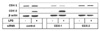
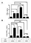
To investigate the relative contribution of COX-1 and COX-2 to the production of PGE2 and PGI2, we carried out siRNA technology to knock down COX-1 and COX-2 proteins in HK cells. HK cells were transfected with siRNA duplexes specific to COX-1 and COX-2 and cultured for 48 h, followed by further cultures in the presence or absence of LPS. The silencing of target proteins was demonstrated by immunoblotting. As shown in Fig. 1, COX-2 protein levels were up-regulated by LPS stimulation in control siRNA-transfected HK cells, whereas COX-1 levels were unaffected by LPS treatment. Transfection with COX-1-specific siRNA resulted in significant reduction of COX-1 protein levels regardless of LPS stimulation. Interestingly, LPS-induced COX-2 levels in COX-1 siRNA-transfected cells were markedly higher compared to control cells. Transfection with COX-2-specific siRNA almost completely prevented induction of COX-2 proteins that was triggered by LPS. COX-2 silencing did not significantly affect COX-1 expression levels. These results indicate that COX-1 and COX-2 proteins were successfully knocked down in HK cells by siRNA duplexes. We next measured the concentrations of PGE2 and 6-keto-PGF1α in the culture supernatants after incubation of siRNA-transfected cells in the presence or absence of LPS for 48 h. 6-keto-PGF1α is the hydrolysis product of unstable PGI2. PGE2 and 6-keto-PGF1α concentrations in control cells were increased 4- and 5-folds, respectively, by LPS stimulation (Fig. 2). Similar levels of enhancement were obtained in COX-1 siRNA-transfected cells. In contrast, COX-2 silencing almost completely prevented the PG production to background levels. We observed that knock down of COX-1 resulted in a significant increase of PGE2 but not PGI2 without LPS stimulation (Fig. 2A). Whether this result reflects a preferential coupling of COX-2 with PGES over PGIS is currently unclear. Based upon these results, we conclude that PGE2 and PGI2 production is coupled with COX-2 but not with COX-1 in HK cells.
DISCUSSION
In this study, we demonstrate that LPS stimulates FDC-like cells to produce PGE2 and PGI2, which depends on the presence of COX-2. Our results imply that FDC is a physiologic source of PG during inflammatory immune responses to bacterial infection. PGs secreted from FDC may exert regulatory roles in the various processes taking place in germinal centers of the secondary lymphoid tissues (7-9).
PGE2 and PGI2 are biosynthesized by mPGES-1 and PGIS. In line with the current data, we previously provided evidence that COX-2 and mPGES-1 are functionally associated. COX-2 and mPGES-1 mRNA and proteins were increased by LPS treatment, which was inhibited by IL-4 (14,15). mPGES-1 induction by LPS stimulation occurred following COX-2 induction in HK cells (14). This coordinated kinetics of COX-2 and mPGES-1 induction may contribute to the efficient production of PGE2. In contrast, PGIS is constitutive in HK cells (13,14). Therefore, the PGI2 production in HK cells may depend on the substrate supply for downstream PGIS from up-stream COX-2, suggesting the coupling of COX-2 with PGIS. This interpretation is compatible with the general belief that inducible COX-2 is required for delayed PG synthesis whereas constitutive COX-1 is necessary for immediate PG production after inflammatory stimulation (2). Further investigation is necessary to determine whether the functional association of COX-2 with mPGES-1 and PGIS results from physical coupling of these enzymes. Confocal microscopy may be a useful method to study their possible colocalization and subcellular localization. However, the coupling between COX-2 and PGIS is not absolute and appears to vary depending on cell types. Different from HK cells, Bolego et al. reported that PGI2 production is coupled with COX-1 but not with COX-2 in human endothelial cells (19). PGIS is constitutively expressed in human endothelial cells (20). Furthermore, stimulus types and the degree of substrate abundance seem to affect the functional coupling between COX and PGIS or mPGES-1.
Considering the accumulating reports on the side effects of selective COX inhibitors (21), the current data may help understand the potential effects of selective COX inhibitors on the humoral immunity.
 XML Download