

 PDF
PDF ePub
ePub Citation
 Print
Print


INTRODUCTION
αβ TCR cells which express clonotypic T cell receptor chain α and β, develop in the thymus and egress out from the thymus after completion of differentiation into helper T cells or CD4+ T cells which express coreceptor CD4 and recognize antigen-MHC class II complex on antigen presenting cells, and cytotoxic T cells or CD8+ T cells which express coreceptor CD8 and recognize antigen-MHC class I complex. Then, CD4+ and CD8+ T cells populate in periphery and undergo further differentiation. CD4+ T cells can differentiate into Th1, Th2, Th9, and Th17 effector cells depending on the micro-environmental milieu including cytokine production. CD8+ T cells can destroy target cells by forming pores with perforin and secreting granzyme A/B (1). Potentially self-reactive T cells can develop because of the generation of random diversity in the T cell repertoire. Self-reactive T cells can be eliminated during the selection process in the thymus, namely central tolerance, and if they escape negative selection, they can be still suppressed by peripheral mechanisms including anergy and/or by CD4+CD25+ regulatory T cells (Tregs). Treg is a specialized subset of CD4+ T cells and able to suppress immune responses (2). Tregs are also generated in the thymus and constitutively express IL-2 receptor α chain, CD25 and transcription factor Foxp3 (3). To note, it is not clear yet what signal triggers Foxp3 expression. TCR signal has been suggested to be important to turn on the expression of Foxp3 in developing Treg precursors in the thymus (4). Although it is known that Treg generation relies on TCR, CD28 and IL-2 signaling during differentiation in the thymus, only a few studies have shown the role of proximal TCR signaling components in intrathymic or natural Treg (nTreg) generation.
OVERVIEW OF TCR SIGNALING
TCR signaling plays a critical role in development, survival and effector cell differentiation and function of T cells. In the thymus, quantitative and qualitative differences in TCR signaling determine the fate of developing thymocytes that subsequently cause to positive and negative selection (5). Interactions between TCR and peptide-MHC lead to the activation of Lck, a member of Src family of tyrosine kinase in T cells, and then activated Lck phosphorylates two tysosine-residues on the ITAMs of CD3 and ζ chains. Then, ZAP-70, another tyrosine kinase, is recruited to phosphorylated ITAMs. ITAM binding to ZAP70 relieves an autoinhibitory conformation of the kinase and the phosphorylation of the specific tyrosine residues that are important for the activity of ZAP-70 (6). Activated ZAP-70 phosphorylates the tyrosine residues on the linker for activation of T cells (LAT), an adaptor molecule, which couples proximal TCR activation to downstream signaling pathways, e.g., PLC-γ1, Ca2+ flux, PKC activation, Ras/ERK activation (7-9). Then these molecules form signalosomes with other proteins containing the Src homology 2 (SH2) domains including PLC-γ1, Grb2, Gads, Grap, 3BP2, and Shb, and indirectly binds Sos, c-Cbl, Vav, SLP-76, and Itk (10,11). In human LAT, one of the four membrane-distal tyrosines, Y132 (Y136 in mouse LAT), associates with PLC-γ1, and further interaction of LAT with PLC-γ1 and Gads-SLP-76 complex is critical for PLC-γ1 activation, which hydrolyzes phosphatidylinositol 4,5 bisphosphate (PIP2) to generate diacylglycerol (DAG) and inositol 1,4,5-triphosphate (IP3). While DAG activates PKC/NF-κB and RasGRP-Ras-Raf-ERK pathways, IP3 mediates Ca2+ flux from the endoplasmic reticulum, leading to the activation of clacineurin, a calcium induced phosphatase, and dephosphorylation of NFAT family transcription factors. Finally, dephosphorylated NFATs are transclocated into the nucleus for their transcriptional activities (12). On the other hand, three tyrosines, Y171, Y191 and Y226, bind to Grb2 upon phosphorylation. Grb2 recruits son of sevenless (Sos), a guanine nucleotide exchange factor (RasGEF), to the plasma membrane for activation of Ras molecule. Two tyrosines, Y171 and Y191 also bind to Gads, which constitutively interacts with SLP-76.
Tyrosine phosphorylation of the N-terminal, positions at 113 and 128 of SLP-76 results in the recruitment of the GEF Vav and the adapter protein Nck and these complexes regulate directly actin cytoskeletal rearrangement following TCR ligation and JNK activation (8,9). Thus, TCR engagement triggers a series of signaling cascades that ultimately lead to T cell development, differentiation and effector function.
TCR SIGNALING AND CENTRAL TOLERANCE IN THE THYMUS
After successful completion of β-selection, where rearranged TCRβ chain associates preTα chain to form preTCR complex, preTCR signaling operates for proliferation and survival of double negative (DN) thymocytes (13). Then, DN thymocytes progress to double-positive (DP) thymocytes stage where thymocytes begin to express both CD4 and CD8 coreceptors. DP thymocytes also re-express RAG1/2 which is required for TCRα rearrangement, resulting αβ TCR complex expression on their surface (14). The development of DP thymocytes has three fates: death by neglect, positive selection or negative selection. During this thymic selection process, random rearrangement of TCRs is useless since these TCRs cannot bind to their cognate ligands (15). The failure of clonotypic αβ TCR expressed on DP thymocytes to engage cognate epitope-MHC could not generate signals required to undergo positive selection and approximately 90% of these cells die. Positive selection occurs when and if the TCR of the thymocytes engages an epitope-MHC complex with low affinity, and then generate signals for differentiation and survival. Positively selected DP thymocytes mature into CD4+ or CD8+ single-positive (SP) T cells. In a way, positive selection enriches for self-reactive cells too, making them a threat for autoimmunity in periphery. On the other hand, negative selection deletes potentially self-reactive thymocytes, thereby generating a repertoire of peripheral T cells that is largely self-tolerant. TCR of thymocytes engaging an epitope-MHC complex with high affinity leads to the apoptotic death of the cells (16).
NTREG DEVELOPMENT IN THE THYMUS
Development of nTregs seems to occur via a two-step process dependent on multiple intracellular pathways activated by a combination of TCR, CD28 and cytokine receptor mediated signals (17-19). The initial step in nTreg development depends on signals generated by TCR engaged with self-peptide-MHC class II complexes (20) and B7/CD28 interactions (21). Tregs emerge from a pool of DP thymocytes which express TCRs with a relatively high affinity for self-antigens to CD4+ SP thymocytes which are CD25+GITRhiFoxp3- (22-24). This requirement for the development of Tregs differs from the fate of conventional CD4+ T cells expressing higher affinity TCRs, which are eliminated by negative selection (25). The subsequent differentiation of Treg precursors into functional Tregs requires IL-2 and/or IL-15 signaling to induce and maintain Foxp3 expression (24).
THE ROLE OF TCR SIGNALING COMPONENTS IN NTREG DEVELOPMENT
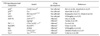
The findings from mutant animal studies have contributed to understand the role of TCR signaling in nTreg development. These animal models include p56Lck, p59Fyn, LAT, ZAP-70, CD5, PLC-γ1, RasGRP-1 and Vav-1 and findings are summarized in Table I.
p56Lck
p56Lck triggers TCR signaling in response to antigenic stimulation to regulate T cell development, survival and differentiation in the thymus. But, conditional ablation of p56Lck in DP and CD4-SP thymocytes has no impact on nTreg generation (26) and inactivation of p59Fyn, another Src family member of tyrosine kinase, also did not affect nTreg development in the thymus (27). Furthermore, inactivation of Lck and Fyn in thymus did not impair nTreg development, suggesting that Lck and Fyn are not directly involved in the development of nTreg. Instead, inactivation of p56Lck seems to compromise Treg homeostasis, which is characterized by impaired turnover, preferential redistribution to lymph nodes, and loss of suppressive function (26).
ZAP-70
Development of nTreg is impaired in mice with a mutation (skg mutation) or tyrosine to phenylalanine mutations of Y315 and Y319 of the ZAP-70 gene (ZAP-70YYAA) (28,29). In addition, these mice are impaired in T cell development and hyporesponsiveness on cells with TCR stimulation as well as defective on positive and negative selection. These two tyrosine residues appear to play an important role in recruitment of downstream molecules, LAT, and auto-inhibition of ZAP-70. Furthermore, ZAP-70YYAA Tregs showed minimal suppressor activity (28), suggesting that these tyrosine residues are critical for nTreg development as well as their function.
LAT
Knockin mutant mouse, where tyrosine 136, a PLC-γ1-binding site in murine LAT, is replaced by phenylalanine (Y136F), develops lymphoproliferative disease (30) and has defective on Treg development (31). These studies suggested that the LAT-PLC-γ1 interaction plays a critical role in Foxp3 expression and the development of nTreg cells. Elegant genetic approach by the same group has revealed that Treg development in LAT mutant mice, where wild-type LAT is used for selection and mutant LAT (Y136F) is replaced after tamoxifen treatment, is intact. However, their suppressor activity is compromised and nonfunctional in periphery, indicating that LAT-PLC-γ1 interaction is essential for the suppressive activity of Tregs not for nTreg development (32).
Raf/SAP-1
The study with mutant mice defective in Raf signaling, i.e., ERK effector SRF accessory protein 1 (SAP-1), and with dominant negative Raf-1 transgenic mice revealed the differential requirement for Treg development (33). To note, Treg development in SAP-deficient animal was intact. Dominant negative-Raf transgenic mice, unlike SAP-1 knockout mice, are defective in Treg development. Nevertheless, neither ablation of SAP-1 nor inactivation of Raf signaling by DN-Raf affected the activity of Treg to suppress naïve T cells in vitro. Furthermore, SAP-1 deficient Tregs are functional to suppress colitits in vivo, suggesting that ERK signaling to SAP-1 is not required for the suppressive activity of Treg (33).
PLC-γ1
Investigation on the function of PLC-γ1 signaling in T cell including Treg development has been impeded by the fact that PLC-γ1 deficiency leads to embryonic lethal during embryonic development. Therefore, the generation and analysis of PLC-γ1 conditional knockout mice allowed the assessment of the role of PLC-γ1 signaling in conventional T cell and Treg development (34). It has been shown that PLC-γ1 ablation affects T cell development and impairs positive and negative selection, resulting in severe reduction of mature T cells in periphery. PLC-γ1 deficiency also leads to impaired TCR-stimulated proliferation and cytokine production, and the activation of multiple signaling mediators and transcriptional factors (e.g., ERK, JNK, AP-1, NFAT and NF-κB). In addition, Treg development is impaired and their suppressor activity is compromised (34). But, it is not clear whether or not defective Treg development in PLC-γ1 mutant mice is due to impaired TCR-induced Foxp3 expression or defective IL-2/IL-2R signaling by impaired IL-2 production.
RasGRP-1
In RasGRP-1 deficient mice, Treg cell development in the thymus was impaired. But in the periphery frequencies of CD4+ Foxp3+ Tregs were significantly increased due to massive proliferation of RasGRP-1 deficient Tregs and increased apoptosis of RasGRP-1 deficient Foxp3-CD4+ T cells. RasGRP-1 deficient Treg cells possessed a more activated cell surface phenotype and RasGRP-1 deficient Tregs are more suppressive than wild-type controls. This study strongly suggested that the intrathymic nTreg development relies on RasGRP-1 signaling pathway, but their peripheral expansion and function do not (35).
CD5
CD5 is a negative regulator of TCR signaling and CD5 deficiency rendered thymocytes hyperresponsive to TCR stimulation, leading to increased proliferation, Ca2+ flux and tyrosine phosphorylation of TCR-ζ, LAT, PLC-γ1 and Vav-1 (36). CD5 deficiency leads to increased nTreg generation in thymus and periphery, and importantly they are functional and suppress naïve T cells. Furthermore, CD5 deficient nTregs showed increased basal level of p-ERK compared with those of wild-type nTreg (37).
Vav-1
Vav-1 is a signal transducer downstream of TCR and is critical for T cell development and activation (38,39). In a genetic study with two rat strains, which have different susceptibility to autoimmune disease, a non-synonymous mutation was identified in the 1st exon of Vav-1, responsible for the substitution of an arginine by tryptophan at position 63. This mutation is associated with increased proportion and absolute numbers of Tregs. This mutation resulted in constitutive activation of Vav-1 protein and increased activity of guanine nucleotide exchange factor (40).
In summary, even considering critical role of Src family kinase and adaptors in T cell development in general, p56Lck, p59Fyn and LAT appear to be dispensable for intrathymic nTreg development but are required for maintenance or suppressive function of Treg in the periphery (28,29,34). ZAP-70, PLC-γ1, RasGRP1 and Raf positively regulate nTreg development in the thymus, since ablation of these mediators impairs nTreg generation in the thymus (30,31,35-37). However, whether IL-2 production or IL-2 signaling is affected in these mutant mice is yet to be defined. Especially, IL-2 production in PLC-γ1 conditional knockout mice is decreased, suggesting that both diminished TCR signal and impaired IL-2 signal may lead to impaired nTreg development.
CONCLUSION
During the last few years, we have seen much progress in our understanding of nTreg generation by TCR signaling. New studies from animal model analysis support the role of TCR signaling in intrathymic Treg development and revealed specific requirement for this process, which is distinct from conventional T cell development. But the challenge remains how to dissect the impact of TCR signaling on negative selection versus Treg generation since these two processes are closely related, yet distinct. We anticipate that more mouse models will be developed and analyzed to understand how TCR signaling pathways regulate nTreg generation in near future.
 XML Download