

 PDF
PDF ePub
ePub Citation
 Print
Print


INTRODUCTION
miRNAs are found in almost all species: virus, plants, nematodes, fly, fish, mouse, human, and are implicated in a wide array of cellular and developmental process (1). There are hundreds of miRNAs encoded in the human genome and thousands of target mRNAs, which illustrates the important regulatory roles of miRNAs in cell development, differentiation, proliferation and apoptosis pathways (2,3). It is not surprising that deregulated miRNAs have been involved in the pathogenesis of many human disease (4-10). miRNAs have recently emerged as important regulators of gene expression. They were formerly thought to mainly repress the translation of target mRNAs, but it has recently been shown that the main function of miRNAs in mammalian system is to decrease target mRNA levels (11). It is estimated that 20~30 % of all human mRNA are miRNA targets (12). And so, it is probable that most mRNAs are controlled by miRNAs to some extent. The expression of miRNAs is highly regulated and they are therefore well placed to function as immunomodulators (13). More recently, miRNA are also proving to be an important link between the innate and adaptive immune systems, and their dysregulation might have a role in the pathogenesis of various diseases (4-7,13,14).
Importantly, it has been increasingly reported that miRNAs are associated with disease (4-6, 14-24). However, the pattern among the miRNA-disease association remains largely unclear. In order to dissect the patterns of miRNA-disease associations, a study performed a comprehensive analysis to the human miRNA disease association data, which is manually collected from publication (14). They built a human miRNA association disease network. Interestingly, miRNAs tend to show similar or different dysfunctional evidence for the similar or different disease clusters, respectively (14). In addition, they also found that there is a negative correlation between the tissue-specificity of a miRNA and the number of disease it associated and that there is an association between miRNA conservation and disease (14). Furthermore, they uncovered that miRNAs associated with the same disease tend to emerge as predefined miRNA groups (14). The effect of inducing or repressing miRNA expression can influence most biological processes, including cell fate specification, cell proliferation, DNA repair, DNA methylation and apoptosis and provide pro-inflammatory or anti-inflammatory stimuli (10,25). Furthermore, with the development of new techniques for genome-wide screening of miRNA expression, abnormal levels of miRNA were identified in various diseases with respect with normal counterpart (4-6,26,27).
All miRNAs are processed and matured through a complex biogenesis process involving multiple protein catalysis accessory proteins, and macromolecular complexes following a coordinated series of event (28-30). They are an abundant class of gene regulatory molecules in multicellular organisms and modulate the expression of many protein-coding genes (28-30). They are transcribed as a huge double-stranded primary transcript (pri-miR) by RNA polymerase II. Subsequently, nuclear enzyme Drosha and Pasha convert this precursor into a double-stranded miRNA precursor of ~70 nulcleotide (pre-miR), which is next transported into the cytoplasm by a mechanisms involving the protein Exportin 5 (7,13,28,31). Finally, Dicer enzyme processes this precursor into the 22-nucleotide double-stranded miRNA. This duplex is then unwinded, and the leading strand ("guide strand"), one of the two strands, is incorporated into the RNA-induced silencing complex (RISC), which is comprise Agonaute and other proteins (7,31,32). miRNAs incorporated in the RISC are able to bind to the 3' untranslated region (UTR) of target mRNAs causing a block of translation or mRNA degradation depending on the level of complementarity (28,29). The other strand so-called "passenger strand" is degraded (7,28,33). Very importantly, miRNAS are altered or induced by both environmentally regulated early life developmental factors through epigenetic and miRNA mechanisms, and genetic polymorphisms including protein or miRNA genes (34).
Importantly, miRNA degradation may contribute to several pulmonary disease (15). In addition, several miRNAs have been shown to be involved in pulmonary allergy and asthma and lung carcinogenesis (15). There are only few reports focused on the role of miRNAs in chronic obstructive pulmonary disease (COPD), namely miR-146a is involved in COPD (16,17,35). Interestingly, changes in miRNA expression are an early event following exposure to cigarette smoke and bronchial airway epithelial cells from current and never smokers differ in the expression of 28 miRNAs in comparison to smokers, whereas the majority of deregulated miRNAs are down regulated in smokers (36). Very interestingly, it has been reported that many miRNAs may play pivotal role in homeostasis and lung development, and other various pulmonary diseases such as idiopathic pulmonary fibrosis (IPF) (15, 37-39) and cystic firbrosis (40). Recent studies provided clear evidence miRNAs are abundant in the liver and modulate a diverse spectrum of liver functions and that deregulation of miRNA expression may be a key pathogenic factor in many liver diseases including viral hepatitis, hepatocellular carcinoma (HCC), and polycystic liver diseases (19). miR-122 is a liver specific miRNA (18). Besides miR-122, many other miRNAs are also abundantly expressed in adult liver tissue (19). Very recently, a large number of genes and signaling mechanisms have been implicated in ethanol's deleterious effects leading to the suggestion that ethanol is a 'dirty drug' (41). It has been known that alcohol-induced gut leakiness is a key factor in alcoholic liver disease (ALD) and it allows endotoxin to enter the circulation and initiate liver damage and that ethanol increases miR-122 expression (42). It is now clear that key miRNAs are highly expressed in the kidney and can act as effector of TGF-β action and high glucose in diabetic kidney disease (20). A striking increase in miR-214 was also detected in monocyte from patients with chronic renal failure and that miR-214 specifically binds to phosphatase and tensin homolog (PTEN) mRNA 3'UTR, implicating PTEN as a target gene of miR-214 (21). Numerous disorders are related to cilia dysfunction, including polycystic kidney disease (PKD), primary ciliary dyskinesia, nephrophthisis (43) Pandey et al. explored the possibility of miRNA-based regulations in PKD (44). The authors found that 935 genes were differentially regulated between PKD and healthy controls.
As mentioned, many viruses have been founded to encode miRNAs that regulate both viral and host mRNA (45). It has been shown that miRNAs encoded in the viral genome have the potential to reshape the cellular environment to maximize viral replication (24,46) and viral miRNAs can suppress host cell genes involved in antiviral immunity (47). Interestingly, some viruses evade immune surveillance by targeting a cellular mRNA with a virally encoded miRNA and viruses use miRNAs not only to regulate their own life cycles but also evade host immune surveillance (48). In contrast, the cellular miRNAs play an important role in the host, defending against virus infection (49). As for bacterial infections, tuberculosis remains a major health issue, causing approximately three million deaths every year (50). Mycobacterium tuberculosis remains one of the most enigmatic bacteria. Currently, Liu et al. performed miRNA expression profiling in peripheral blood mononuclear cells (PBMCs) from pulmonary tuberculosis patients and health controls (51). They showed that expression of 30 miRNAs was significantly altered during active tuberculosis as compared with healthy controls and 28 miRNAs were up-regulated and 2 miRNAs down-regulated (51). They also showed that miR-144* was one of the miRNAs that were over-expressed in active tuberculosis patients. Helocobacter pylori is the main cause of peptic ulceration and gastric adenocarcinoma in human (46,52). H. pylori was able to increase miR-155 expression in gastric epithelial cell lines and gastric mucosal tissue (46). Currently, a study showed that H. pylori infections alter the expression of oncogenes, tumor suppressor genes and miRNAs (52). Surprisingly, Salmonella significantly induces several miRNAs and these miRNAs chiefly induced miR-155 and miR-146a, as well as miR-21 (53). Treatment of immune cells with bacterial lipopolysaccharide (LPS) from Salmonella and Escherichia coli led to the induction of miR-155, miR-132 and miR-146 expression (54).
Since mature erythrocytes are terminally differentiated cells without nuclei and organelles, it is commonly thought that they do not contain nucleic acids (55). Interestingly, however, human mature erythrocytes contains diverse and abundant miRNAs (56). Increased expression of these miRNAs in primary erythroid progenitor cells results in elevated fetal and embryonic hemoglobin gene expression (57). Interestingly, it has been shown that during the menstrual cycle, human endometrium undergoes extensive cyclic, morphologic and biochemical modifications in preparation for embryo implantation and that endometrial expression of miRNAs and their potential regulatory functions are under normal and pathologic conditions such as endometeriosis, dysfunctional uterine bleeding, and endometrial cancer (58).
miRNAs also have an essential role in both the innate and adaptive immune system. Proper miRNA expression is required for correct differentiation of immune cells (22). Immune responses are symphonies of molecular and cellular interactions, with each player doing its part to produce the composite behavior we see as effective host defense, or when discoordinated, as immunopatholgy or immunodeficiency (6,59). It is therefore not surprising that they have been implicated in various human diseases, including lung diseases (15-17,35,60), liver diseases (18,19,61-63), kidney diseases (20,21,43,44,64), infectious diseases (22-24,61,65-69), sickle cell disease (55-57), and endometrium disease (58,70). Here I briefly summarize the newly discovered roles of miRNAs in various human diseases including infectious diseases, sickle cell disease and enodmetrium diseases as well as lung, liver and kidney diseases.
miRNAs IN LUNG DISEASES
A recent study showed that miRNAs have a strong potential to regulate fundamental biological processes also in the lung compartment and at least 900 different miRNA genes have been discovered in the human genome (15). As shown in Fig. 1, the lung has a very specific miRNA expression profile. However, the knowledge of the role of miRNAs in physiolgocial and pathological conditions in the lung is still limited. miRNA deregulation may contribute to several pulmonary diseases (15). Interestingly, several miRNAs such as miR-148a/b, miR-152, miR-21, miR-126, let-7, miR-29a, miR-155 and miR-133a have been shown to be involved in pulmonary allergy and asthma (15). And, several miRNAs such as miR-155, let-7, miR-17~92 cluster, miR-212, miR-34 families, miR-210 and miR-218 have been shown to be involved in lung carcinogenesis (15). There are only few reports focused on the role of miRNAs in chronic obstructive pulmonary disease (COPD): miR-146a is involved in COPD (16,17,35). COPD shares a common environmental risk factor in cigarette smoke exposure (17). Recent studies on fibroblasts from COPD subjects stimulated in vitro with pro-inflammatory cytokines released less miR-146a than smokers without COPD (35). More recently, a study demonstrated that eight miRNAs were expressed at a significantly lower level in current-smoking patients with COPD compared with never-smokers without airflow limitation and expression of let-7c and miR-125 was reduced in patients with COPD compare with healthy subjects and that target genes of let-7c were significantly enriched in the sputum of patients with severe COPD (16). This study also showed that the concentration of TNF receptor type II (TNFR-II) was inversely correlated with the sputum levels of let-7c (16). This study indicated that let-7c is significantly reduced in the sputum of currently smoking patients with COPD and is associated with increased expression of TNFR-II. Interestingly, Izzotti et al. analyzed the expression of 484 miRNAs in the lungs of rats exposed to environmental cigarette smoke (ECS) for 28 days and found that ECS down-regulated 126 miRNAs (60). The most remarkably down-regulated miRNAs belonged to the families of let-7, miR-10, miR-26, miR-30, miR-34, miR-99, miR-122, miR-123, miR-124, miR-125, miR-140, miR-145, miR-146, miR-191, miR-192, miR-219, miR-222, and miR-223, which regulate stress response, apoptosis, proliferation, angiogenesis, and expression of genes (60). In contrast, the authors also showed that miR-294, an inhibitor of transcriptional repressor genes, was up-regulated by ECS and that there was a strong paralleism in dysregulation of rodent miRNAs and their human homologues (63). Importantly, the authors reported that changes in miRNA expression were an early event following exposure to cigarette smoke. In another study, bronchial airway epithelial cells from current and never-smokers differed in the expression of 28 miRNAs in comparison to smokers, whereas the majority of deregulated miRNAs were down-regulated in smokers (36). Furthermore, expression of miR-218 (a miRNA that is strongly affected by smoking) is reduced in primary bronchial eipthelium exposed to cigarette smoke condensate (36). These data indicate that miR-218 levels modulate the airway epithelial gene expression response to cigarette smoke. Similar observation was observed in lung squamous cell carcinoma, where down-regulation of miR-218 was associted with a history of cigarette smoking (71).
Very interestingly, it has been reported that many miRNAs may play pivotal role in homeostasis and lung development, and other various pulmonary diseases such as idiopathic pulmonary fibrosis (IPF) (15,37-39) and cystic fibrosis (40). IPF is a chronic, progressive, and usually lethal fibrotic disease characterized by profound changes in epithelial cell phenotype and fibroblast proliferation (27,38,39). Recently, Liu et al. showed that eighteen miRNAs including let-7d were significantly decreased in IPF and prevent lung fibrosis which is a key regulatory role for this miRNA in IPF (38). They also demonstrated that TGF-β, a central pathologic mediator of fibrotic diseases, enhanced miR-21 expression in primary pulmonary fibroblasts and that increasing miR-21 levels promoted the pro-fibrogenic activity of TGF-β in fibroblast (38). The authors suggested an important role for miR-21 in fibrotic lung diseases and also suggested a novel approach using miRNA therapeutics in treating clinically refractory fibrotic diseases like IPF.
A current study presented evidence that, downstream of TGF-β signaling, miRNAs of the miR-23a cluster and transcription factor Zeb1 could have roles in mediating an epithelial to mesenchymal transition and the resultant persistence of mesenchymal cells in interstitial lung disease (72). Currently, as shown in Fig. 1, Pandit et al. reported that approximately 10% of the miRNAs are significantly changed in IPF lungs (73). They also showed that among the significantly down-regulated miRNAs are member of let-7, miR-29, and miR-30 families as well as miR-17~92 cluster among the up-regulated miR-155 and miR-21 and that down-regulation of let-7 family members leads to changes consistent with epithelial cells both in vitro and in vivo, whereas inhibition of miR-21 modulates fibrosis in the bleomycin model of lung fibrosis (73). A recurrent global theme is that many miRNAs studied in IPF are both regulated by TGF-β1 and regulate TGF-β1 signaling pathway by their target genes (73). The changes in miRNA expression in the IPF lung and the evidence for their role in the fibrosis suggest that miRNA should be evaluated as therapeutic targets in IPF. It is interesting to note that acute interstitial pneumonia (AIP) clinically and radiologically resemble IPF and is considered to represent the small subset of patients with idiopathic acute respiratory distress syndrome (ARDS) and it has a grave prognosis with >70% mortality in 3 months (74). It is synonymous with Hamman-Rich syndrome, occurring in patients without preexisting lung disease (74). As regards cystic fibrosis, cystic fibrosis is one of the most common lethal genetic diseases (40). A recent study showed miR-126 is involved in this disease and TOM1 mRNA is a miR-126 target that may have an important role in regulating innate immune response in the cystic fibrosis lung (40).
miRNAs IN LIVER DISEASES
Recent studies have shown the involvement of miRNAs in te regulation of HCV infection (18,19,63). It has been reported that miR-122 is first identified liver-specific cellular miRNA and is specifically expressed and highly abundant in the human liver, which has been shown to enhance the replication of HCV by targeting the viral 5' non-coding region and that the sequestration of miR-122 in liver cells results in marked loss of autonomously replicating hepatitis C viral RNAs (18). It appears that HCV replication is associated with an increase in expression of cholesterol biosynthesis genes that are regulated by miR-122 and hence is considered as potential target of antiviral intervention (65,75). Lecellier et al. also showed a cellular miRNA effectively restricts the accumulation of the retrovirus primate foamy virus type 1 (PFV-1) in human cells, indicating that cellular miRNAs have direct antiviral effects in addition to their regulatory functions (76). Therapeutic strategies based on modulation of miRNAs activity hold great promise due to the ability of these small RNAs to potently influence cellular behavior. Kota et al. investigated the efficacy of a miRNA replacement therapy for liver cancer (62). Interestingly, they found that HCC cells exhibits reduced expression of miR-26a, a miRNA that is normally expressed at high levels in diverse tissues and they also found that expression of this miRNA in liver cancer cells in vitro induces cell-cycle arrest associated with direct targeting of cyclins D2 and E2 (62). In addition, the authors found that systemic administration of this miRNA in a mouse model of HCC using adeno-associated virus results in inhibition of cancer cell proliferation, induction of tumor-specific apoptosis, and dramatic protection from disease progression without toxicity (62). These findings provide a general strategy for miRNA replacement therapies and proof-of-concept support for systemic delivery of tumor-suppressing miRNA as a powerful and highly specific anticancer therapeutic modality (62). As mentioned, it is now clear that miRNA can potentially regulate every aspect of cellular activity, including cellular differentiation and development, metabolism, proliferation, apoptotic cell death, viral infection and tumorigenesis (19,75,77). Recent studies provided clear evidence that miRNAs are abundant in the liver and modulate a diverse spectrum of liver functions and that deregulation of miRNA expression may be a key pathogenic factor in many liver diseases including viral hepatitis, HCC and polycystic liver diseases (PCLDs) (19). As discussed, miR-122 is a liver specific miRNA (18). Besides miR-122, many other miRNAs, such as miR-1, miR-16, miR-27b, miR-30d, miR-126, miR-133, mir-143, and the let-7 family, are also abundantly expressed in adult liver tissues (19). While miR-122 appears as the most highly expressed miRNA in adult liver, miR-92a and miR-483 seem to be more specifically expressed in the fetal liver (19). Thus, the liver displays a differential miRNA expression profile during its development. Recently, Chen reported that miRNAs are abundant and finely regulated in the liver and that miRNAs may be key players in the regulation of live functions (19). Furthermore, recent studies have shown the involvement of miRNAs in liver disease caused by various factors, such as HCV, HBV, metabolic disorders, and by drugs (19,63). Viral gene encodes miRNAs and these miRNAs have a regulatory effect on the viral protein-coding genes (75). HBV infection is a global problem and it can cause acute or chronic hepatitis B, liver cirrhosis, and, in some instances HCC (63). However, little is known about the involvement of miRNAs during HBV infection. Considering the fact that cellular miRNAs play an important role in viral pathogenesis (61), it is likely that they have a role in HBV infection. Using computational approach, HBV has been found to encode a candidate pre-miRNA, suggesting that HBV has the capacity to use viral miRNA to regulate its own gene expression (78). miRNA from the host cells may also play a role in regulating viral genes (75-77).
Importantly, Lee et al. found that the cholangiocyte cell line PCK-CCL, which is derived from the PCK rats, a model of autosomal recessive polycystic kidney disease (ARPKD), displayed global changes in miRNA expression compared with normal rat cholangiocytes (79). Interestingly, their more specific analysis revealed decreased levels of miR-15a, both in PCK-CLL cells and in liver tissue from PCK rats and patients with PCLDs. They also found that the decrease in miR-15a expression was associated with up-regulation of its target, the cell-cycle regulator cell division cycle 25A (Cdc25A). Moreover, they found that over-expression of miR-15a in PCK-CCL cells decreased Cdc25A levels, inhibited cell proliferation and reduced cyst growth (79). Furthermore, in contrast, they reported that suppression of miR-15a in normal rat cholangiocytes accelerated cell proliferation, increased Cdc25A expression, and promoted cyst growth (210). These results suggest that suppression of miR-15a contributes to hepatic cystogenesis through dysregulation of Cdc25A (80).
Drug-induced liver injury is a frequent side effect of many drugs and constitutes a significant threat to patient health. Numerous efforts have been made to identify reliable and predictive markers to detect the early signs of drug-induced injury to the liver (81). Using acetaminophen overdose-induced liver injury in the mouse as a model system, Wang et al. have observed highly significant differences in the spectrum and levels of miRNAs in both liver tissues and in plasma between control and overdosed animals (81). The authors have also demonstrated that specific miRNA species, such as miR-122 and miR-192, both are enriched in the liver tissue and exhibit dose- and exposure duration-dependent changes in the plasma that parallel serum aminotransferase levels and the histopathology of liver degeneration, but their changes can be detected significantly earlier (81). These findings suggest that potential of using specific circulating miRNAs as sensitive and informative biomarkers for drug-induced liver injury.
Very recently, a large number of genes and signaling mechanisms have been implicated in ethanol's deleterious effects leading to the suggestion that ethanol is a "dirty drug" (41). Ethanol-sensitive miRNAs have been recently identified as master switch (41). Accumulating evidence showed that miRNAs mediate several ethanol pathologies, including disruption of neural stem cell proliferation and differentiation in the exposed fetus, gut leakiness that contributes to endotoxemia and alcoholic liver disease, and possibly also HCC and other gastrointestinal cancer (41). It has been known that alcohol-induced gut leakiness is a key factor in alcoholic liver disease (ALD) and it allows endotoxin to enter the circulation and initiate liver damage and that Zonula occludens 1 (ZO-1) protein is a major component of tight junctions that regulates intestinal permeability (42). Tang et al. investigated the effect of alcohol on miR-212 and on expression of their target gene, ZO-1 (42). The authors found that ethanol increased miR-212 expression, decreased ZO-1 levels, disrupted tight junctions, and increased the permeability of monolayerss of Caco-2 cells and that an miR-212 over-expression is correlated with hyperpermeability of the monolayer barrier (42). In addition, they also found that miR-212 levels were higher in colon biopsy samples in patients with ALD than in healthy controls and ZO-1 protein levels were lower (42). These studies suggest a novel mechanisms for alcohol-induced gut leakiness, one in which ethanol induces miR-212 over-expression which causes gut leakiness by down-regulating ZO-1 translation. Alcoholic and nonalcoholic steatohepatitis are leading causes of liver diseases worldwide (41,82). While of different etiology, these share common pathopysiological mechanisms and feature of abnormal fat metabolism, inflammation and fibrosis (41). Dolganiuc et al. reported etiology-specific changes in miRNA expression profile during steatohepatitis models, which opens new avenues for research in the pathophysiology of alcoholic and nonalcoholic fatty liver disease (82). The authors reported that the both Lieber-DeCarli alcohol diet and methionine-choline-deficient (MCD) diets lead to development of liver steatosis, liver injury, indicated by increased alanine aminotrasnferase (ALT), and elevated levels of serum TNF, suggesting that animal models portray the pathophysilogical features of alcoholic and nonalcoholic fatty liver, respectively (82). Interestingly, the authors also reported that after 5 weeks of either diet, expression of both miR-705 and miR-1224 was increased, whereas expression of miR-182, and miR-199a was down-regulated during Lieber-DeCarli feeding and up-regulated during MCD feeding (82). They proposed that changes in miRNAs contribute to impaired hepatic lipid homeostasis and the inflammatory cascade in alcoholic steatohepatitis and nonalcoholic steatohepatitis (82).
miRNAs IN KIDNEY DISEASES
It is now clear that key miRNAs are highly expressed in the kidney and can act as effectors of TGF-β actions and high glucose in diabetic kidney disease (20). Moreover, it is interesting that podocyte-specific deletion of Dicer, a key enzyme involved in miRNA biogenesis, led to proteinuria and severe renal dysfunction in mice (20). Overwhelming evidence implicating miRNAs in the pathology of key human diseases has sparked tremendous interest in development of modalities to block specific miRNAs and their function in vitro and in vivo (20). miRNAs, such as miR-192 and miR-377, may be novel targets for diabetic nephropathy and other diabetic complications.
Advanced glycation end products (AGEs) delay spontaneous apoptosis of monocytes and continue to the development of inflammatory response (21). Currently, a study investigated the role of miR-214 and its target gene in AGE-induced monocytic apoptosis delay (21). The authors reported that a striking increase in miR-214 was also detected in monocyte from patients with chronic renal failure and that miR-214 specifically binds to phosphatase and tensin homolog (PTEN) mRNA 3'UTR, implicating PTEN as a target gene of miR-214. The study revealed also that PTEN expression is inversely correlated with miR-214 level in monocytes and that compared with normal monocytes, AGE-treated monocytes and monocytes from chronic renal failure patients exhibited lower PTEN levels and delayed apoptosis (21).
Cilia and flagella are ancient, evolutionary conserved organelles that project from cell surface to perform diverse biological roles, including whole-cell locomotion, movement of fluid, chemosensation, mechanosensation, and photosensation, and sexual reproduction (64). Recently, the biological importance of primary cilia has been recognized in a number of human genetic diseases (43,64). Numerous disorders are related to cilia dynsfunction, including PKD, primary ciliary dyskinesia, nephronophthisis (43). Pandey et al. explored the possibility of miRNA-based regulations in PKD (44). The authors analyzed the simultaneous expression changes of miRNAs and mRNAs by microarrays. They found that 935 genes, classified into 24 functional categories, were differentially regulated between PKD and control animals. In parallel, 30 miRNAs were differentially regulated in PKD rats (44). Their results suggest that several miRNAS might be involved in regulating genetic switches in PKD. Furthermore, they described some newly detected miRNAs, miR-31 and miR-217 (44). Their results suggest an important role of miRNAs in specific pathways underlying PKD (44).
miRNAs IN INFECTION
Emerging evidence suggest that miRNA-mediated gene regulation may serve as a defence mechanism against viral infections in vertebrate cell (69). Pedersen et al. have shown that the IFN-signaling system, the key defence mechanism against viral infection, works in concert with miRNAs to control viral infection (69). They have shown that IFN-β can induce the expression of numerous cellular miRNAs. Specifically, eight of the IFN-β-induced miRNAS (miR-1, miR-30, miR-128, miR-196, miR-296, miR-351, miR-431 and miR-448) have sequenced-predicted targets within the HCV genomic RNA (69). In addition, they demonstrated that IFN-β treatment leads to a significant reduction in the expression of the liver-specific miR-122, an miRNA that has shown to be essential for HCV replication (69). These findings strongly support the notion that mammalian organisms too, through the IFN system, use cellular miRNAs to combat viral infections (69). Several viruses, especially herpesviruses, also encode miRNAs and over 225 viral miRNAs have now been identified (83,84). Current evidence indicates that viruses use these miRNAs to manipulate both cellular and viral gene expression (83,84). Furthermore, viral infection can exert a profound impact on the cellular miRNA expression profile, and several RNA viruses have been reported to interact directly with cellular miRNAs and/or to use these miRNAs to augment their replication potential. (83,84). Recently, some reports have shown that a limited number of mammalian miRNAs are also involved in anti-viral defense (83-86).
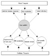
Increasing evidence supports that miRNAs also have important functions in viral replication and may be used by host cells to control viral infection (85). Expression of miRNAs has been reported for various groups of viruses including herpesviruses, small DNA viruses and retroviruses (85). The recent identification of target genes regulated by some of these viral miRNA suggests that they may function in the control of lytic and latent viral replication, in the limitation of antiviral responses, in the inhibition of apoptosis, and in the stimulation of cellular growth (85). Importantly, from the viral perspective, miRNAs encoded in the viral genome have the potential to reshape the cellular environment to maximize viral replication (24,87). In this context they have several potential advantages. Namely, they can specifically down-regulate host cell genes. They also occupy less than 200 nucleotides of the viral genome and are not antigenic (24). One can predict that viral miRNAs probably interfere with aspects of the host adaptive immune responses, including antigen presentation, or innate immune responses, and the interferon system as shown in Fig. 2 (24). It is increasingly clear that miRNAs of both viral and cellular origin can positively or negatively influence viral replication (86). Viral miRNAs can directly alter host physiology, including component of the immune system, and host miRNAs can directly alter the virus life cycle (86). Indeed, there is evidence that viral miRNAs can suppress host cell genes involved in antiviral immunity (47). For instance, a miRNA encoded in the genome of herpes simplex virus-1 (HSV-1), miR-LAT, inhibits apoptosis by targeting TGF-β and SMAD-3, two components of the TGF-β pathway (47). Thus, viral miRNAs can maintain latent infections by protecting infected cells from undergoing apoptosis. Viral miRNAs can also interfere with host antiviral responses by targeting viral genes (87). Taken together, viral miRNAs may contribute to the viral immune escape.
Interestingly, Jopling et al. have shown that IFN-β also induces the down-regulation of the expression of miR-122 (18). Not surprisingly, such a gene regulatory mechanism may also be exploited by virus to facilitate their infections (45). Many viruses have been founded to encode miRNAs that regulate both viral and host mRNA (45). Virus that encode such miRNAs include the Epstein-Barr virus (EBV), cytomegaloviurs (CMV) and Kaposi's sarcoma-associated herpes virus (88-90). Interestingly, a human cytomegaloviurs (HCMV)-encoded miRNA, miR-UL112, repress the expression of MHC-class-1 polypeptide-related sequence-B (MICB) and MICB is a stress-induced ligand of the NK-cell-activating receptor NKG2D, which is required for NK-cell-mediated killing of virus-infected cells (47,48). Interestingly, these results demonstrated that HCMV evades immune surveillance by targeting a cellular mRNA with a virally encode miRNA, and suggest that viruses use miRNAs not only regulate their own life cycles but also evade host immune surveillance (48). It is interesting to note that most viral miRNAs identified so far have no substantial homology to one another or to any known animal miRNAs and that miRNAs are only found in DNA viruses and not in RNA viruses or retroviruses (89,91).
In order to investigate the global and dynamic host miRNAs/mRNAs expression alteration during in vitro acute HCV infection, Liu et al. recently performed a comprehensive microarray analysis using human hepatoma cell (61). They identified, totally, 108 human miRNAs and 1247 mRNAs whose expression levels changed for more than 2.0-fold in response to HCV infection. They identified the differentially expressed miRNAs such as miR-24, miR-149, miR-638, and miR-1181. In addition, the authors showed the miRNAs were involved in HCV entry, replication and propagation (61). These results suggest that combined miRNA and mRNA profiling may have superior potential as a diagnostic and mechanistic feature in HCV infection. Ura et al. also reported that miRNAs are important mediators of HBV and HCV infection as well as liver disease progression, and therefore these miRNAs could be potential therapeutic target molecules (68). Importantly, acute retroviral infection provokes rapid and striking innate immune responses in what has been termed a cytokine storm and IFN-β production is an inaugural event in the innate immune response to viral infections (65). A recent study showed that miRNAs, including miR-26a, miR-34a, miR-145, and let-7b, may directly regulate IFN-β in human and macaque cells and that in primary primate macrophages, the main cell type implicated in human immunodeficiency virus (HIV) and simian immunodeficiency virus infection in the CNS, specific miRNAs reduce, whereas miRNA inhibitors enhance, IFN-β production (65).
The role of miRNAs in regulating gene expression has been increasingly appreciated in a wide spectrum of species including plants, animals and viruses (22,50,76,92,93). It has been shown that miRNAs extensively mediate antiviral defenses in plants and animals (50). For instance, human miRNA hasmiR-32 restricts the replication of retrovirus primate foamy virus type 1 (PFV-1) in human cells (76). Viruses also use miRNAs to regulate host cellular environment. For instance, miR-K12-11 encoded by Kaposi's-sarcoma-associated herpes virus down-regulates an extensive set of common host mRNAs (94). As already mentioned, a cellular miRNA effectively restricts the accumulation of the PFV-1 in human cell and through fortuitous recognition of foreign nucleic acids, cellular miRNAs have direct antiviral effects in addition to their regulatory functions (76). More interestingly, the latency of HIV type 1 (HIV-1) in resting primary CD4+T cells is the major barrier for the eradication of the virus in patients on suppressive highly active antiretroviral therapy (HAART) (95). Even with optimal HAART, replication-competent HIV-1 still exists in resting primary CD4+ T cells. Huang et al. demonstrated that cellular miRNAs potently inhibit HIV-1 production in resting primary CD4+ T cells (95). Interestingly, they have found that the 3' ends of HIV-1 mRNAs are targeted by a cluster of cellular miRNAs including miR-28, miR-125b, miR-150, miR-223 and miR-382, which are enriched in resting CD4+ T cells as compared to activated CD4+ T cells (95). Their data indicate that cellular miRNAs are pivotal in HIV-1 latency and suggest that manipulation of cellular miRNAs could be a novel approach for purging the HIV-1 reservoir (95). In addition to HIV, the H1N1 influenza virus continue to pose serious threat to public health. Recently, Song et al. reported that miR-323, miR-491 and miR-654 inhibit replication of the H1N1 influenza A virus through binding to the PB1 gene (49). Intriguingly, despite the fact that the miRNAs and PB1 mRNA binding sequences are not a perfect match, the miRNAs down-regulate PB1 expression through mRNA degradation instead of translation repression (49). This may be the first demonstration that cellular miRNAs regulate influenza viral replication by degradation of the viral gene. These findings support the notion that any miRNA has antiviral potential, independent of its cellular function, and that the cellular miRNAs play an important role in the host, defending against virus infection (49). To determine whether differential expression of cellular miRNAs plays a role in the host response to the reconstructed 1918 influenza virus (r1918), Li et al. compared the lung cellular "microRNAome" of mice infected by r1918 virus with that of mice infected by a nonlethal seasonal influenza virus, A/Texas/36/91 (23). The authors found that a group of miRNA, including miR-200a and miR-223, were differentially expressed in response to influenza virus infection and that r1918 and A/Texas/36/91 infection induced distinct miRNAs expression profiles (23). Moreover, they observed significant enrichement in the number of predicted cellular target mRNAs whose expression was inversley correlated with the expression of these miRNAs (23). Interestingly, gene ontology analysis revealed that many of these mRNAs play roles in immune response and cell death pathway, which are known to be associated the extreme virulence of r1918 (23). These data strongly suggested that cellular gene expression patterns in influenza virus-infected mice may be attributed to miRNA regulation and that such regulation may be a contributing factor to the extreme virulence of the r1918 (23). More recently, Zhang et al. reported suppression of HBV replication by miR-199a-3p and miR-210 (67). It is interesting to note that accumulating evidence suggest that miRNA control the replication of both RNA and DNA virus (67). Currently, Potenza et al. reported that the analysis of the HBV genome by computer program MiRanda led to the identification of seven sites that are potential targets for human liver miRNAs (96). These site were found to be clustered in a 995-bp segment within the viral polymerase ORF and the overlapping surface antigen ORF, and conserved among the most common HBV subtypes. One of the selected miRNAs, hasmiR-125a-5p, was found to interact with the viral sequence and to suppress the reporter activity markedly (96). Interestingly, the miRNA was then shown to interfere with the viral translation, down-regulating the expression of the surface antigen (96). Overall, these results support the emerging concept that some mammalian miRNAs play a role in virus-host interaction. Furthrmore, the authors provided the basis for the development of new strategies for anti-HBV intervention (96).
Transmissible spongiform encephalopathies (TSEs) or prion disease, are an invariably fatal class of neurodegeneartive disorders (97). However, little is known about their role of miRNAs in neurodegeneration. Using microarrays and RT-PCR, Saba et al. investigated profile of miRNA expression changes in the brains of mice infected with mouse-adapted scrapie (97). The authors found that 15 miRNA were deregulated during the disease process; miR-342-3p, miR-320, let-7b, miR-328, miR-128, miR-139-5p, and miR-146a were over 2.5 fold up-regulated and miR-338-3p, and miR-337-3p over 2.5 fold down-regulated (97). Only one of these miRNAs, miR-128, has been shown to be deregulated in neurodegenerative disease. Taken together, they presented the first comprehensive analysis of miRNA expression in the brain during prion-induced neurodegeneration and reported that differential expression of 15 miRNAs, majority of which are up-regulated during prion disease (97).
As for bacterial infections, tuberculosis remains a major health issue, causing approximately three million deaths every year (50). Mycobacterium tuberculosis, the causative agent for tuberculosis, remains one of the most enigmatic bacteria. The interactions between M. tuberculosis and its environment have been extensively studied. However, our knowledge about potential M. tuberculosis-host interaction at the RNA level is still very limited. Guo et al. presented finding that suggests possible interactions of human miRNAs with M. tuberculosis transcripts and speculated possible roles of human miRNAs in regulating macrophage-M. tuberculosis interactions (50). Using an miRNA target prediction software miRanda followed by analyses of cross-species conservation and miRNA expression profile, they predicted 26 candidate M. tuberculosis genes that may be targeted by human miRNAs expressed in the lung or macrophages, including genes related to M. tuberculosis drug resistance, virulence and growth (50,92). Central to the pathogenic virulence of M. tuberculosis is its ability to persist and multiply within alveolar macrophages after being phagocytosed, forming the primary lesion or tubercle (50,92). Currently, to understand whether miRNAs play roles in regulating immune responses to M. tuberculosis infection in humans, Liu et al. performed miRNA expression profiling in PBMCs from patients with pulmonary tuberculosis and health controls (51). They showed that expression of 30 miRNAs was significantly altered during active tuberculosis as compared with healthy controls and 28 miRNAs were up-regulated and 2 miRNAs down-regulated. They also showed that miR-144* was one of the miRNAs that were overexpressed in active tuberculosis patients. Interestingly, real-time RT-PCR analysis showed that miR-144* was mainly expressed in T cells (51). Additionally, transfection of T cells with miR-144* precursor demonstrated that miR-144* could inhibit TNF-α and IFN-γ production and T cell proliferation (51). They concluded that miR-144* might involve in regulation of anti-tuberculosis immunity through modification of cytokine production and cell proliferation of T cells. As for another bacteria, Helicobacter pylori is a Gramnegative microaerophilic bacterium estimated to colonize more than half the world's population (46,98). It typically establishes a life-long infection with the stomach, where it induces a marked immune response with local infiltration of neutrophil, lymphocytes and macrophages (46,98). H. pylori is the main cause of peptic ulceration and gastric adenocarcinoma and it has been shown to cause B cell mucosa associated lymphoid tissue lymphoma, while several mechanisms involved in adenocarcinoma induction have been described, mechanisms underlying the onset of other H. pylori-implicated lymphomas remain obscured (46,98). Currently, a study showed that H. pylori infections alter the expression of oncogenes, tumor suppressor genes and miRNAs (52). Xiao et al. reported that H. pylori was able to increase the miR-155 expression in gastric epithelial cell lines and gastric mucosal tissues, and nuclear factor-κB (NF-κB) and activator protein-1 pathway were required for the induction of miR-155 (46). They also found that miR-155 may down-regulate IκB kinase ɛ, Sma- and Mad-related protein (SMAD2), and Fas-associated death domain (FADD) protein. Furthermore, the authors showed that the over-expression of miR-155 negatively regulated the release of IL-8 and growth-related oncogene-α (46). This study may provide the first description of increased expression of miR-155 in H. pylori infection, and miR-155 may function as novel negative regulator that help to fine-tuning the inflammation of H. pylori infection (46). Interestingly, Belair et al. reported that miR-21 was also over-expressed in chronically H. pylori-infected gastric epithelium tissue, as opposed to noninfected tissue (66). Recently, Fehri et al showed that miR-155 is induced by H. pylori (98). Currently, Matsushima et al. determined expression pattern of miRNAs in gastric mucosa infected with H. pylori (99). Using endoscopic biopsy specimens, the authors determined miRNA expression patterns in H. pylori-infected gastric mucosa by microarray (99). They quantitated the differentially expressed miRNAS by real-time RT-PCR. An in vitro infection model was assessed to monitor the regulation of miRNAs in gastric epitherlium in response to H. pylori. The comprehensive method unraveled the expression profile. Namely, among 470 human miRNAs loaded, 55 were differentially expressed between H. pylori-positive and -negative subjects. They showed that expression levels were significantly decreased in 30 miRNAs, whereas hsa-miRNA-223 was the only miRNA to be overexpressed on quantitative RT-PCR and that eight miRNAs enabled discrimination of H. pylori status with acceptable accuracy. They reported that gastritis scores of activity and chronic inflammation according to the updated Sydney system correlated significantly with the expression levels of diverse miRNAs (99). Interestingly, they showed that cure of the infection with an anti-H. pylori regimen restored decreased expression of 14 of the 30 miRNAs and that expression levels of some miRNAs, including let-7 family members, were significantly altered following infection with a virulent H. pylori strain carrying intact cag pathogenicity island including cagA but not isogenic mutant (99). These results may provide insight into miRNA involvement in the pathogenesis of H. pylori-associated gastritis.
As regards Salmonella infection, Tili et al. showed treatment of immune cells with bacterial lipopolysaccharide (LPS) from Salmonella and Escherichia coli led to the induction of miR-155, miR-132 and miR-146a expression (54). Most recently, Schulte et al. investigated the role of miRNAs in mounting defences against such an intracellular bacteria, using mouse and human in vitro systems that recapitulate infection by Salmonella enterica, the agent of gastroenteritis (53). Deep sequencing revealed that Salmonella significantly induces several miRNAs within 4 h of mouse macrophage infection (53). Perhaps not surprisingly, these miRNAs chiefly induced miR-155 and miR-146a, as well as miR-21 (53). Strikingly, several members of the let-7 miRNA gene family were, by contrast, rapidly down-regulated. Mutant Salmonella defective in cell invasion and/or intracellular replication also triggered let-7 down-regulation, suggesting that the process involves sensing of extracellular bacterial antigen (53). This study identifies the let-7 family as the common denominator of Salmonella-regulated miRNAs in macrophages and epithelial cell, and suggests that repression of let-7 relieves cytokine IL-6 and IL-10 mRNAs from negative post-transcriptional control. Intriguingly, while IL-6 is pro-inflammatory, IL-10 prevents the detrimental consequences of systemic inflammation, suggesting that the observed response is tightly balanced and hence biologically relevant (100). These results establish a paradigm of miRNA-mediated feed-forward activation of inflammatory factors when mammalian cells are targeted by bacterial pathogens (53).
miRNAs IN SICKLE CELL DISEASE (SCD)
Since mature erythrocytes are terminally differentiated cells without nuclei and organelles, it is commonly thought that they do not contain nucleic acids (55). Chen et al. investigated this issue by analyzing the transcriptome of a purified population of human mature erythrocytes from individuals with normal hemoglobin (HbAA) and homozygous sickle cell disease (HbSS) (56). They found a dramatic difference in their miRNA expression pattern and also reported that miR-320 played an important role for the down-regulation of its target gene, CD71 during reticulocyte terminal differentiation (56). Importantly, they also provided several lines of evidence that human mature erythrocytes, although lacking in ribosomal and large-sized RNAs, contain diverse and abundant miRNAs (56). Further investigation revealed that poor expression of miR-320 in HbSS cells was associated with their defective down-regulation CD71 during terminal differentiation (56). Thus, the global analysis of miRNA expression in circulating erythrocytes can provide mechanistic insights into the disease phenotypes of erythrocyte disease.
In human trisomy 13, there is delayed switching and persistence of fetal hemoglobin (HbF) and elevation of embryonic hemoglobin in newborns. Currently, using partial trisomy cases, Sankaran et al. mapped this trait to chromosomal band 13q14; by examining the genes in this region, two miRNAs, miR-15a and miR-16-1, appear as top candidates for the elevated HbF levels (57). Indeed, increased expression of these miRNAs in primary human erythroid progenitor cells results in elevated fetal and embryonic hemoglobin gene expression (57). Moreover, they showed that direct target of these miRNA, MYB, plays an important role in silencing the fetal and embryonic hemoglobin genes (56). These data suggest that miR-15a, miR-16-1, and MYB may be important therapeutic targets to increase HbF levels in patients with sickle cell disease and β-thalassemia (57).
miRNAs IN ENDOMETRIUM DISEASES
During the menstrual cycle, human endometrium undergoes extensive cyclic, morphologic and biochemical modifications in preparation for embryo implantation that are remarkably consistent during each cycle throughout the reproductive years (58). This process, which begins with degenerative signals resulting in menstrual bleeding and endometrial shedding, integrates many overlapping and dynamic events to regenerate and become receptive (58). Pan and Chegini et al. provided evidence showing the endometrial expression of miRNAs and their potential regulatory functions under normal and pathologic conditions such as endometeriosis, dysfunctional uterine bleeding, and endometrial cancer (58). Recently, a study assessed miRNA expression by microarray analysis in paired ectopic and eutopic endometrial tissues (70). They identified 14 up-regulated (miR-145, miR-143, miR-99a, miR-99b, miR-126, miR-100, miR-125b, miR-150, miR-125a, miR-223, miR-194, miR-365, miR-29c, and miR-1) and 8 downregulated (miR-200a, miR-141, miR-200b, miR-142-3p, miR-424, miR-34c, miR-20a and miR-196b). In addition, functional analysis suggested that the 673 miRNA targets constitute molecular pathways previously associated with endometriosis, including c-Jun, CREB-binding protein, protein kinase B (Akt), and cyclin D1 (CCND1) signaling (70). Accordingly, miRNAs are potential therapeutic targets for treating this disease.
CONCLUSION
Emerging evidence has demonstrated that miRNAs play a major role in a wide range of developmental process including cell proliferation, cell cycle, cell differentiation, metabolism, apoptosis, developmental timing, neuronal cell fate, neuronal gene expression, brain morphogenesis, muscle differentiation and stem cell division. Recent studies provided clear evidence that miRNAs are abundant in the lung, liver and kidney and modulate a diverse spectrum of their functions. Moreover, a large number of studies have reported links between alterations of miRNA homeostasis and pathological conditions such as infectious diseases, sickle cell disease and endometrium diseases as well as lung, liver and kidney diseases. Interestingly, miRNA deficiencies or excess have been correlated with a number of clinically important diseases ranging from infectious diseases to liver diseases. Particularly, accumulating evidence indicates that viruses use their own miRNAs to manipulate both cellular and viral gene expression. Furthermore, viral infection can exert a profound impact on the host cellular miRNA expression profile, and several RNA viruses have been reported to interact directly with cellular miRNAs and/or to use these miRNAs to augment their replication potential. It is therefore unsurprising that due to their non-immunogenic nature, viral miRNAs represent an elegant tool for the virus to evade the host immune system and likely play a key role in the latent/lytic switch during the viral life cycle. Here I briefly summarized the newly discovered roles of miRNAs in various human diseases including lung, liver and kidney diseases as well as infectious diseases, sickle cell disease and enodmetrium diseases. In the future, distinct miRNA signature involved in diseases should define the role of miRNA in biochemical and immunobiological processes. Furthermore, identification of miRNAs of significance in disease could provide rationale for the design and implementation of disease classification, early detection, disease prognosis and successful therapeutic decision making.

 XML Download