

 PDF
PDF ePub
ePub Citation
 Print
Print


INTRODUCTION
Malaria is the most prevalent parasitic disease in the world, with an estimated 500 million cases arising annually, and 1 to 3 million deaths is attributable to this disease (1). Furthermore, most of victims are under 7-yr old. Of the 4 species of Plasmodium that infect humans, P. falciparum has been studied most extensively, partly due to the severity of the disease and partly due to the successful in vitro cultivation of this organism during its erythrocytic stages.
Much progress has been made since the first report of the continuous cultivation of this parasite, leading to its long-term cultivation in vitro (2, 3). The Petri dish method is the most popular and the easiest to carry out (4). The optimal range for each culture condition of the Petri dish method has been determined (5-7). When the Petri dish method was established, it was reported that these organisms grow well in aged erythrocytes compared with freshly obtained erythrocytes (4). However, there has been no subsequent experimental evidence that clarifies the relationship between the age of the erythrocytes and the multiplication of P. falciparum.
In addition, despite the apparent simplicity of the Petri dish method, it is labor-intensive because the medium should be changed everyday. It would be convenient if the interval for changing media could be prolonged. In the present study, we investigated the rate of in vitro multiplication of P. falciparum with regard to the "age" of erythrocytes; i.e., the length of time in storage following collection of the blood from which these erythrocytes were obtained and the interval at which the medium was changed.
MATERIALS AND METHODS
Parasite strain, serum, culture medium, and erythrocytes
P. falciparum FCR-3/FMG strain (No. 30932) was obtained from the American Type Culture Collection (ATCC) (Manassas, Va). The culture medium consisted of RPMI medium 1640 (GIBCO BRL, Rockville, MD, U.S.A.) supplemented with 25 mM HEPES, 2 g/L NaHCO3, 50 µM hypoxanthine, 2 mM L-glutamine, 30 mM D-glucose, 1 mg/L glutathione, and 25 mg/L gentamicine (hereafter abbreviated as RP). Each blood of 5 different malaria-naive adult volunteers was collected in a Vacutainer® tube (Becton Dickinson, Franklin Lakes, NJ, U.S.A.) containing 0.057 mL of 15% EDTA or 1.5 mL of ACD, respectively, and stored at 4℃ for up to 4 weeks. After being washed with RP twice, the erythrocytes were suspended in RP supplemented with 10% human AB+ serum (RP-10S); this was referred as the uninfected blood cells (UIBC) suspension. Infected erythrocytes were obtained from a previous culture. The cultured cells were washed with RP twice, suspended in the RP-10S, and referred as the infected blood cëlls (IBC) suspension.
Culture procedure
The initial culture and subculture were performed as described previously (8). For routine subculture, the IBC suspension obtained from a previous culture was mixed with the UIBC suspension to make a final erythrocyte suspension of 5% that reached in a parasitemia of 0.1%. The cell suspension was maintained in a medium with the depth of 4 mm. Culture plates were incubated with shaking at 60 rpm and at 37℃ in a 3-way incubator with a gas mixture of 5% CO2, 5% O2, and 90% N2. The culture medium, in which the erythrocytes in an ACD tube were used, was exchanged every 12 hr, and the other culture medium, in which the erythrocytes in an EDTA tube were used, was exchanged every 12, 24, and 48 hr. Blood films were normally prepared from each culture every day and stained with Giemsa's stain to determine the parasitemia level. Studies were performed on erythrocytes obtained from 5 different individuals under the same condition and the results were read by one expert. The final parasitemia level was calculated as a percentage of infected erythrocytes out of the total number of erythrocytes counted.
RESULTS
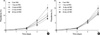
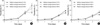
Fig. 1 shows the multiplication rate of P. falciparum using erythrocytes with various ages. In vitro multiplication rates of P. falciparum decreased as the age of erythrocytes increased. This trend was consistent regardless of the anticoagulant or the interval of medium change. On the fourth day after inoculation (2 rounds of asexual erythrocytic cycle), the parasitemia level in the culture using 28-day old erythrocytes in an ACD tube reached approximately 70% of that using freshly collected erythrocytes. On the other hand, the parasitemia level in the culture using 28-day old erythrocytes in an EDTA tube reached approximately half of that using freshly collected erythrocytes. To determine the relationship between the interval of medium change and the rate of in vitro multiplication of P. falciparum, we tested 3 different intervals, 12, 24 and 48 hr, with using the anticoagulant of EDTA. As shown in Fig. 2, we found similar parasitic multiplication in cultures in which the media had been changed every 12 to 24 hr, irrespective of the age of the erythrocytes used in each culture. In cultures in which the media had been changed every 48 hr, the multiplication rate decreased by 23% to 50% compared with the culture of which medium was changed every 12 or 24 hr, particularly at the fourth day after inoculation. By contrast, the parasite multiplication rate seemed not to differ during the first 3 days regardless of the medium change interval.
DISCUSSION
Since its initial success, many investigators have concentrated their efforts to determine optimal conditions of the Petri dish method, including the depth of the culture medium, the hematocrit in the medium, and the condition of gas mixture (4, 5, 9-11). A previous study established that outdated erythrocytes, which had been stored for longer than 4 weeks, were better for cultivation of P. falciparum than freshly collected erythrocytes (4). Their result was shown to somewhat contradictory to the common sense that fresh cells can provide intracellular organisms with more favorable environment for multiplication than old ones, although many studies were performed using aged erythrocytes for a while based upon the result of Jensen and Trager (5, 12, 13).
However, freshly collected erythrocytes were used in several recent studies (14, 15), which raised the necessity to verify the difference of in vitro multiplication rate according to the age of erythrocytes used for cultivating P. falciparum. In a previous study performed by Jensen and Trager, the parasitemia of the first inoculation differed for each culture (4). Since a starting parasitemia is one of the main factors that determine multiplication rates, the differences shown by their study was hard to evaluate their findings regarding parasite multiplication (6). In the present study, the starting parasitemia level was adjusted to 0.1%, and we investigated the influence of erythrocyte age on parasite multiplication for a much longer period of time, up to 28 days after blood collection. Our results demonstrate that freshly collected erythrocytes provide the multiplication of P. falciparum better than aged erythrocytes regardless of the anticoagulant and that the multiplication rate of this parasite was inversely correlated with the age of erythrocyte, which is opposed to the result of a previous study (4). A previous study demonstrated that a parasitemia level >10% with a hematocrit of 5% could be achieved when the medium was changed every 12 hr. In the present study, however, supplement of freshly collected erythrocytes in the media resulted in parasitemia exceeding 12% by the fourth day (2 rounds of asexual erythrocytic cycle) after inoculation when the medium was changed every 12 or 24 hr. This parasitemia is 2.5 to 6.0 times greater than was seen in a previous study (6).
P. falciparum had been known to invade erythrocytes at any stage, while P. vivax preferentially invades reticulocytes. Previous investigations have found that P. falciparum also invades younger blood cells more efficiently, including reticulocytes (16, 17). As the length of blood storage increases, erythrocytes become older and a subpopulation of young erythrocytes is reduced, thereby reducing the ability of the parasite to invade erythrocytes. Furthermore, schizogony cycle, which is essential for parasitic multiplication, is completed in erythrocytes (7). Along with the time lapse after blood collection, erythrocyte membrane becomes more fragile, which results in unfavorable environment for the completion of schizogony; this may be one of the ascribable factors that reduce the multiplication rate for the parasites in aged erythrocytes. On the other hand, results of the present study showed that the anticoagulant of ACD was better for the erythrocyte invasion of P. falciparum than EDTA especially when old erythrocytes stored longer than one week were used. It strongly indicates that ACD is less cytotoxic than EDTA for the long-term preservation of cells, which is consistent with the previous study (18). However, freshly collected erythrocytes provided the multiplication of P. falciparum better than aged erythrocytes even when ACD was used as an anticoagulant, which was the direct opposition of the result of a previous study (4).
In spite of its simplicity, the Petri dish method is labor-intensive because the medium should be changed everyday (11). It is believed that frequent medium changes can result in removal of the lactic acid that is produced by the parasites and can cause the medium pH to fall to an unfavorable level (7). A previous study indicated that daily medium change was not necessary only when the hematocrit was low (1%), while the medium was in a shaking flask (12). In the present study, the rate of the parasite growth did not differ much regardless of the medium change interval when the parasitemia level was lower than 8% with a hematocrit of 5%. When the parasitemia level exceeded 8% and the media was changed every 48 hr, the multiplication rate showed great decrease. This difference might be attributed to the large amount of lactic acid produced in culture by this organism and the subsequent fall in pH. Nevertheless, our results suggest that the medium may be changed every other day when a high (5%) hematocrit is used until the parasitemia level exceeds 8%. Use of this protocol can result in parasitemia level increasing 100-fold in 4 days (2 rounds of asexual erythrocytic cycle), which corresponds with an approximately 10-fold increase during in each cycle. This yield is approximately twice that achieved in previous studies and with a less frequent medium change schedule. The obvious improvement in yield in the present study might be due to the use of freshly collected erythrocytes in the culture (5).
In conclusion, the results of the present study suggest that the rate of in vitro multiplication of P. falciparum was higher in freshly collected erythrocytes than in aged erythrocytes regardless of the anticoagulant used. In cultures containing freshly collected erythrocytes with a hematocrit of 5%, the medium may be changed every other day at a parasitemia level below 8%. Through this way, the most troublesome matter of the Petri dish method can at least partially be overcome.
 XML Download