Abstract
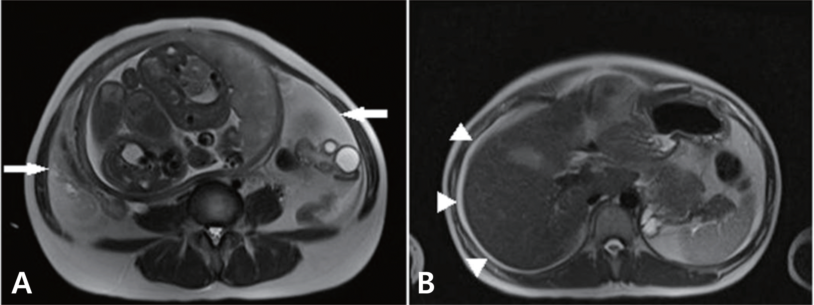
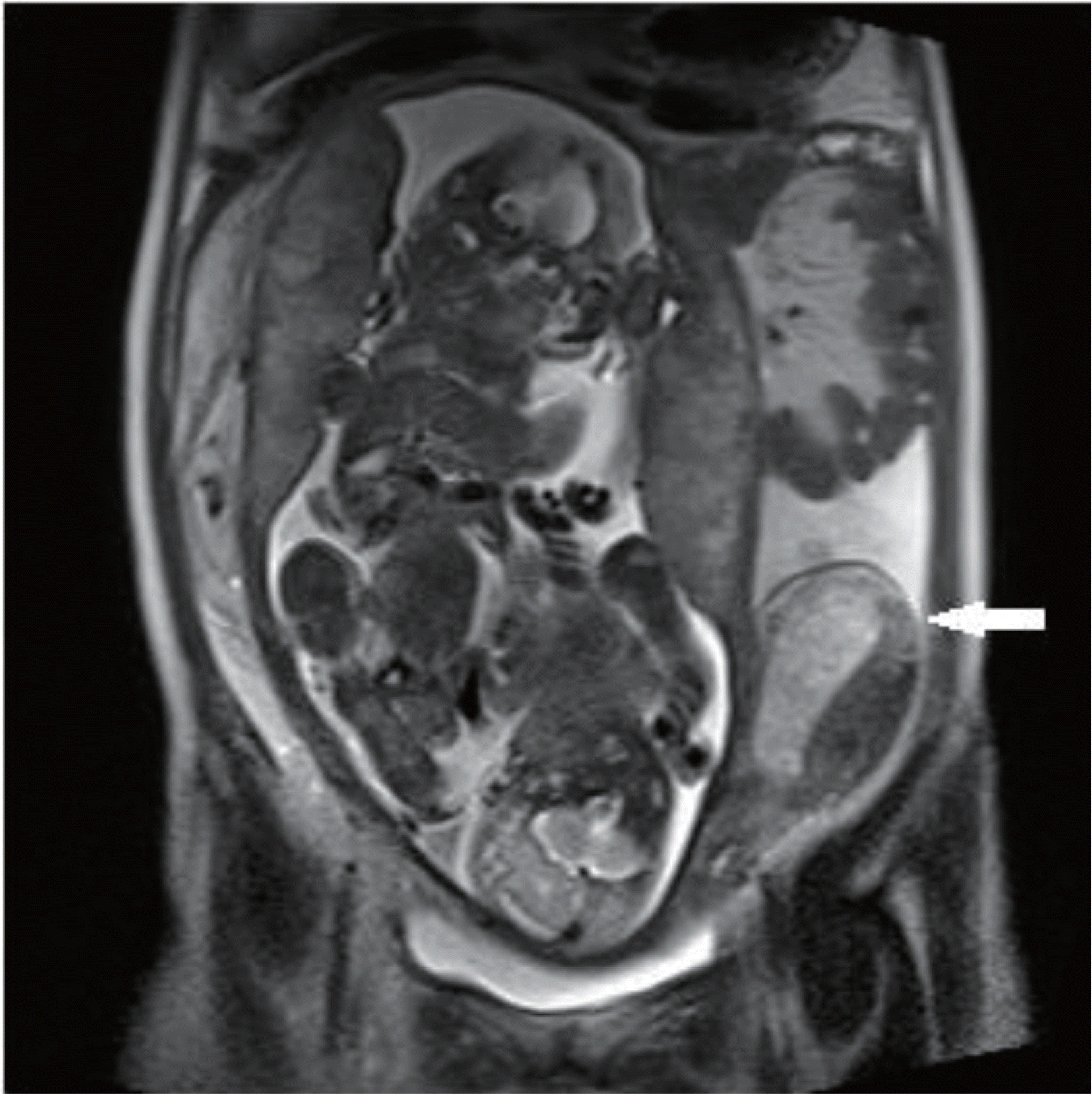
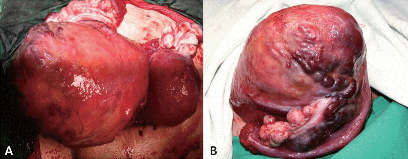
At early stage of pregnancy, hemoperitoneum often occurs in heterotopic ectopic pregnancy or bleeding of hyperstimulated ovary and can be managed easily by laparoscopic surgery while maintaining pregnancy. But in the 3rd trimester pregnancy, surgical management without delivery is very difficult and preterm birth is inevitable because of life-threatening complications not only for mother but fetus. We present a woman with 31 weeks and 3 days’ gestation and spontaneous hemoperitoneum that was treated by conservative management without preterm delivery successfully. A review of the literature was undertaken.
REFERENCES
1.Aziz U., Klkarni A., Lazic D., Cullimore JE. Spontaneous rupture of the uterine vessels in pregnancy. Obstet Gynecol. 2004. 103(5, Pt2):1089–91.

2.Williamson H., Indusekhar R., Clark A., Hassan IM. Spontaneous severe haemoperitoneum in the third trimester leading to intrauterine death: Case report. Case Rep Obstet Gynecol. 2011. 173097. DOI: doi: 10.1155/2011/173097.

3.Muhihir JT., Nyango DD. Massive haemoperitoneum from endometriosis masquerading as ruptured ectopic pregnancy: case report. Niger J Clin Pract. 2010. 13:477–9.
4.Lee HJ., Hong JS., Im ES., Jeon YT., Park KH. Fatal spontaneous utero-ovarian vessel rupture during pregnancy in a patient with endometriosis. J Womens Medicine. 2009. 2:121–4.
5.Vyshka G., Capari N., Shaqiri E. Placenta increta causing hemoperitoneum in the 26th week of pregnancy: a case report. J Med Case Rep. 2010. 4:412. DOI: doi: 10.1186/1752-1947-4-412.

6.Esmans A., Gerris J., Corthout E., Verdonk P., Declercq S. Placenta percreta causing rupture of an unscarred uterus at the end of the first um Reprod. 2004. 19:2401–3.
7.Swaegers MC., Hauspy JJ., Buytaert PM., De Maeseneer MG. Spontaneous rupture of the uterine artery in pregnancy. Eur J Obstet Gynecol Reprod Biol. 1997. 75:145–6.

8.Giulini S., Zanin R., Volpe A. Hemoperitoneum in pregnancy from a ruptured varix of broad ligament. Arch Gynecol Obstet. 2010. 282:459–61.

9.Hashimoto K., Tabata C., Ueno Y., Fukuda H., Shimoya K., Murata Y. Spontaneous rupture of uterine surface varicose veins in pregnancy. J Reprod Med. 2006. 51:722–4.
10.Park YJ., Ryu KY., Lee JI., Park MI. Spontaneous uterine rupture in the first trimester: a case report. J Korean Med Sci. 2005. 20:1079–81.

11.Shiau CH., Hsieh CC., Chiang CH., Hsieh TT., Chang MY. Intrapartum spontaneous uterine rupture following uncomplicated resectoscopic treatment of Asherman's syndrome. Chang Gung Med J. 2005. 28:123–7.
12.Hua M., Odibo AO., Longman RE., Macones GA., Roehl KA., Cahill AG. Congenital uterine anomalies and adverse pregnancy outcomes. Am J Obstet Gynecol. 2011. 205:558. .e1-5.

13.Jayaprakash S., Muralidhar L., Sampathkumar G., Sexsena R. Rupture of bicornuate uterus. BMJ Case Rep. 2011. pii: bcr08 20114633. DOI: doi: 10.1136/bcr.08.2011.4633.

14.Koifman A., Weintraub AY., Segal D. Idiopathic spontaneous hemoperitoneum during pregnancy. Arch Gynecol Obstet. 2007. 276:269–70.

15.Maya ET., Srofenyoh EK., Buntugu KA., Lamptey M. Idiopathic spontaneous haemoperitoneum in the third trimester of pregnancy. Ghana Med J. 2012. 46:258–60.
16.Brosens IA., Fusi L., Brosens JJ. Endometriosis is a risk factor for spontaneous hemoperitoneum during pregnancy. Fertil Steril. 2009. 92:1243–5.


 PDF
PDF ePub
ePub Citation
 Print
Print


 XML Download