

 PDF
PDF ePub
ePub Citation
 Print
Print


Introduction
The prevalence of antibiotic resistance is a continual problem due to the evolution of a potent defense mechanism against antibiotics [1]. One of the main concerns is spread of various diseases by fish and food-related pathogenic bacteria, which can affect both the aquaculture and agriculture sectors leading to economic losses. Therefore, it is necessary to exploit and develop a novel inhibitory agent against those bacteria. An increasing number of studies have considered plant extracts for immune enhancement and antimicroorganism activities [2,3]. Essential oils as well as ethanol and methanol extracts of plants possess antimicrobial activity [4,5], which can be used as antibacterial agents.
The appearance and development of fish and food-related diseases are the result of the interaction among pathogens, hosts, and the environment. Prophylactic chemo-therapeutants have been commonly used in the fish and food industries for controlling opportunistic bacteria. Hence, nonchemotherapeutic methods for microbial control, such as the use of vaccine, probiotics, immune-stimulants, and natural therapeutics from plants have been considered [6,7].
Finding new naturally active components from plants or plant-based agricultural products has been of interest to many researchers. Hence, a great deal of attraction has been paid to the antibacterial activity of citrus as a potential and promising source of pharmaceutical agents [8-11]. Furthermore, citrus fruit peel has been used in traditional Asian medicines for centuries to treat indigestion and to improve bronchial and asthmatic conditions [12]. Ghasemi et al. [13] and Johann et al. [14] have shown that citrus varieties are considered and containing a rich source of secondary metabolites with the ability to produce a broad spectrum of biological activities. Yao et al. [15] reported that seven individual polymethoxylated flavones can be obtained from Citrus sinensis peel. Moreover, different parts of Citrus reticulate (peel, pith, endocarp, pulp, and seeds) have been used to identify five kinds of flavonoids, including hesperidin, naringin, didymin, tangeretin and nobiletin by high performance liquid chromatography-photodiode array [16]. In addition, shoots of germinated citron seeds (Citrus junos Sieb. ex Tanaka) have significant antioxidant and antilipidemic activities due to their high content of flavonoids in methanol extracts [17]. Some significant components are abundantly available in citrus peel, including ascorbic acid, phenolic acids, polyphenols, and dietary fiber [18]. Constituents with antioxidant, antiviral, antibacterial, antifungal, and anticancer activities have also been reported in citrus [19-21]. Numerous studies have described the inhibitory activities of citrus against human pathogens, fungi, and yeasts. However, only a few studies have reported on the antibacterial effects against fish [22] and food pathogens [23].
Our previous studies have revealed that citrus by-products (CBPs) from citrus peel, which are comprised of flavonoid compounds, might be a useful antimicrobial agent [24,25]. Senevirathne et al. [26] have shown that enzymatic digests from CBPs dried by high speed drying (HSD) or freeze drying (FD) are responsible for the antioxidant properties in the in vitro assays conducted. In addition, citrus essential oils, including citrullene and limonene from CBPs have antimicrobial effects against a broad range of microorganisms [27]. Therefore, the aim of the present study was to investigate the antibacterial activity of a dried CPC methanol extract by HSD and far-infrared radiation drying (FIR) against pathogenic bacteria by comparing the results with those of FD dried CPC. We used 11 fish and five food-related pathogenic bacteria to evaluate the effectiveness of the HSD and FIR drying systems compared to that of the qualified FD method using maximum inhibitory concentrations (MIC), minimum bactericidal concentration (MBC), and scanning electron microscopy (SEM) techniques in accordance with the antibacterial activity of dried CPCs, and to describe the alternative beneficial value of processing and utilizing CPCs.
Materials and Methods
Material and chemicals
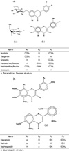
CPCs were obtained from Jeju Provincial Development Co. in Korea. Nutrient agar (NA), nutrient broth (NB), brain heart infusion agar (BHIA) and brain heart infusion (BHI) were purchased from Difco (Sparks, MD, USA). Authentic reference compounds, including narirutin, quercetagetin, sinesetin, 3',4',7,8-tetramethoxyflavone, 5,6,7,3',4',5'-hexamethoxyflavone, and scutelarin tetramethyl ether were purchased from Extrasynthase (Genay, France). Hesperidin and neohesperidin were purchased from Sigma Co. (St. Louis, MO, USA). Nobiletin and tangeritin were purchased from Wako Pure Chemicals (Osaka, Japan), and 3,5,6,7,8,3'4', heptamethoxyflavone was provided by the Pharmacy Department of Tokyo University. All other chemicals used were of analytical grade.
Microorganisms and culture
Fish and food-related pathogenic bacteria were kindly provided by the Marine Microbes Laboratory at Jeju National University, Korea. The bacterial strains were cultured at an appropriate temperature as given in Table 1. The optical densities of each working culture were adjusted to 0.1 mg/mL at 625 nm using fresh broth to give standard inoculums of 1 × 10⁶ colony-forming units (cfu) per mL.
HSD or FIR of CPCs
CPCs stored at -50℃ were converted to a dried form by HSD (Okadora, Incheon, Korea) or FIR drying (TOURI-Q, Seoul, Korea) as explained in our previous studies [24,28]. Then, the dried CPCs were pulverized into fine powder using a grinder (MF 10 basic mill, GMBH & Co., Staufen, Germany) and passed through a 300 mm standard testing sieve (no. 50).
Extraction of bioactive compounds from dried CPC
A 20 gram portion of the ground dried CPC powder was mixed with 100% methanol and kept in a shaking incubator at 25℃ for 1 day, and then it was filtered in a vacuum using Whatman no. 1 (Whatman Ltd., Maidenstone, England) filter paper. The methanol was evaporated in a rotary evaporator, and the extracts were dissolved in 1% dimethyl sulfoxide (DMSO).
Disc diffusion assay
The CPC extract was screened for antibacterial activity against 16 bacterial strains using agar diffusion technique [29,30]. The bacterial strains were grown in nutrient agar (NA) and brain heart infusion agar (BHIA) (Difco Laboratories, Detroit, MI). Sterilized paper disks (Whatman no. 1, 6 mm diameter) containing 50 µL of each extract were applied to the surface of agar plates that were previously seeded by spreading 0.1 mL of culture overnight. Ampicillin and 1% DMSO was used as positive and negative controls, respectively. The plates were incubated overnight at respective temperatures, and the diameter of the clear zone was measured in millimeters. The measurement scale was the following: > 20 mm clear zone was strong inhibitory activity; < 12-20 mm clear zone was moderate/mild inhibitory activity; and < 12 mm was low inhibitory activity.
Determination of MIC
MIC was defined as the minimum concentration of the test extract that was required to limit turbidity (growth) to < 0.05 absorbance units. The MICs of the extracts from the dried CPCs were assessed by a modified microdilution broth method described by Cai and Wu [31]. Briefly, bacteria from overnight cultures were adjusted to 1 × 10⁶ CFU/mL. The extracts were serially diluted with broth to give concentrations of 0.5, 1, 2, 4, 8, and 16 mg/mL. A 100 µL aliquot of the diluted extracts was added to wells containing 100 µL of bacterial suspension. A parallel series of uninoculated broth samples were used as blanks. After 24 h incubation at an appropriate temperature under anaerobic conditions, optical density was measured at 600 nm using a microtiter plate reader (Tecan, Vienna, Austria).
Determination of MBC
The MBC was defined as the lowest concentration of the extract that allowed < 0.1% of the original inoculum treated with the extracts to survive and grow on the surface of the medium used. A method described by Patrick [32] in the ASM Pocket Guide to Clinical Microbiology was slightly modified to determine MBC in this study. Briefly, a 50 µL aliquot of the mixture was withdrawn from the MIC assay wells where no visible turbidity (growth) was observed and spread on freshly prepared BHIA/NA plates. The plates were incubated at an appropriate temperature for 24 h to determine the MBC.
SEM observations
Bacterial cells were suspended in NA or BHI broth and incubated overnight at 37℃ or 30℃ respectively. Both suspensions were divided equally into four tubes accordingly. The extracts from the dried CPCs (8 mg/mL) were added to the tubes and incubated at 30℃ for 4 h. One tube was used as a control for each (no extract). Later, bacterial cells were harvested and prefixed with 2.5% glutaraldehyde solution at 4℃ overnight followed by resuspension in 0.1 M Na-cacodylate buffer. The mixture was dehydrated rapidly in an ethanol series (30, 50, 70, 90, and 100%). Then, bacterial cells were dried with liquid CO2 at "critical point" (Balzers CPD 030) under 95 bars of pressure and gold-covered by cathodic spraying (Edwards S 150 B). Finally, bacterial cell morphology was observed by SEM (JEOL, JSM 6700F, Tokyo, Japan).
Statistical analysis
All experiments were conducted in triplicate (n = 3) and an analysis of variance (using SPSS 11.5 statistical software, Chicago, IL, USA) was used to compare the mean values of each treatment. Significant differences among the means of parameters were determined using Duncan's test. A P < 0.05 was considered significant.
Results
Growth inhibitory activity
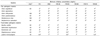
Eleven fish pathogenic bacteria (nine Gram-negative and two Gram-positive) and five food-related pathogenic bacteria (two Gram-negative and three Gram-positive) were tested for their sensitivity against methanol extracts from CPCs prepared using different drying methods. Antibacterial potency was initially determined by the disc diffusion assay. Table 2 presents the inhibition zone diameters (clear zones around discs) formed by the various extracts against the bacteria. The CPC extracts dried by HSD or FIR showed strong inhibitory activity against fish pathogenic bacteria strains, as > 21 mm clear zones were observed with respect to Streptococcus iniae, Vibrio ichioenteri, and S. parauberis. In addition, those extracts showed higher inhibitory activities against other fish and food-related bacterial strains, and the activities were almost the same as with the CPC extract dried by FD (inhibition zone, 9-20 mm). The inhibition zones were very prominent when compared with that of ampicillin, which is a commercial antimicrobial agent (positive control). Vibrio salmonicida was more susceptible to the extracts and showed the smallest inhibition zones (9-11 mm). However, the methanol extracts from CPCs dried by HSD or FIR showed antibacterial activity in both Gram-negative and Gram-positive bacteria.
Susceptibility test
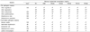
A quantitative evaluation of the antibacterial activity of the extracts from the dried CPCs was carried out against selected bacteria according to the method described by Cai and Wu [31]. The MICs of the extracts to the fish and food-related pathogenic bacteria are shown in Table 3. The MIC values were 2-8 mg/mL for the fish pathogenic bacteria and both extracts from CPCs dried by HSD or FIR showed strong activities against S. iniae, V. anguilarum, S. parauberis, and V. parahemolyticus. These values were almost the same as those of CPC extracts dried by FD. Furthermore, all extracts showed moderate MIC values against the V. alginolyticus and V. ichyienter strains tested. In contrast to the food-related pathogenic bacteria, MIC values were 0.5-8 mg/mL. Strong activity was evident against L. monocytogens, S. aureus, and Bacillus subtilis at 0.5-2 mg/mL, which was significant. These values were also the same as those of the HSD and FIR extracts from dried CPCs with the extract from CPCs dried by FD. All extracts showed moderate MIC values against Salmonella thyphimurium and E. coli. The MBC results against fish and food-related pathogenic bacteria are shown in Table 4. The MBC results were 1-16 mg/mL. In contrast to the food-related pathogenic bacteria, strong inhibitory activities were shown against Listeria monocytogens and S. aureus at 1-4 mg/mL. Moreover, moderate MBC values were exhibited at 4-16 mg/mL against all other tested fish and food-related pathogenic bacteria. These MBC values were the same for all extracts dried by HSD and FIR compared to that of those dried by FD. Therefore, these results further confirmed the potential activities of the CPC extracts.
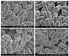
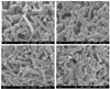
SEM observations
The CPC extracts dried by HSD, FIR, and FD were added to S. iniea and B. subtilis bacteria strains and observed by SEM (Figs. 1 and 2 respectively). The SEM results suggested that the CPC extracts caused morphological changes in the surface of the treated bacteria compared to those of the control. Untreated bacterial cells (control) remained intact and showed a smooth surface, whereas the treated bacterial cells showed damage or distinctive changes in cell membranes.
Discussion
Efficient, fast, and economically reliable drying methods were used to prepare HSD and FIR extracts from CPCs and was compared with that of FD in our previous studies [24,25]. Furthermore, total flavonoid contents in the methanol extracts from the CPCs, dried by HSD, FIR, and FD were determined to be 477.4 mg/100 g, 463 mg/100 g, and 624 mg/100 g, respectively. All extracts from dried CPCs had hesperidin and narirutin in large quantities as compared to other flavonoids [24,25]. We focused on the antibacterial activity of some of the different flavonoid compounds found in the extracts (Table 5). Hence, the antibacterial activities shown in this study might be due to the presence of a variety of flavonoid or phenolic compounds. The synergistic effects of those flavonoids may have resulted in higher antibacterial activities, and they are thermostable, as they showed activity even after being dried at very high temperatures during HSD compared to those of FIR. However, the FIR drying process is shorter drying time than that of FD [26]. Moreover, differences in the activities of the CPC extracts could be partially explained by variations in flavonoid content and strain sensitivity. Yi et al. [9] showed that hesperidin is more active against bacterial growth than that of polymethoxylated flavones such as tangeritin and nobiletin, whereas various flavanones such as hesperitin and naringenin are active against Helicobactor pylori [33]. The antimicrobial activity of purified flavonoids has also been described [34].
The reason for the lower sensitivity of the Gram-negative bacteria compared to that of Gram-positive bacteria could be due to differences in their cell wall composition. Gram-positive bacteria contain an outer peptidoglycan layer, which is an effective permeability barrier [35], whereas Gram-negative bacteria have an outer phospholipidic membrane that makes the cell wall impermeable to lipophollic solutes, and porins act as a selective barrier to hydrophilic solutes [36].
The SEM results showed that the active components of the extracts destroyed the bacterial cell walls and changed their morphology. Several mechanisms are responsible for those changes. The phospholipid bilayer of the cytoplasmic membrane in the bacterial cell wall may be sensitized and inhibit membrane-bound enzymes by the biologically active compounds in the CPC crude extracts. In fact, those bind to the surface of bacterial cells, preventing target points, and changing biochemical processes in the bacterial cells. These processes may lead to uncoupling of oxidative phosphorylation, restraints from on active transport, reductions in metabolites, and disruption of nucleic acid, protein, lipid, and polysaccharide synthesis [37-39]. The bacterial cells loses its structural integrity, and membrane permeability due to the damage to the cell wall and cytoplasmic membrane [40]. This has been shown in many cases with an increase in altered membrane fluidity in the hydrophilic and hydrophobic regions. Therefore, a reduction in membrane fluidity of bacterial cells may have led to the reduced number of CFUs in the viable counts [41]. In addition, perturbing the lipid bilayer by membrane fusion by leakage of intramembranous materials, including ions, ATP, nucleic acid, and amino acids that cause aggregation, may have led to a disruption in activity [42,43].
There may be a relationship between antibacterial activity and the chemical structures of the major phenolic compounds in the extracts [44]. However, the different groups of phytochemicals may target different components and functions of bacterial cells [45]. The inhibitory action on DNA and RNA synthesis could be affected by creating a hydrogen bond with the B ring of flavonoids [46]. Moreover, inhibiting the enzyme's ATPase activity which is related to DNA gyrase, has been coupled in cases of antibacterial activity [47].
In conclusion, we showed that the major active components from dried CPCs possessed significant in vitro antibacterial properties against 11 fish and five food-related pathogenic bacteria. CPCs as a citrus peel by-product from the citrus processing industry can be recovered as a value-added source. The HSD and FIR drying systems were confirmed as economical and time effective alternative techniques compared to the qualified FD technique. Taken together, our results demonstrate that drying CPCs by HSD and FIR could provide beneficial antibacterial components for the fish and food preservative industries. However, further studies on improving food safety by controlling or eliminating these pathogenic bacteria is needed for future prospective applications.
 XML Download