

 PDF
PDF Citation
 Print
Print


Introduction
Stroke is the second leading cause of death globally and is related to high social and clinical burdens [1-3]. The incidence of cerebrovascular diseases is expected to increase because of increasing life expectancy [4]. Moreover, pathologies of the cerebral circulation are involved in the pathophysiology of various types of dementia. In 2010, 36.5 million people suffered from dementia, and this number is expected to triple by 2050 [5]. An area for the development of cerebrovascular diseases that needs further exploration is the system of the deep cerebral perforating arteries—arteries originating directly from the large intracranial arteries (the middle cerebral artery [MCA] and the basilar artery [BA], among others) [6-8]. A small diameter of well below 1 mm contrasts the vital clinical significance of these arteries: its pathologies underlie lacunar infarcts, intracerebral hemorrhage, dementia, and complications after subarachnoid hemorrhage and neurovascular procedures [9-14]. Clinically available imaging methods can visualize only a small portion of these arteries and are insufficient for studying their courses and hemodynamics [15-18]. Additionally, animal models are limited by differences in neurovascular anatomy [19-21].
The aim of this study was to analyze the morphology of the origins of the deep cerebral perforating arteries branching from the MCA and the BA (the anterolateral central arteries and the pontine arteries) 22 —connection points between high-pressure intracranial arteries and microcirculation—with the use of micro-computed tomography and the histology of human brain specimens.
Go to : 
Methods
The study was conducted using a collection of properly prepared anatomical specimens of the human brain that were scanned using a microtomography scanner. Radiologic and histological results were subsequently analyzed. All procedures performed in the study were in accordance with the ethical standards of the institutional research committee and with the 1964 Helsinki Declaration and its later amendments. The study protocol was approved by the Ethics Committee of the Medical University of Warsaw, Poland (number 138/2020).
Collection of the specimens
The study is based on a collection of brain specimens from the Department of Descriptive and Clinical Anatomy, Medical University of Warsaw, Poland (17 cadavers from the years 2020–2021). For every case, we excluded a cause of death related to the central nervous system as well as any significant intracranial abnormalities (tumor, cerebral infarction, neurodegeneration, etc.). Twenty-three unfixed specimens of the human basal ganglia with the MCA and ten specimens of the brainstem with the BA were prepared using an OPMI Pico microsurgical microscope (Carl Zeiss, Jena, Germany) as described previously [23]. The arteries were filled with contrast medium containing barium sulfate, and the specimens were fixed with a 10% buffered formalin solution and scanned with a Nikon/Metris XT H 225 ST microtomography scanner (Nikon Corp., Tokyo, Japan; voxel size approximately 25 μm, 225 kV, 4 million pixels detector, 0.5 mm copper filter). The clinical characteristics were collected (age at death, sex, presence of atherosclerosis).
Radiological measurements
The results of the radiological studies were analyzed with Mimics 23.0 software (Materialise NV, Leuven, Belgium). The vascular tree of every specimen was reconstructed using the Threshold tool and cleared of artifacts with the use of Region Grow and Edit Mask tools (Figure 1). The perforating arteries were counted, and the segments of origin were noted. Additionally, for the pontine arteries, data on the vessel type (long circumflex, short circumflex, paramedian, composition of paramedian and short circumflex, median branches) [7] and origin quadrant (clival, pontine, left and right lateral quadrants) were collected.
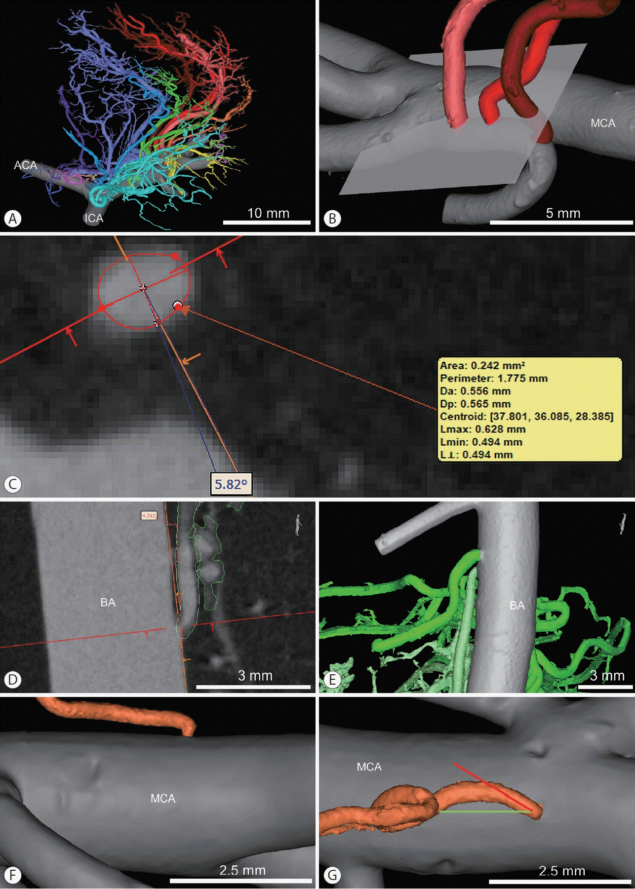 | Figure 1.Models of the cerebral arteries and radiological measurements. (A) Posterior view of bifurcation of the right ICA and the perforating arteries of the MCA. (B) Example of the measurement plane determined for the perforator first from the left. The plane is tangential to the parent vessel at the level of arterial wall of the parent artery. (C) Measurements of the perforating artery ostium shown in (B) (area, long and short axes lengths, the angle between the projection of the parent artery axis and origin short axis). (D and E) Measurements of the angle between the measurement plane and perforator axis and determination of the perforator course type (parallel, the brighter perforator, representative images). (F and G) Illustrations of the angle between projections of the parent artery axis (green line) and perforator axis (red line). Side view (F) and top view (G). Initial segment of the perforating artery is perpendicular to the parent artery (MCA). ICA, internal carotid artery; MCA middle cerebral artery; ACA, anterior cerebral artery; BA, basilar artery.
|
For every perforating artery of every specimen, several measurements were taken using semiautomated built-in tools. Before starting the measurements, individual measurement planes were selected with the use of a multiplanar reconstruction tool. The measurements were made in the tangential plane at the level of the parent artery wall (Figure 1A and B).
The measurements consisted of the following steps:
(1) Surface area measurement: The surface area of the perforator origin was measured with the use of the Area tool. Long and perpendicular short axes were calculated automatically (Figure 1C).
(2) Determination of the perforator course type: parallel versus perpendicular (Figure 1D and E). The course was considered perpendicular if the angle between the perforator axis and the measurement plane ranged from 45° to 135°. In all other cases, the course was considered parallel. The angle was acute in cases of recurrent perforator course.
(3) Measurements of angles: (a) The angle between the projection of the parent artery axis on the measurement plane and the origin short axis was measured (Figure 1C). (b) The angle between the projection of parent artery axis and perforator axis on the measurement plane was also measured (applies only to parallel perforators) (Figure 1F and G). The angle was acute in cases of recurrent perforators. In cases where the origin short axis and projection of the perforator axis were tilted in opposite directions, the (b) angle was positive, and the (a) angle was negative.
Statistical analysis
The statistical analysis was performed with SAS 9.4 software (SAS Institute Inc., Cary, NC, USA). Continuous variables are presented as medians and interquartile ranges; comparisons were made using the two-tailed t or Wilcoxon tests depending on the distribution of the variables (tested using the Anderson-Darling test). Due to the exposure of groups of perforators branching from one artery to similar biological and hemodynamic factors, linear mixed models were deemed appropriate for studying relationships between the deviation angles of the origin short axis and perforator axis from the parent artery axis, as well as the origin short-to-long axis length ratio and age and atherosclerosis severity. The calculations were performed using the restricted maximum likelihood method. Random effects had to be removed from the model because too little variation remained for the random effects after controlling for the fixed effects and non-convergence [24-26].
Histological studies
Cross-sections were made in the long axis of the MCA, BA, internal carotid artery (ICA), and vertebral artery, containing the ostia of the perforating arteries as well as other small branches (e.g., the anterior inferior cerebellar artery and the anterior choroidal artery). Hematoxylin and eosin stain, Mallory’s trichrome stain, orcein stain, and immunohistochemical staining for alpha-smooth muscle actin (NCL-L-SMA, ASM-1 clone, dilution 1:200; Leica Biosystems, Newcastle upon Tyne, UK) and desmin (NCL-L-DESDERII, DE-R-11 clone, dilution 1:50; Leica Biosystems) were performed. The slides were analyzed with an Olympus BX53 microscope, and photographs were taken using an Olympus UC90 camera and cellSens Dimension software without additional processing (Olympus Corp., Tokyo, Japan).
Go to :
Results
Characteristics of the studied sample
Twenty-three anatomical specimens of the MCA with the basal ganglia and 10 specimens of the BA with the brainstem were prepared, scanned, and analyzed (Figure 1A). The donors were between 21 and 78 years old, and one-third of donors were females (3/10 BA, 8/23 MCA). We identified 172 anterolateral central arteries and 162 pontine arteries (334 perforating arteries in total); seven and four arteries, respectively, were excluded from the statistical analysis for technical reasons (incomplete filling due to the presence of air bubbles). The median (first–third quartiles) numbers of perforating arteries of the MCA and BA were 7 (6–9) and 17 (14–19), respectively. Notably, in the case of the perforators of both the MCA and BA, the ostia were asymmetrical (in the shape of an ellipse, short-to-long axis length ratio less than 1), and the asymmetry was more pronounced in the group of perforators with a parallel course (significantly lower short-to-long axis ratio: MCA perforators: mean±standard deviation [SD] 0.77±0.11 vs. 0.81±0.07, degrees of freedom [DF]=63.9, t=2.47, P=0.016, t-test; BA perforators: median [Q1–Q3] 0.78 [0.72–0.86] vs. 0.83 [0.79–0.89], Z=2.73, P=0.007, Wilcoxon test). Careful investigation of the perforating arteries ostia with the microsurgical microscope and comparison with the results of radiological studies revealed that the ostium asymmetries correspond to local stenoses (Figure 2). The detailed characteristics and morphometry of the arteries are presented in Table 1.
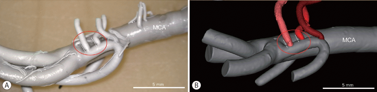 | Figure 2.Comparison of the (A) microscopic view and (B) three-dimensional model of the middle cerebral artery (MCA). Local narrowings of ostia of the lenticulostriate arteries visualized by microcomputed tomography correspond with thickening of the arterial wall. Dashed, white lines represent vessel wall surface.
|
Table 1.
Characteristics and morphometry of BA and MCA specimens
| BA specimens (n=10) | MCA specimens (n=23) | |
|---|---|---|
| Number of the perforating arteries | 158 | 165 |
| Age (yr) | 53.5 (42.0–62.8) | 48.0 (38.0–65.5) |
| Atherosclerosis* – sum | 3.5 (2.0–4.8) | 2.0 (0.5–4.5) |
| Circle of Willis | 0 (0–0) | 0 (0–0) |
| Coronary arteries | 2 (1–2) | 1 (0–2) |
| Aorta | 1.5 (1–2) | 1 (0.5–2) |
| Right side | 82† (52.6) | 95 (57.6) |
| Segment of origin of perforating artery | ||
| Proximal BA | 75 (47.8) | n/a |
| Middle BA | 57 (36.1) | |
| Distal BA | 26 (16.5) | |
| M1 | n/a | 89 (53.9) |
| M1/M2 | 35 (21.2) | |
| M2 | 33 (20.0) | |
| Anterior temporal artery | 8 (4.9) | |
| Quadrant of origin of pontine artery | ||
| Right pontine | 51 (32.3) | n/a |
| Left pontine | 49 (31.0) | |
| Right lateral | 32 (20.3) | |
| Left lateral | 25 (15.8) | |
| Right clival | 1 (0.6) | |
| Left clival | 0 (0) | |
| Type of pontine artery‡ | ||
| Paramedian | 36 (22.8) | n/a |
| Short circumflex | 31 (19.6) | |
| Composition of 1&2 | 33 (20.9) | |
| Long circumflex | 23 (14.6) | |
| Median | 24 (15.2) | |
| Branch to PPS | 11 (7.0) | |
| Parallel course | 115 (72.8) | 133 (80.6) |
| Short-to-long axis length ratio§ | 0.80 (0.73–0.87); 0.52–0.99 | 0.77 (0.71–0.86); 0.44–1 |
| Parallel course | 0.78 (0.72–0.86); 0.52–0.98 | 0.76 (0.70–0.86); 0.44–1 |
| Perpendicular course | 0.83 (0.79–0.89); 0.63–0.99 | 0.81 (0.75–0.87); 0.66–0.95 |
| Area of ostium (mm2) | 0.11 (0.07–0.18); 0.02–1.15 | 0.10 (0.04–0.23); 0.01–1.80 |
| Length of ostium long axis (mm) | 0.43 (0.35–0.56); 0.20–1.30 | 0.41 (0.29–0.62); 0.12–1.64 |
| Length of ostium short axis (mm) | 0.34 (0.27–0.44); 0.14–1.15 | 0.33 (0.21–0.48); 0.08–1.49 |
| Parent artery-ostium short axis angle (°) | 29.5 (10.8–63.6); -35.0–194.9 | 17.2 (4.6–39.6); -64.4–194.6 |
| Parent artery-perforator axis angle (°) | 36.7 (12.9–89.8); 1.9–176.3 | 30.7 (16.1–48.0); 0.6–178.9 |
| Common ostium of 2 perforators | 2 (1.3) | 6 (3.6) |
Values are presented as median (interquartile range), n (%), or median (interquartile range); min–max.
BA, basilar artery; MCA, middle cerebral artery; PPS, posterior perforated substance.
![]()
The statistical analysis revealed that 1° increases in deviation of the perforator axis from the parent artery axis were related to 0.95° or 0.74° increases in the deviation of the origin short axis from the parent artery axis for the MCA and BA perforators, respectively (standard error [SE]=0.071°, DF=125, t=13.41, P<0.001 and SE=0.056°, DF=113, t=13.03, P<0.001, respectively) (Figure 3A). In the group with parallel MCA perforating arteries 10-year increase in age was related to a 0.011 decrease in the short-to-long ostium axis ratio (SE=0.005, DF=125, t=-2.10, P=0.038) (Figure 3B), and an increase of 1 unit in atherosclerosis (semiquantitative scale) was associated with a 0.008 decrease in the short-to-long ostium axis ratio (SE=0.04, DF=125, t=-2.17, P=0.032). The estimated level-1 residual variances were 979.04, 999.89, 0.1104, and 0.1102 (P<0.001).
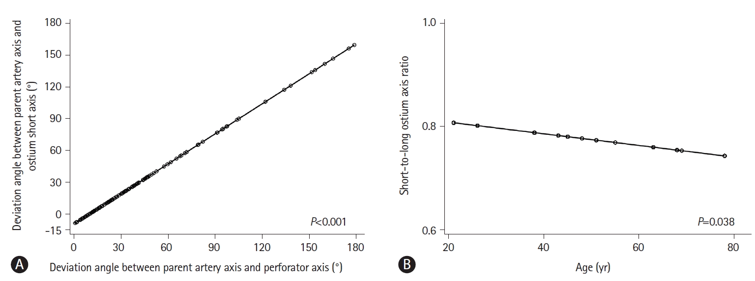 | Figure 3.Results of the statistical analysis (linear mixed models). (A) Model of the relation between deviations of the ostium short axis and perforator axis from the parent artery axis (MCA perforators). (B) Model of the relation between age and the short-to-long ostium axis ratio (MCA perforators with a parallel course).
|
Histological studies
Histological studies of the perforating artery ostia of the ICA, the MCA, the BA, and the vertebral artery revealed local disruption of the internal elastic lamina with multilayer thickening of the intima stenosing the ostia (Figure 4). The media and the adventitia were intact. A similar structure was found at the origin of a secondary perforator branching in the subarachnoid space from one of the deep perforating arteries of the MCA (Figure 4H). The cells present in the intimal pads stained positive for smooth muscle actin and negative for desmin (Figure 5).
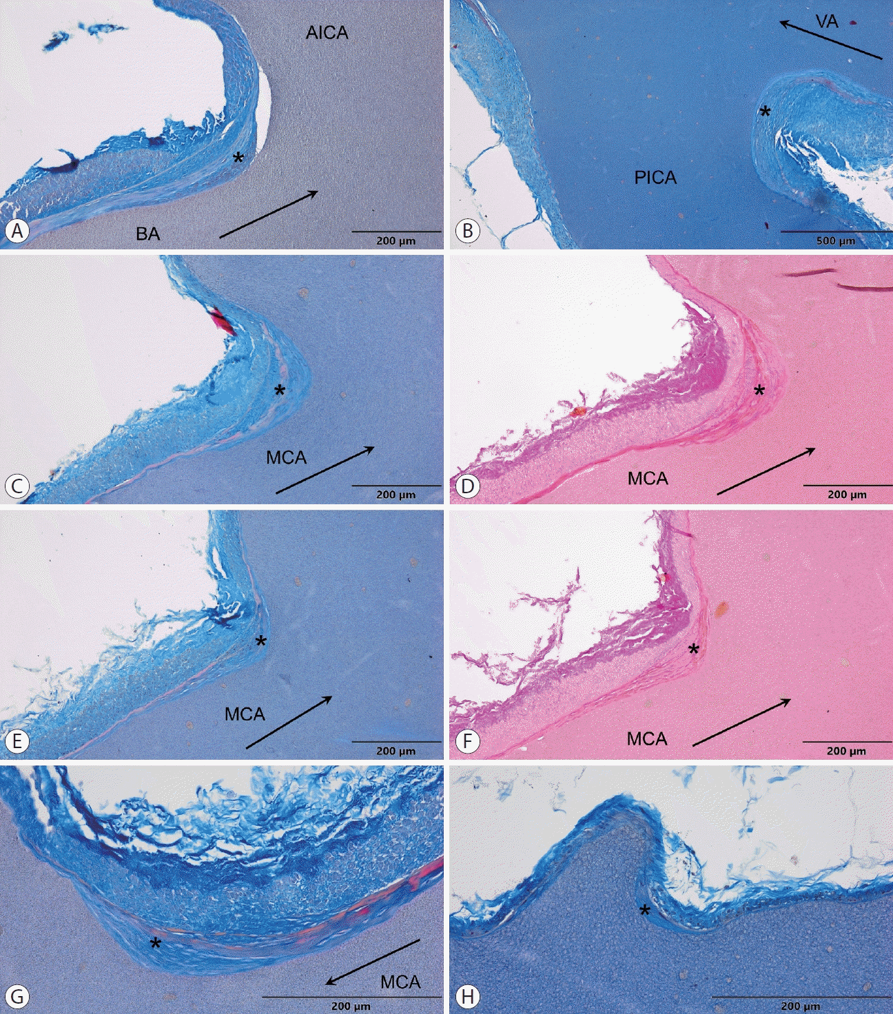 | Figure 4.Results of the histological studies. (A and B) Origins of the AICA and the PICA, respectively, showing intimal thickening with a disruption of the internal elastic lamina (Mallory’s trichrome staining). (C, D and E, F) Comparison of Mallory’s trichrome staining and orcein staining of perforators of the right MCA. Orcein staining confirmed multilayer separation of the internal elastic lamina. (G and H) Examples of intimal thickening of the left MCA perforator ostium (G) and a similar structure at origin of a small perforator (H) branching in the subarachnoid space from a deep perforating artery of the right MCA (Mallory’s trichrome staining). The arrows show blood flow direction in the parent artery. The asterisks indicate neointimal hyperplasia. AICA, anterior inferior cerebellar artery; BA, basilar artery; MCA, middle cerebral artery; PICA, posterior inferior cerebellar artery; VA, vertebral artery.
|
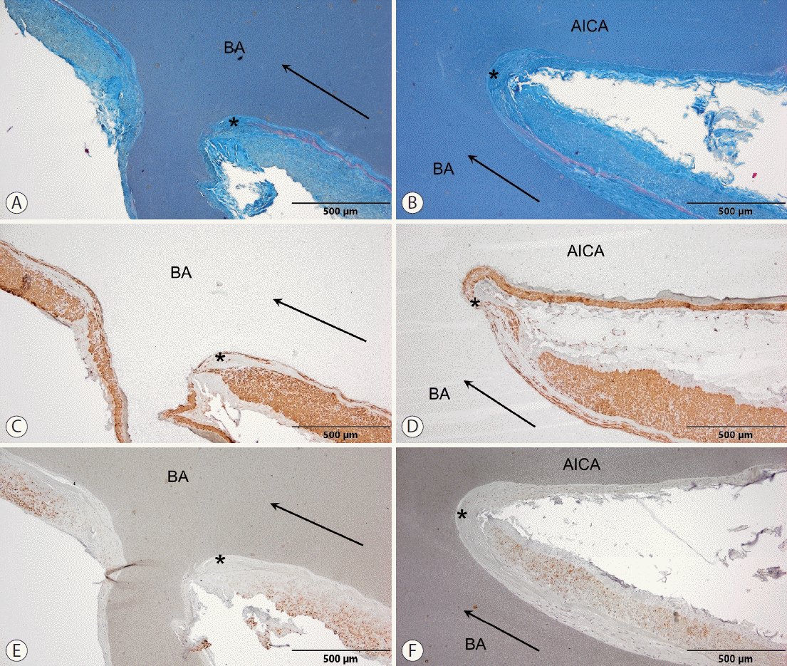 | Figure 5.Results of immunohistochemical staining and comparison with Mallory’s trichrome staining of origins of the pontine artery (A, C, E) and the anterior inferior cerebellar artery (B, D, F). (A and B) Mallory’s trichrome staining, (C and D) smooth muscle actin (SMA), (E and F) desmin. Cells present in the thickened intima stained positive for SMA and negative for desmin, which are characteristic of the secretory phenotype of the smooth muscle cells. Normal smooth muscle cells forming the media stained positive for both SMA and desmin, confirming their contractile phenotype. The arrows show blood flow direction in the parent artery. The asterisks indicate neointimal hyperplasia. AICA, anterior inferior cerebellar artery; BA, basilar artery.
|
Go to :
Discussion
The uniqueness of the deep cerebral perforating arteries in terms of their direct origination from high-pressure intracranial arteries and their clinical significance motivated us to study the microanatomy of the lenticulostriate and pontine arteries. Contrast-enhanced microtomography of anatomical specimens revealed stenoses of the perforators’ ostia, which led us to study histological samples that revealed local remodeling of the vessel walls. The main finding of the study is that the ostia of the deep cerebral perforating arteries are not round, but are ellipsoidal in shape, which results from the local neointimal remodeling of the arterial wall.
Shape and orientation of the perforating artery ostia
The results of the radiological studies revealed that the shapes of the deep cerebral perforating artery ostia were elliptical. This finding is neither an artifact nor a normal situation because of (1) the precision of the method used [15,23,27], (2) Murray’s law [28,29], and (3) the orientation of the ellipses. The method used visualizes perforating arteries precisely enough to study the precapillary arterioles and complex anatomical variations. For every perforating artery, we confirmed the fact that its initial segment (sometimes very short) is perpendicular to the parent vessel. According to Murray’s law, if the diameter of the branching artery is significantly smaller than that of the parent artery, the angle between the branches equals to approximately 90°, and thus the cross-section of the ostium should be round. Even if the perforator has a recurrent parallel course, its first segment is perpendicular to the parent artery. Moreover, if the elliptical shape results from oblique branching, the long axis of the ostium should be parallel to the parent vessel, while the opposite is true.
Contrast-enhanced tomography visualizes a cast of the arterial lumen, which, in the case of local stenosis, suggests the presence of intraluminal structures. Additional microsurgical microscopy observations revealed filling defects corresponding to the asymmetrical ostia of the perforating arteries, which led us to conclude that the narrowings resulted from local thickening of the vessel wall. Interestingly, similar ostial narrowings can be observed in the figures of casts of the lenticulostriate arteries in the article by Marincovic et al. [30] However, they were neither described nor discussed.
The orientation of the perforating artery origins was closely associated with spatial relationships between the parent artery and the perforator. When the perforating artery is parallel to the parent vessel, the short axis of the ellipsoidal origin is also parallel to the parent artery, and the more the perforator’s axis deviates from the parent artery’s axis, the more the ellipse rotates. Considering the results of extensive research on hemodynamic conditions, e.g., around the origins of intercostal arteries, revealing complex patterns of wall shear stress [31] and patterns of arterial remodeling in response to disturbed flow [32], our observations suggest that the phenomenon observed may be related to local hemodynamic conditions that directly result from the geometry of the arteries. Moreover, stenoses of the ostia may act as pressure reducers at the junctions of the high-pressure intracranial arteries (the MCA, BA, etc.) with the perforating arteries so that the blood pressure is adjusted to the microcirculation [33]. Hemodynamic conditions are expected to be complex; the analogy with the intercostal arteries may not be ideal because they are of larger caliber and supply different, high-resistance vascular bed, and the flow is completely different in the aorta than in the large intracranial arteries [34,35]. Therefore, thorough hemodynamic studies on the perforating arteries are necessary.
Histological structure of the ostia
After discovering the narrowings of the perforating artery ostia, the next step was to study their structures. Histological studies confirmed the presence of the narrowings and revealed local arterial wall hypertrophy internally from the internal elastic lamina that was discontinuous and multilayered. Additional immunohistochemical staining helped us to identify the smooth muscle cells with a secretory phenotype [36-38] that caused the thickening of the intima. The structure is characteristic of neointimal hyperplasia, a well-described form of arterial wall remodeling in which the smooth muscle cells disrupt the internal elastic lamina, migrate to the intima, and proliferate [32]. Similar regions of neointimal hyperplasia were identified around the ostia of the small branches of the ICA, the BA, and the vertebral artery other than the perforating arteries, namely, the anterior choroidal artery, the posterior communicating artery, the anterior inferior cerebellar artery, and the posterior inferior cerebellar artery. This result is not surprising, as their calibers and branching schemes are similar to those of larger perforators.
Neointimal hyperplasia develops in regions where the arterial wall is exposed to abnormal hemodynamic conditions characterized by low wall shear stress and reciprocating flow [32]. The narrowings were more pronounced on the inflow sides of the ostia, and the rotation of the ellipse was dependent on the deviation of the perforator from the parent vessel. The presence of areas of low shear stress on the inflow side of the ostia can be expected; however, considering the complex vascular geometry, it should be the subject of thorough hemodynamic studies.
Recent excellent reviews addressing the topic of cerebral small vessel disease (SVD) do not mention neointimal hyperplasia as a type of vessel remodeling and indicate arteriolosclerosis as the major neuropathological finding [10,39-42], It is worth noting that the vast majority of the studies analyze radiological markers of SVD and neuropathological examinations are usually limited to the intraparenchymal vessels. Although some authors point out the role of the connection of the high-pressure MCA or BA with the deep penetrating arteries, the structure of the perforating artery origins has not yet been studied. Marincovic ´ et al. [43] described the histology of the extraparenchymal course of the lenticulostriate arteries; however, branching points from the MCA were not studied. Interestingly, in some cases “subendothelial smooth muscle cells of secretory phenotype” were noticed, which suggests the presence of neointimal remodeling. The role of the perforating artery origins in SVD is thought to be limited to parent artery atherosclerosis covering the perforator origins, but the concept is based on case series of patients with lacunar infarction [44-47]. In this study, after analysis of 334 perforating arteries we were able to describe normal microanatomy of the perforating artery ostia. So far, neointimal hyperplasia has only been mentioned in the context of remodeling after stenting and moyamoya disease (both in humans and animal models) [36,48-51].
Relationships with age, cardiovascular risk factors, and clinical significance
The statistical analysis revealed that the severity of stenosis of the ostia is associated with both age and the severity of atherosclerosis (a pathological marker of cardiovascular health); the ostia were narrower in older patients and those with more severe atherosclerosis. These findings suggest that the narrowings may increase with age and may be dependent on cardiovascular risk factors, such as hypertension. Interestingly, the ostia were already stenosed among young adults. This finding is consistent with the proposed explanation of pressure reducers: even normal arterial blood pressure is too high for the perforating arteries, whereas elevated blood pressure (typical of elderly individuals and those with atherosclerosis) must be lowered more to adjust it to the microcirculation; therefore, a more pronounced reducer is needed. The association discussed was found only in the subgroup of MCA perforators with a parallel course, characterized by a recurrent course that may aggravate adverse hemodynamic conditions, which is confirmed by more severe stenoses in this subgroup. Other explanations include the limited sample size of BA perforators or different hemodynamic conditions. This issue should be the subject of future studies. An analysis of the level-1 residual suggested the existence of other factors explaining the change in the severity of perforator ostia narrowing with age [52], which supports further research.
The existence of stenoses of the origins of the perforating arteries is of particular interest in the context of cerebral SVD and neurovascular procedures. Cerebral blood flow is postulated to be a marker of brain health and a predictor of future cognitive function [53]. The median short-to-long axis length ratio was approximately 0.8 (which corresponds with 20% stenosis), a value considered to indicate insignificant stenosis in the coronary circulation; however, for cerebral circulation, ischemia does not have to occur for dementia to develop, and chronic hypoperfusion is sufficient [39,53-58]. Notably, fewer perforators and slower blood flow velocities in the perforators are detected among elderly individuals and those with SVD [59-61]. Moreover, dementia develops faster and earlier among people with hypertension diagnosed at a younger age [53,62]. These observations may be explained by the presence of novel narrowings—pressure reducers—that increase with age and cause a reduction in blood flow, eventually contributing to cognitive impairment. The supply area of the perforating arteries and other branches where neointimal narrowing occurs (e.g., the anterior choroidal artery) includes structures crucial for brain health and cognition: the hypothalamus, the Papez circuit, the basal ganglia, the thalamus, etc [6]. Notably, the effectiveness of rivastigmine in patients with Alzheimer’s disease is associated with improved perfusion in the perforators supply area, because of their cholinergic innervation [63,64]. Narrowings of the perforator ostia may also lead to a poststenotic flow profile in the distal perforating arteries, which may impair the function of the glymphatic system (and waste clearance) because arterial pulsations drive the flow of cerebrospinal fluid through the perivascular spaces [65].
Study sample
The study was conducted using a sample of more than 160 perforating arteries of both the MCA and BA obtained from human anatomical specimens. Although the availability of unfixed specimens is limited, only human brain specimens adequately reflect real vessel geometry. The disproportion in the size of the MCA and the deep perforating arteries is a result of phylogenesis—very large MCA in humans is in fact a dominant perforator of the anterior cerebral artery that hypertrophies in response to an enlarged cortical volume [19,66]. Moreover, some mammals show the presence of rete mirabile [20], whereas, in rats, the diameter of the MCA is an order of magnitude smaller [21], which modifies hemodynamic conditions. The numbers of different branch types were similar to those known from the literature [6,7,13], and thus despite a slight overrepresentation of men, we considered the sample to be representative. Further studies on larger sample sizes are needed to investigate the relationship of the perforating artery ostium narrowings with cardiovascular risk factors etc.
Perspectives and limitations of the study
This study has several limitations. The arteries have been investigated ex vivo; however, an in vivo method for imaging the human brain with sufficient resolution is unavailable. The method used is limited by the availability of unfixed human brain specimens; on the other hand, it does not require expensive reagents. We did not have access to full medical histories; therefore, we assessed the severity of atherosclerosis, which is a marker of cardiovascular health and has the advantage of reflecting the impact of unrecognized and poorly controlled diseases. The statistical analysis was cross-sectional, and the results must be confirmed [67,68].
This study revealed problems that need to be studied in future projects: moment of development and the exact cause of stenoses, the hemodynamic conditions in the deep perforating arteries, the role of the phenomenon in brain health, and the pathophysiology of SVD. The study identifies a new potential area for the search for therapeutic interventions to slow brain aging. The majority of the studies on cerebral blood flow have focused on cortical flow [53]; therefore, we must emphasize that deep perforating arteries require an independent analysis because the pressure drop across cortical branches is bypassed [69,70].
Go to :
Conclusions
The ostia of the deep cerebral perforating arteries are stenosed locally by neointimal hypertrophy, as are the origins of the small branches of the circle of Willis: the anterior choroidal artery, the posterior communicating artery, the anterior inferior cerebellar artery, and the posterior inferior cerebellar artery. The cross-sections of the ostia are ellipsoidal, and the rotation of the ellipse axes depends on the geometry of the perforating artery. The severity of the narrowing, which usually reaches approximately 15%–30%, may increase with age and depends on cardiovascular risk factors. The formation of stenoses may be a protective mechanism to adjust high blood pressure in the MCA and BA to the microcirculation. However, their roles in brain health and the pathophysiology of cerebral SVD require further research.
Go to :
 XML Download