

 PDF
PDF Citation
 Print
Print


INTRODUCTION
Congenital absence of the portal vein (CAPV) is a rare venous malformation in which the mesenteric venous blood drains directly into the systemic circulation. The majority of CAPV patients show no signs or symptoms of portosystemic encephalopathy. They only show slightly abnormal liver function test results. Liver transplantation (LT) is indicated for patients with symptomatic CAPV refractory to medical treatment [1-4].
Congenital portocaval shunt (PCS) drains the entire mesenteric venous blood either directly into the inferior vena cava (IVC) or through the left renal vein via the splenorenal shunt, therefore preventing portal hypertension and collateral circulation [3-5]. Since a liver with CAPV does not have sufficient portal inflow, the hepatic arterial flow is the main blood inflow. Such deprivation of portal blood inflow results in various liver pathologic diseases. If a patient does not respond to medical treatment, LT should be taken into account. Liver function profiles of patients with CAPV are not severely impaired, thus usually show low Pediatric End-stage Liver Disease scores. Because of very low chances of deceased donor liver transplantation in the current Korean setting, patients with CAPV need to be prioritized for living donor liver transplantation (LDLT). We herein present a case of pediatric LDLT using dextrorotation of a left liver graft for CAPV with direct drainage into the suprahepatic IVC.
Go to : 
CASE PRESENTATION
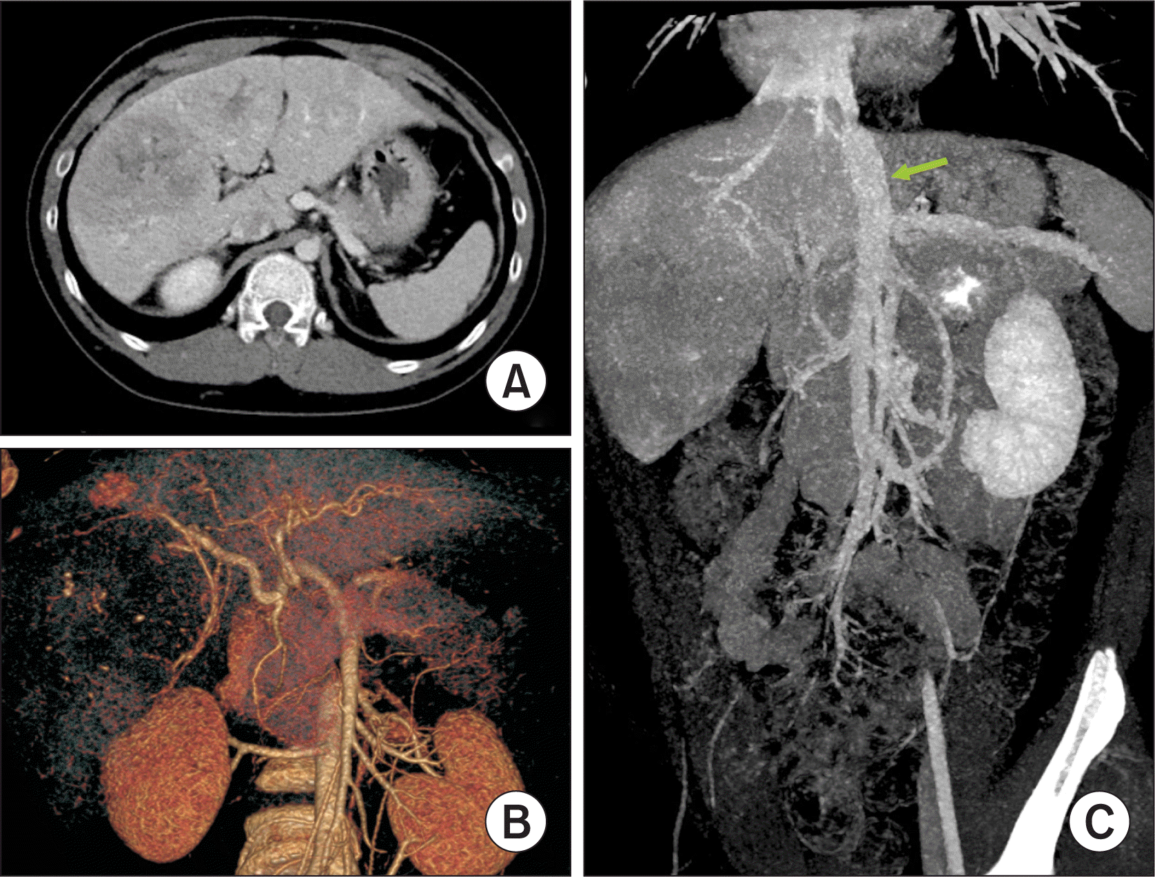
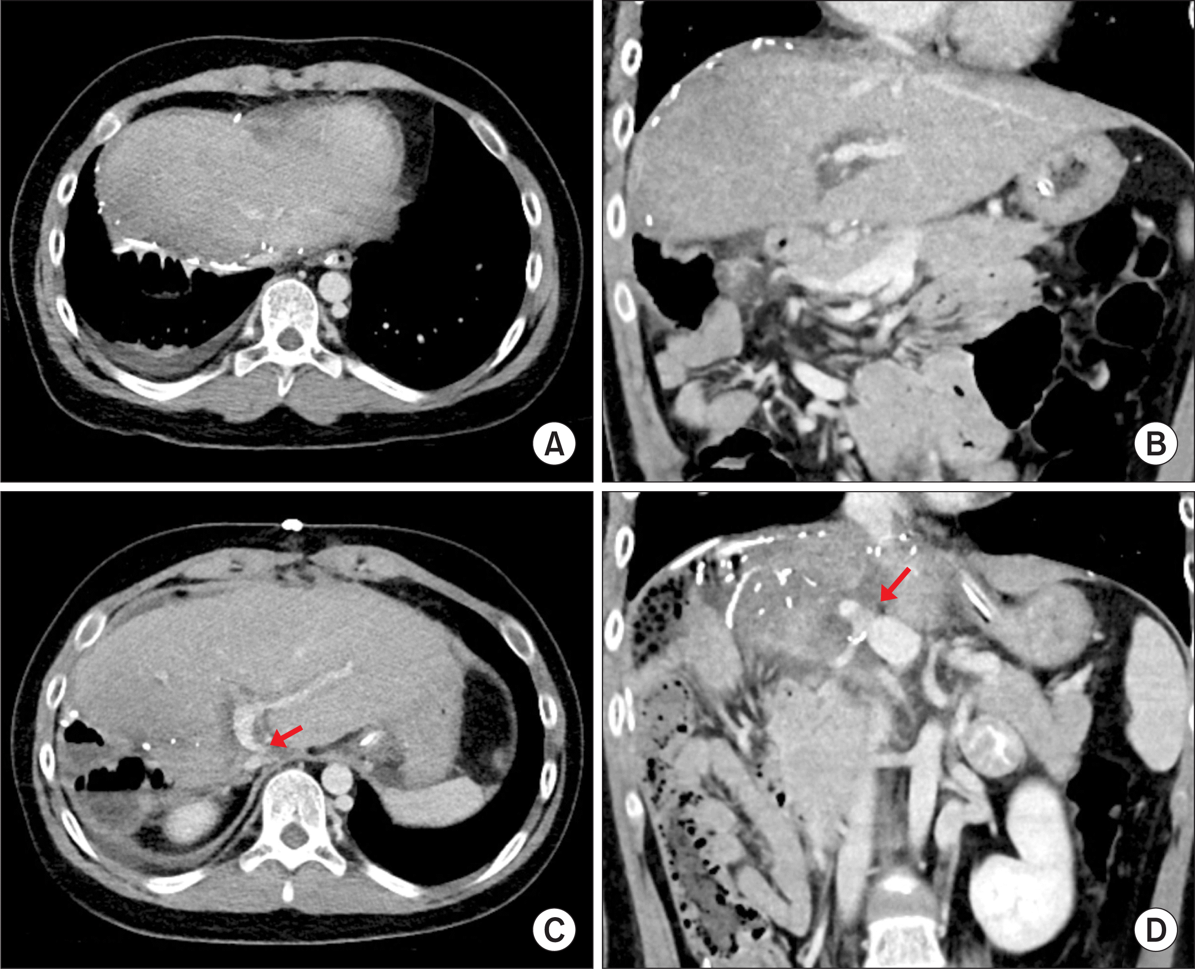
A 9-year-old boy was referred to our hospital due to precocious puberty with liver mass and abnormal liver function. He was born in full-term cesarean section delivery. He had been doing well after birth except for operation for scrotal hydrocele at the age of 3 years. Imaging studies showed CAPV with multiple focal nodular hyperplasia (FNH) nodules. There was a large PCS vein from the mesenteric vein to the suprahepatic IVC (Fig. 1). He was also diagnosed with hepatopulmonary syndrome grade 2.
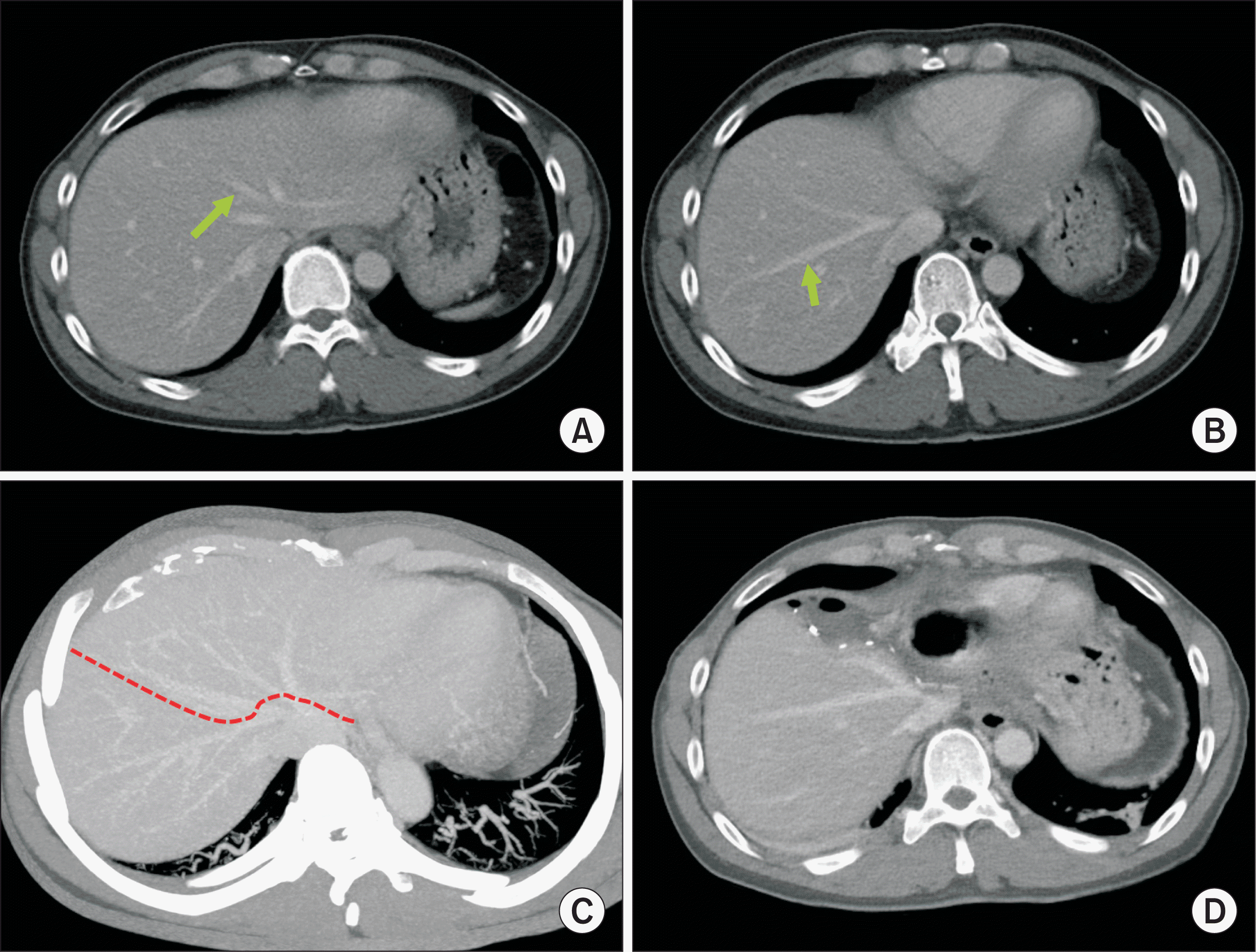
 | Figure 1Preoperative and postoperative computed tomography findings of the donor. (A) A fissural vein was identified at the medial section (arrow). (B) A large segment VIII vein was identified (arrow). (C) Parenchymal transection plane was designed to preserved the segment VIII vein (dotted line). (D) Postoperative computed tomography taken at 7 days after surgery showed no significant hepatic venous congestion at the remnant right liver.
|
While waiting over one year, multiple liver tumors progressively increased in size and number. Hepatopulmonary syndrome also progressed. Allocation for deceased donor liver transplantation was not expected due to very low Pediatric End-stage Liver Disease score (—9 points). Thus, we decided to perform LDLT at the age of 10 years with a bodyweight of 45 kg.
The donor was the 45-year-old father of the patient. The left liver volume was determined as 460 mL on computed tomography volumetry. The anatomy of the middle hepatic vein (MHV) had large segment VIII (V8) and segment V veins at the right liver with separate ventral segment IVa and dorsal segment IVb. Transection at the level of the MHV trunk would induce huge hepatic venous congestion at the remnant right liver. To protect the donor’s remnant liver from excessive hepatic venous congestion, we decided to preserve the large V8 branch at the donor remnant liver (Fig. 2).
 | Figure 2Pretransplant computed tomography findings of the recipient. (A) Multiple liver nodules were identified. (B) Hepatic arteries were enlarged without anatomical variation. (C) Congenital absence of the portal vein was identified with development of a large portocaval shunt in to the suprahepatic inferior vena cava (arrow).
|
The recipient’s operation was performed according to standard procedure of pediatric LDLT. During hepatic hilar dissection, the recipient’s native portal vein (PV) was totally absent, and a large PCS vein was identified along the caudate lobe (Fig. 3). The recipient hepatic artery branches were meticulously dissected.
A V8-preserving whole left liver graft was harvested, with a graft weight of 420 g at the back table, equivalent to a graft-to-recipient weight ratio of 0.96%. There were two separate outflow veins at the graft liver cut surface. We adopted the technique of MHV branch reconstruction developed for a modified extended right liver graft. A 10-cm-long cold-preserved iliac vein graft was prepared at the institutional tissue bank. The internal semilunar valves at the common iliac vein level were excised. The transected MHV trunk stump was anastomosed with the iliac vein side of the conduit in an end-to-side fashion. The IVC side of the conduit was anastomosed with the left hepatic vein stump. Because this conjoined outflow vein orifice was relatively small, an incision was made at the left lateral wall of the graft left hepatic vein, and patch venoplasty was performed. These procedures resulted in a single outflow vein orifice of 3 cm in the transverse diameter (Fig. 4). The length of the recipient PCS vein appeared to be too short to perform direct anastomosis, thus the remnant iliac vein graft was anastomosed with the graft portal vein stump at the back table (Fig. 5).
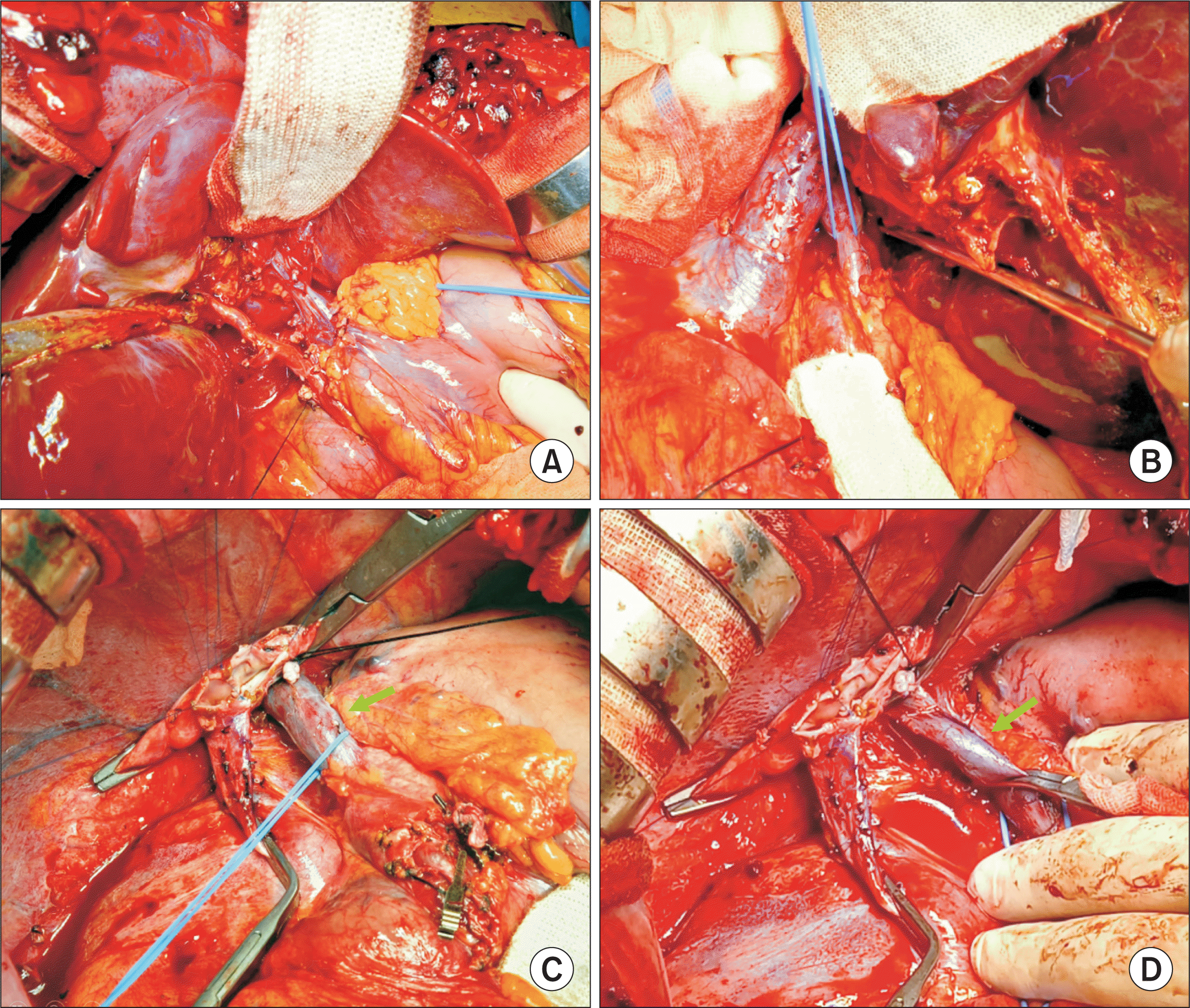
 | Figure 4Intraoperative photographs of bench work for a left liver graft. (A) A small-sized left hepatic vein orifice was identified (arrow). (B) Transected middle hepatic vein trunk was exposed at the liver cut surface (arrow). (C–D) An iliac vein segment was anastomosed with the middle hepatic vein trunk. (E) The inferior vena cava side of the conduit was anastomosed with the left hepatic vein stump. (F–H) An incision was made at the left lateral wall of the graft left hepatic vein, and patch venoplasty was performed to make a wide single outflow vein orifice.
|

After the native liver was removed, the hepatic vein orifices at the recipient inferior vena cava were unified. The axis of the PCS vein was not well matched with that of the graft PV in the classical orthodox position, which can result in buckling deformity of the PV anastomosis. To prevent such risk of vascular complication, the left liver graft was fallen into the right subphrenic fossa to make graft dextrorotation, and the axes of the outflow vein and PV anastomoses were determined. The graft outflow vein orifice was reconstructed by continuously suturing with 5-0 Prolene (Fig. 6A–E). The interposed iliac vein conduit at the graft PV was obliquely resected to match with the length and axis of the excised PCS vein. A 1 cm-long wedge-shaped iliac vein segment was inserted between the graft PV and the PCS vein stumps, which made the anastomosis not redundant (Fig. 6F–H). Thereafter, graft reperfusion was initiated. Surgical microscopy was used for the reconstruction of the graft left hepatic artery. Roux-en-Y hepaticojejunostomy was used for biliary reconstruction.
 | Figure 6Intraoperative photographs of the hepatic vein and portal vein reconstruction. (A) The graft hepatic vein was enlarged through unification venoplasty. (B) The isolated portocaval shunt vein was transected. (C–E) The liver graft was placed into the right subphrenic fossa, thus inducing dextrorotation and hepatic vein reconstruction was performed at that position. (F–H) The interposed iliac vein conduit at the graft portal vein was obliquely resected to match the length and axis of the excised portocaval shunt vein. A 1-cm-long wedge-shaped iliac vein segment (arrows) was inserted between the graft portal vein and the portocaval shunt vein stumps to prevent redundancy.
|
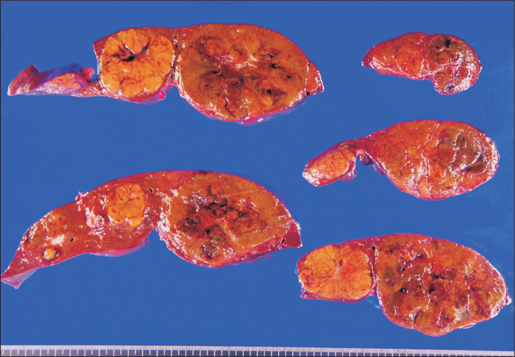
The pathology report of the explant liver showed two nodules of hepatocellular carcinoma (each 10 cm and 6 cm) in the background of multiple hepatic adenomas of unclassified type. There were collapse of intralobular portal veins, with proliferation of hepatic arterioles and nodular regenerative hyperplasia. These findings were consistent with the pathologic changes associated with PCS (Fig. 7).
The patient recovered from the LDLT operation. The reconstructed graft hepatic vein and portal vein were maintained well without hemodynamic abnormality (Fig. 8, 9). This patient has been doing well for 2 months after the LDLT.
 | Figure 8Posttransplant computed tomography scan taken at 4 days after transplantation. (A, B) Uneventful reconstruction of the graft hepatic vein was identified. (C, D) Portal vein anastomosis showed a slight anastomotic stenosis (arrows), with unusual location of the inflow portocaval shunt vein.
|
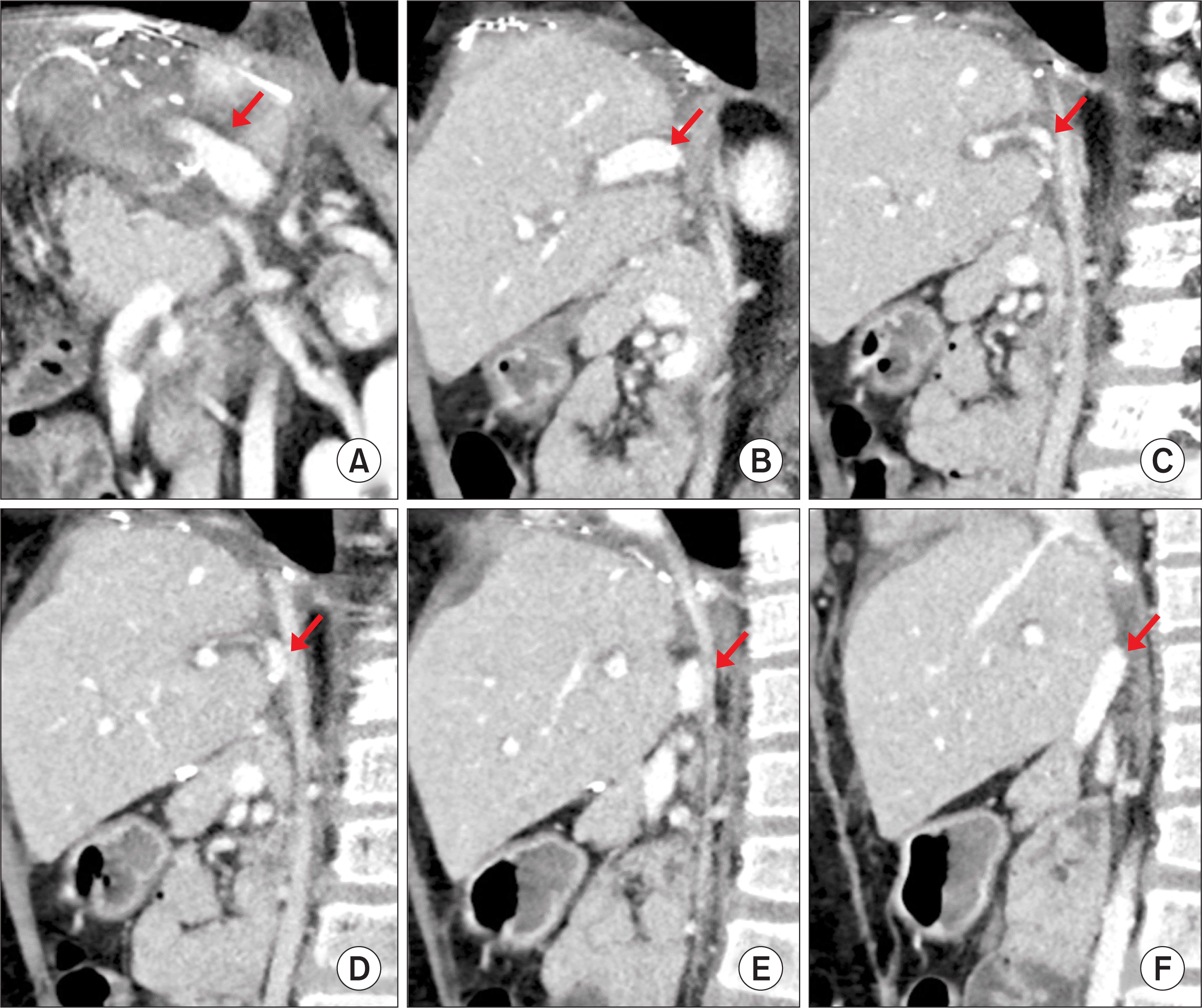 | Figure 9Posttransplant computed tomography scan taken at 14 days after transplantation. (A) Three-dimensional reconstruction of the portal vein shows stream-lined configuration (arrow). (B–F) The running course of the reconstructed portal vein shows the unusual location behind the liver graft (arrows).
|
Go to :
DISCUSSION
CAPV is a rare venous malformation in which the mesenteric venous blood drains directly into the systemic circulation. There are two types of congenital PCS: intrahepatic PCS and extrahepatic PCS. Intrahepatic PCS is localized between the PV and hepatic veins [6]. Extrahepatic PCS is divided into type I and type II according to intrahepatic portal venous supply [7]. Type I PCS is an extrahepatic shunt without a patent intrahepatic portal vein. Thus, the entire mesenteric venous blood flow drains directly into the systemic veins such as the IVC and the left renal vein. This type is called CAPV. Type II PCS is an extrahepatic shunt with a patent intrahepatic PV. Our present patient had type I PCS without noticeable variceal collateral veins.
The standard treatment for CAPV has not yet been established. Although PCS can be accompanied by hyperammonemia, the majority of the patients with PCS show no signs of encephalopathy. Such patients only show slightly abnormal liver function test results. Our present patient showed only elevation of liver enzymes. The majority of patients with CAPV receive conservative medical treatment for hyperammonemia, while only a small portion of patients with CAPV require surgical treatments, including LT. Surgical treatment is indicated when hyperammonemia or portosystemic encephalopathy is refractory to medical treatment. Surgical treatment for CAPV is also indicated for hepatopulmonary syndrome [8,9].
Liver neoplasm was observed in 11 of the 25 reported CAPV patients [10]. FNH was observed in 4 patients, hepatoblastoma in 2, nodular regenerative hyperplasia in 2, hepatocellular carcinoma in 1, hepatocellular adenoma in 1, and nodular hyperplasia in 1. The underlying causes of development of these tumors are not well known, but insufficient portal blood flow to the liver might have caused the hyperplastic nodules.
Pretransplant imaging studies in CAPV patients demonstrate a large communication vein to the IVC through the splenorenal shunt, thus no evidence of portal hypertension has been observed in the imaging studies or intraoperative findings. However, we have previously reported two atypical cases of CAPV showing portal hypertension with gastric and esophageal varix [11,12]. In the present case, like our previous case, no portal hypertension and collaterals were observed [13].
Since surgical reconstruction of the portal vein structures of the native liver is impossible, LT is indicated for most patients with type I PCS. Although LT for symptomatic CAPV has been reported in the literature [1-3,5,9,12-16], techniques for portal vein reconstruction have not yet been well established. There are two methods of portal vein reconstruction in LT for CAPV. The first method is to anastomose the PCS directly to the graft portal vein in an end-to-end fashion [2,14]. The second method is to use a venous interposition graft through an end-to-side anastomosis to the PCS [3,5]. In our four CAPV cases, the portal vein stump was absent [11-13], thus direct anastomosis was technically impossible. Therefore, we used vein conduit interposition as an end-to-side anastomosis to the PCS. The prerequisite for reconstruction with a vein conduit is the availability of an adequate vein homograft. In the present case, the length of the PCS shunt was too short for direct anastomosis, so interposition of a short iliac vein segment was performed, as a mixed form of two reconstruction techniques mentioned above.
In the present case, we did not perform intra-operative cine-portography because collateral veins were absent due to presence of a large PCS vein. Intra-operative cine-portography is an effective tool for secure PV reconstruction [12]. Intraoperative splanchnic venogram works as an accurate roadmap to design the PV reconstruction and interruption of collateral drainage. Intra-operative cine-portography is a useful method for identifying and embolizing residual portosystemic collateral veins in young pediatric patients who undergo LT [17].
The concept of graft dextro-plantation was applied in the present case because the axis of the PCS vein was not well matched with that of the graft PV in the classical orthodox position. Because such mal-alignment can result in buckling deformity of the PV anastomosis, the left liver graft was fallen into the right subphrenic fossa to make graft dextrorotation, and then the axes of the outflow vein and PV anastomoses were determined. Such dextro-plantation requires meticulous axis adjustment of the hepatic vein and portal vein anastomoses [18].
In conclusion, since CAPV patients show various types of PCS, individualized PV reconstruction with or without homograft vein interposition should be performed after thorough anatomical assessment before and during the LT operation.
Go to :
 XML Download