

 PDF
PDF Citation
 Print
Print


INTRODUCTION
Vascular smooth muscle cells (VSMCs) are the predominant cell type in the blood vessels, and are crucial for healthy vasculatures by providing a contractile function. Under physiological conditions, VSMCs showing a contractile phenotype facilitate the contraction and relaxation of the vasculatures [1]. In response to biological stress signals, VSMCs differentiate towards a synthetic phenotype expressing proteins that mediate proliferation and migration [2]. The conversion of VSMCs from a contractile to a synthetic phenotype has been implicated in the pathogenesis of proliferative vascular diseases including vascular remodeling in the injured vasculatures [3].
VSMC migration into the intima in response to a vascular injury or pro-atherosclerotic factors is known as a key event in developing various proliferative vascular diseases [4]. When exposed to biomechanical and chemical stresses, cells release high-mobility group box 1 (HMGB1), which is considered one of the most well-characterized damage-associated molecular patterns (DAMPs) mediating the progression of proliferative cardiovascular diseases [5,6]. HMGB1 has been proposed as a pivotal mediator involved in the development of proliferative vascular diseases [7]. Abundant HMGB1 within atherosclerotic plaques was suggested as a significant mediator inducing the increased proliferation and migration of VSMCs [8]. A previous study [9] also indicated that HMGB1 is a regulator of VSMC phenotypic switching and vascular remodelling. Although HMGB1 is indicated as a novel therapeutic target for vascular remodeling, little is known about the signals mediating cell migration in HMGB1-stimulated VSMCs, a key feature of vascular remodeling diseases.
VSMC migration is initiated by multiple environmental cues, including biochemical compounds and biophysical conditions. Increased osteopontin (OPN) levels have been observed in atherosclerotic plaque and neointima after experimental angioplasty [10,11]. Previous studies also indicated that OPN was expressed strongly in the synthetic phenotype of VSMCs [12-14] and indicated as a pivotal player in VSMC migration induced by HMGB1 [15]. As mechanisms, OPN plays a vital role in the development of atherosclerosis and vascular injury responses via an increase in the extracellular matrix invasion, migration and proliferation of VSMCs [16-18]. Similarly, the vascular remodeling effects of OPN have attracted considerable research interests [19], but the effectors regulating OPN expression in VSMCs are undeveloped.
Recently, echinochrome A (Ech A, 6-ethyl-2,3,5,7,8-pentahydroxy-1,4-naphthoquinone), a bioactive red pigment isolated from sea urchins, has been introduced as a potential cardioprotective effector [20]. Through it's antioxidant, anti-inflammatory and chelating abilities, Ech A clearly showed cardioprotective effects against ischemia/reperfusion injury [21]. In addition, Ech A corrected the immune imbalances through the inhibitory effects on the production of pro-inflammatory cytokines including IL-8, TNF-α and INF-α [22]. Although these activities of Ech A have led to its evaluation as a therapeutic candidate in various cardiovascular diseases, there are still many unresolved questions regarding the effects and precise mechanisms of Ech A on VSMC migration, a characteristic feature of vascular remodeling diseases.
Considering the importance of OPN in the progression of vascular remodeling diseases [11], this study investigated the molecular mechanisms of OPN expression and its precise role in the migration of VSMCs stimulated with HMGB1. Importantly, this study evaluated the effects of Ech A on the transcription signals related to OPN expression and cell migration induced in HMGB1-stimulated VSMCs to identify the Ech A-targeted signals underlying OPN expression in HMGB1-stimulated VSMCs.
Go to : 
METHODS
Cell culture
A10 cell derived from the thoracic aorta of embryonic rats is a commonly used VSMC line because of its well-characterized electrophysiological properties [23]. A10 cell was purchased from the American Type Culture Collection (ATCC, LGC Standards GmbH) and cultured in Dulbecco's modified Eagle medium (DMEM) (Gibco BRL) supplemented with antibiotic-antimycotics (Gibco BRL) and 10% fetal bovine serum (FBS) (Gibco BRL). The cells were maintained in a humidified atmosphere of 5% CO₂ at 37°C.
Antibodies and chemicals
Human recombinant HMGB1 (1690-HMB-050) was obtained from R&D System Inc. The antibodies for OPN (sc-21742) and β-actin (sc-47778) were purchased from Santa Cruz Biotechnology Inc. The activator protein 1 (AP-1) (10024-2-AP) antibody was purchased from Proteintech (Proteintech Group). Horseradish peroxidase (HRP)-conjugated IgG secondary antibody and thymidine (T9250) were purchased from Santa Cruz Biotechnology Inc. and Sigma-Aldrich, respectively.
Assay for cell migration
A wound-healing assay was applied in this experiment to evaluate the migration of A10 cells. Briefly, VSMCs were seeded and cultured onto six-well plates containing DMEM supplemented with 10% FBS. A wound was applied to the cultured cells by scraping the cells with a pipette tip to produce a gap in the central region of the plate. After the cellular debris was washed out with phosphate-buffered saline (PBS), VSMCs were incubated with the FBS-restricted fresh DMEM containing 0.5% FBS and 0.1% thymidine. The migrated cells in the wound area were photographed with a microscope (AxioVision Software 200; Carl Zeiss), and measured using a cell count and Image J program (National Institutes of Health).
Assay for OPN mRNA expression
The cellular level of OPN mRNA was determined using a reverse transcription polymerase chain reaction (RT-PCR). GAPDH was used as an internal standard. The total RNA was isolated from A10 cells using Qiazol (Qiagen) and reverse transcribed into cDNA using the Improm-II Reverse Transcription System (Promega). The specific primers were as follows: Forward, 5'-AGA CTG GCA GTG GTT TGC TT-3'; Reverse, 5'-ATG GCT TTC ATT GGA GTT GC-3'. After the cDNA was amplified for 23 cycles, equal amounts of the RT-PCR products were separated on an agarose gel (1%). The stained ethidium bromide (5%) signals were quantified using the UNSCAN-IT GEL 7.1 program. The data were expressed as the relative intensity to the GAPDH densities.
Western blot analysis
The cell lysates were prepared using ice-cold lysis buffer (Thermo Fisher Scientific). Equal amounts of protein were then separated on polyacrylamide gels (8%–10%) under reducing conditions. The separated gels were transferred to nitrocellulose membranes (Amersham-Pharmacia Biotech). The transferred membranes were blocked with 5% skim milk in Tris-buffered saline containing Tween-20 (TBST) for two hours. The membranes were then incubated overnight with 5% skim milk containing a primary antibody at 4°C. These membranes were washed with TBST and incubated with the HRP-conjugated secondary antibody for two hours at room temperature. The blots were developed using the enhanced chemiluminescence Western blotting detection reagents (Amersham-Pharmacia Biotech), and the blot densities were captured using image capturing software (Imager 680 version. 2.0; Amersham). The band signals were quantified using the UNSCAN-IT CEL 7.1 program. For β-actin analysis as an internal control, the membranes were reblotted with the anti-β-actin antibody.
Preparation of the OPN promoter constructs and luciferase assay
A series of OPN promoter constructs were prepared using the luciferase expression vector pGL3-basic (Promega), as described elsewhere [11]. The OPN promoter was amplified from the genomic DNA using the following PCR primers: Forward, 5'-AGT GTA GGA AGC AGT CAG TCC TGT CAG-3'; Reverse, 5'-TAC CTT GGC TGG CTT CTC GAG CAT GCT-3'. The amplified OPN promoter was cloned into pGL3-basic to generate a pLuc-OPN-2284 construct. Additional deletion constructs denoted as pLuc-OPN-538 and pLuc-OPN-234 lacking the distal promoter sequences were prepared by digesting pLuc-OPN-2284 with their specific restriction enzymes (NheI, Sac1 or Xho1). In this study, all plasmids were prepared using a QIAprep spin kit (Qiagen Inc.). Lipofectamine 2000 transfection reagent (Invitrogen) was used to transfect the cells with plasmids according to the manufacturer’s instructions. The cell lysates were prepared using the passive lysis buffer from the Promega assay system, and the luciferase activity was analyzed using the dual luciferase reporter assay system (Promega).
Chromatin immunoprecipitation (ChIP) assay
ChIP analysis was performed using a Chip assay kit (Sigma) using a slight modification of the manufacturer's instructions. The cells (8 × 10⁷ cells) on a 15 cm dish were stimulated with HMGB1 (100 ng/ml), and formaldehyde (1%) was then added to the culture medium to crosslinks the protein to DNA. The cells were washed with PBS, and the scraped cells were centrifuged (2,000 rpm) and resuspended in SDS lysis buffer containing protease inhibitors. Chromatin was sonicated with a Misonix sonicator 3000 (Misonix), and diluted in a ChIP dilution buffer.
After the nonspecific background was reduced with protein A agarose/salmon sperm DNA beads, the chromatin samples were hybridized overnight at 4°C with the AP-1 antibody or IgG as control. The sample was incubated with agarose beads for immunoprecipitation and then rinsed with a wash buffer. The washed agarose beads were eluted and reverse crosslinked with RNase in 5M NaCl at 65°C. After treating the immunoprecipitated chromatin with Proteinase K and purifying by the GenElute Binding Column G, the immunoprecipitated chromatins were analyzed by PCR using the following OPN gene promoter primers: Forward, 5'–AGA AGG TCT CAC TCT GTT GCC CAT-3'; Reverse, 5'–AGA ATC CTG GAA GAG CAT CAG GGA-3'. The cycling parameters were as follows: 63°C for 1 min, 95°C for 30 sec, followed by 40 amplification cycles.
Statistical analysis
The results were reported as the means ± SEMs. A student’s t-test or one-way analysis of the variance followed by a Dunnett multiple comparison test was used to determine the significant differences. Statistical significance was accepted for p-values less than 0.05.
Go to :
RESULTS
Time-course of cell migration in HMGB1-stimulated VSMCs
The cell migration of A10 cells stimulated with HMGB1, a major DAMP released in the injured tissues, was examined to identify the pathogenic molecules mediating vascular remodeling in the injured vasculatures. The cultured A10 cells derived from the thoracic aorta of embryonic rats were wounded and then stimulated with HMGB1 (100 ng/ml) for varying duration from 0 to 48 h. At 24 and 48 h after HMGB1 stimulation, the area of migrated cells expressed as % changes in the wound area was increased in a time-dependent manner. In addition, the number of migrated cells at 24 and 48 h after HMGB1 stimulation was increased significantly (Fig. 1).
 | Fig. 1Time-dependent increases in VSMC migration induced by HMGB1.(A) A10 cells were stimulated with HMGB1 (100 ng/ml) for 0 to 48 h, and the cell migration was determined using a wound-healing assay. Photomicrographs of the cell monolayers at 0 to 48 h post-wounding are representative of 5 independent experiments. (B) The area of migrated cells in the wound area was calculated and expressed as % changes in the wound area. The area of migrated cells was quantified and expressed as the means ± SEM of 5 independent experiments. **p < 0.01 vs. corresponding value at 0 h, #p < 0.05 and ##p < 0.01 vs. value in the corresponding control. (C) The number of cells migrated into the wound area was counted at the indicated time. The data were quantified and expressed as the means ± SEM of 5 independent experiments. **p < 0.01 vs. corresponding value at 0 h, ##p < 0.01 vs. value in the corresponding control. VSMCs, vascular smooth muscle cells; HMGB1, high-mobility group box 1.
|
Functional roles of OPN in VSMC migration induced by HMGB1
Considering the importance of OPN in the progression of vascular remodeling diseases [11], it has been suggested that OPN plays a pivotal role in the VSMC migration induced by HMGB1 released in the injured vasculatures. The functional role of OPN in VSMC migration was examined by pretreating the A10 cells with MPIIIB10 (100 ng/ml), a neutralizing monoclonal antibody for OPN, and inducing cell migration HMGB1 (100 ng/ml). As shown in Fig. 2, VSMC migration was increased markedly in HMGB1-stimulated cells. Interestingly, the HMGB1-induced VSMC migration was attenuated significantly in cells pretreated with MPIIIB10, but not in the control cells treated with IgG at a concentration of 1 μg/ml concentration, suggesting the vital role of OPN in the increased cell migration induced by HMGB1 stimulation.
 | Fig. 2Functional role of OPN in VSMC migration induced by HMGB1.(A) A10 cells were stimulated with HMGB1 (100 ng/ml) for 48 h in the presence of MPIIIB10 (100 ng/ml), a neutralizing monoclonal antibody for OPN or IgG (1 μg/ml), and cell migration was then determined using a wound-healing assay. Representative photomicrographs of 4 independent experiments show cell monolayers at 48 h post-wounding. (B) The area of migrated cells in the wound area was calculated and expressed as % changes in the wound area. The area of migrated cells (%) was quantified and expressed as the means ± SEM of 4 independent experiments. **p < 0.01 vs. control, ##p < 0.01 vs. vehicle. OPN, osteopontin; VSMCs, vascular smooth muscle cells; HMGB1, high-mobility group box 1.
|
Increased OPN mRNA and protein expression in HMGB1-stimulated VSMCs
The VSMCs were treated with HMGB1 for 0 to 48 h to determine the effect of HMGB1 on OPN expression at the mRNA and protein levels. In cells treated with HMGB1 (100 ng/ml), the cellular expression of OPN mRNA began to increase at 3 h of HMGB1 stimulation and peaked at 6 h. The increased OPN mRNA expression was decreased gradually until 24 h of HMGB1 stimulation (Fig. 3A). Compared to β-actin expression, Western blot analysis revealed a time-dependent increase in OPN protein expression in HMGB1-stimulated cells over the 48 h period (Fig. 3B). Considering many regulatory changes might be independent between the mRNA and protein level with respect to their timing or direction, the differential dynamics of mRNA and protein expression might be possible. However, the relative regulation at the mRNA versus protein level to set steady-state protein concentrations has to be examined in the future study.
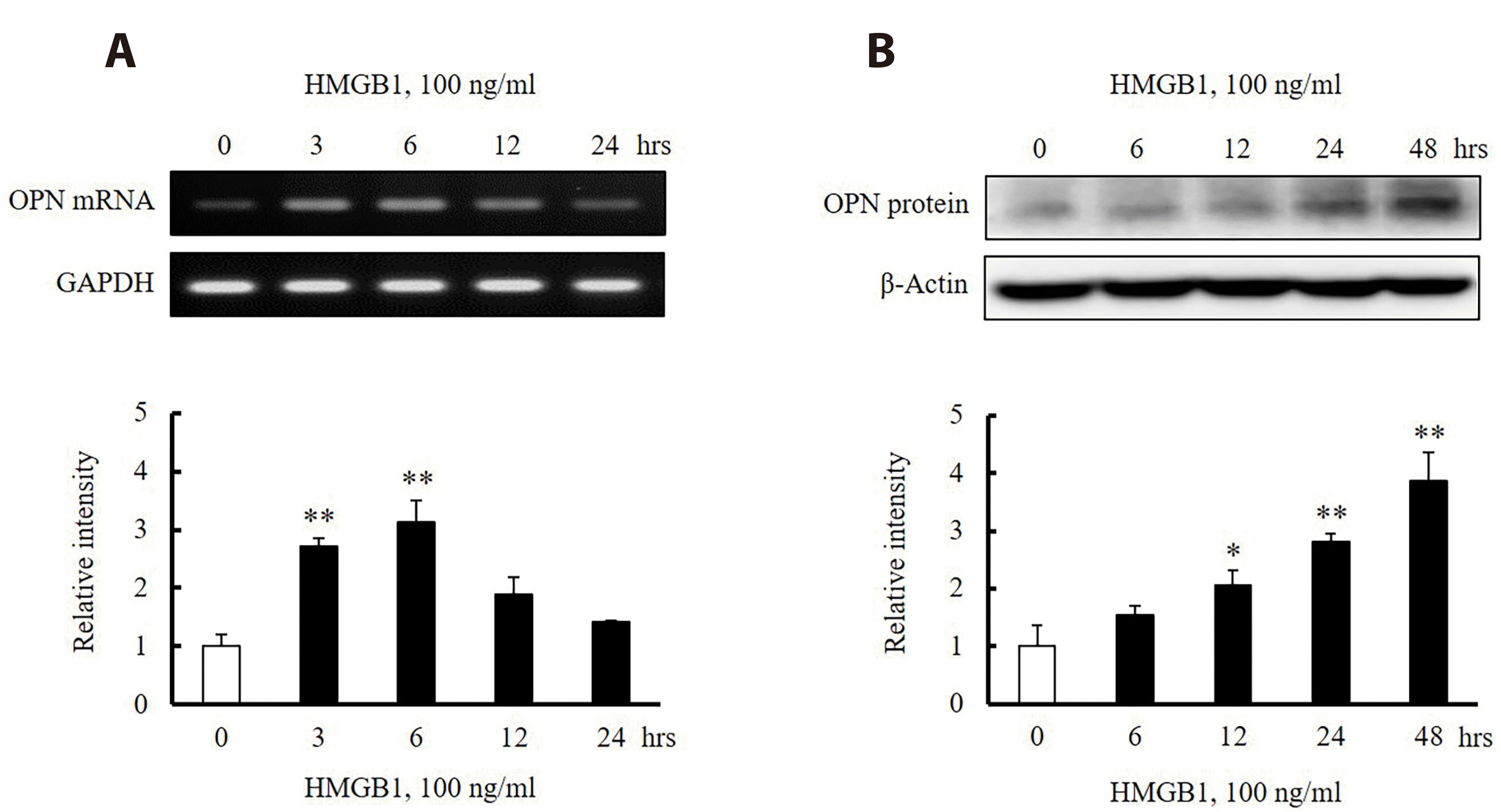 | Fig. 3Characteristics of OPN expression in HMGB1-stimulated VSMCs.(A) VSMCs were stimulated with HMGB1 (100 ng/ml) for 0 to 24 h, and then the expression of OPN mRNA was determined by RT-PCR. GAPDH was used as an internal control. The relative intensities of OPN mRNA to GAPDH were quantified and expressed as the means ± SEMs of 4 independent experiments. **p < 0.01 vs. value at 0 h. (B) In VSMCs treated with HMGB1 (100 ng/ml) for 0 to 48 h, the expression of OPN protein was determined using a Western blot assay. β-Actin was used as an internal control. The relative intensities of OPN protein to β-actin were quantified and expressed as the means ± SEMs of 4 independent experiments. *p < 0.05 and **p < 0.01 vs. value at 0 h. OPN, osteopontin; VSMCs, vascular smooth muscle cells; HMGB1, high-mobility group box 1; RT-PCR, reverse transcription polymerase chain reaction.
|
Promoter deletion assay of OPN in VSMCs stimulated with HMGB1
Several lengths of OPN promoter segments were cloned into a luciferase-based reporter vector, pGL3, to identify the regions responsible for HMGB1-induced OPN transcription within the OPN promoter in HMGB1-stimulated VSMCs. The A10 cells were transiently transfected with three constructs containing different promoter lengths including pLuc-OPN-2284, pLuc-OPN-538 and pLuc-OPN-234. As shown in Fig. 4A, the luciferase reporter activity of pLuc-OPN-2284 in VSMCs exposed to 100 ng/ml HMGB1 was 1.49 ± 0.12 times higher than that in untreated control cells. These relative luciferase activities were markedly increased up to 1.98 ± 0.33 times in cells transfected with pLuc-OPN-538, which was attenuated in cells transfected with the pLuc-OPN-234 construct (1.25 ± 0.10). Therefore, it was suggested that the -538 ~ -234 region of the OPN promoter might be responsible for HMGB1-induced OPN transcription in VSMCs. The putative binding sites for AP-1 and C/EBPβ in this region were suggested using the TF Search program (Fig. 4B).
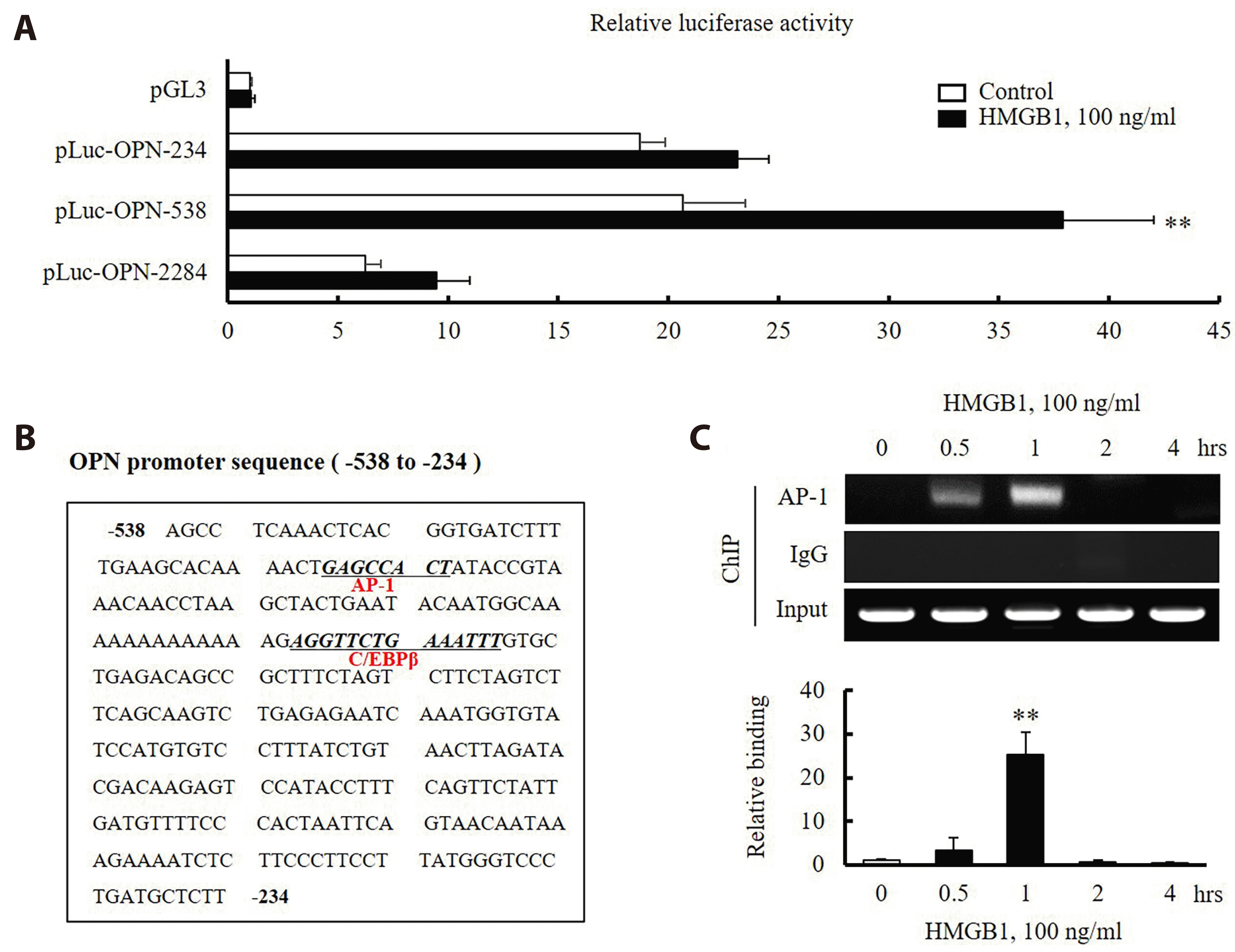 | Fig. 4Identification of the transcription factors mediating OPN transcription in HMGB1-stimulated VSMCs.(A) Three lengths of the OPN promoter were individually constructed in a luciferase-based reporter vector to produce pGL3-OPN-2284 (full-length), pGL3-OPN-538 and pGL3-OPN-234 promoter constructs. The VSMCs were transiently transfected with these promoter constructs and an empty luciferase vector (pGL3) for 24 h, and then stimulated with HMGB1 (100 ng/ml) for 1 h. The differences in promoter activity between the control and HMGB1-stimulated cells were presented as the means ± SEMs of 6 independent experiments. **p < 0.01 vs. non-treated control. (B) Nucleotide sequence of the -538 ~ -234 region of the OPN promoter. The transcription factor binding sites were identified using TF Search software. The sequences of the potential binding sites for AP-1 and C/EBPβ in pLuc-OPN-538 were underlined. (C) The binding activity of AP-1 on the OPN promoter segment in HMGB1-treated VSMCs was assessed using a ChIP assay. IgG was used as the negative control. The relative binding intensities to the input signals were quantified and presented as the means ± SEMs of 6 independent experiments. **p < 0.01 vs. the value at 0 h. OPN, osteopontin; VSMCs, vascular smooth muscle cells; HMGB1, high-mobility group box 1; AP-1, activator protein 1; ChIP, chromatin immunoprecipitation.
|
Increased AP-1 binding on the OPN promoter in HMGB1-stimulated cells
A potential role for AP-1 in OPN expression in HMGB1-stimulated cells was suggested in our previous study using AP-1-deficient cells by siRNA [15], but the direct molecular mechanism was unclear. The binding activity of AP-1 on the OPN promoter segment in HMGB1-stimulated cells was evaluated by incubating the chromatin samples from HMGB1-stimulated cells with an AP-1 antibody, followed by hybridizing for 4 h. The eluted DNA was collected and analyzed as shown in Fig. 4C. The results showed a significant increase in the levels of AP-1 binding in HMGB1-stimulated cells compared to those of the IgG control (Fig. 4C), demonstrating a pivotal role for AP-1 in regulating OPN expression in VSMCs stimulated with HMGB1.
Effects of Ech A on OPN transcription in HMGB1-stimulated VSMCs
To determine the effect of Ech A on HMGB1-induced OPN transcription, the VSMCs were transfected with pLuc-OPN-538 for 24 h and then stimulated with HMGB1 in the prpesence of various concentrations of Ech A (0, 3 and 10 μM). As shown in Fig. 5A, the increased binding activity of AP-1 on the segment of the OPN promoter in HMGB1-stimulated cells was attenuated in the cells pretreated with Ech A in a dose-dependent manner. Similarly, HMGB1-enhanced OPN promoter activity expressed as luciferase activity in the cells transfected with pLuc-OPN-538 was attenuated in Ech A-pretreated cells in a dose-dependent manner (Fig. 5B).
 | Fig. 5Effects of echinochrome A (Ech A) on OPN expression in HMGB1-stimulated VSMCs.(A) The effects of Ech A on the binding activity of AP-1 on the OPN promoter were assessed using a ChIP assay. IgG was used as the negative control. The relative binding intensity to input signals was quantified, and expressed as the means ± SEMs of 6 independent experiments. **p < 0.01 vs. non-treated control, #p < 0.05 and ##p < 0.01 vs. vehicle in the HMGB1-treated group. (B) Cells were transiently transfected with pLuc-OPN-538 constructs for 24 h, and then stimulated with HMGB1 (100 ng/ml) for 1 h in the presence of the indicated concentrations of Ech A (3 or 10 μM). The relative luciferase activities were presented as the means ± SEM of 6 independent experiments. **p < 0.01 vs. control, #p < 0.05 and ##p < 0.01 vs. vehicle in the HMGB1-treated group. (C) VSMCs were pretreated with Ech A (3 or 10 μM) for 24 h and then stimulated with HMGB1 (100 ng/ml) for 6 h and 48 h to determine mRNA and protein expression, respectively. GAPDH and β-actin were used as the internal controls for mRNA and protein expression, respectively. The relative band intensities were quantified, and expressed as the mean ± SEM of 6 independent experiments. **p < 0.01 vs. corresponding control, #p < 0.05 and ##p < 0.01 vs. corresponding value in vehicle of the HMGB1-treated group. OPN, osteopontin; VSMCs, vascular smooth muscle cells; HMGB1, high-mobility group box 1; AP-1, activator protein 1; ChIP, chromatin immunoprecipitation.
|
To further determine the effects of Ech A on OPN mRNA and protein expression in HMGB1-stimulated VSMCs, cells were stimulated with 100 ng/ml of HMGB1 for 6 h and 48 h for mRNA and protein assay, respectively, in the presence of Ech A at concentrations of 0, 3 and 10 μM. As shown in Fig. 5C, both the increased OPN mRNA and protein expression in the HMGB1-stimulated cells were significantly and concentration-dependently reduced by Ech A pretreatment, suggesting the important role for Ech A in regulating OPN expression in HMGB1-stimulated VSMCs.
Preventive effects of Ech A on VSMC migration induced by HMGB1
The role of Ech A in VSMC migration induced by HMGB1 was evaluated by pretreating the cultured A10 cells with Ech A at 0, 3 and 10 μM, and measuring the extent of cell migration using a wound-healing assay in cells stimulated with HMGB1 (100 ng/ml). As shown in Fig. 6, the area of migrated cells expressed as the % changes in the wound area was increased markedly in the cells stimulated with HMGB1 for 48 h. This increase was attenuated dose-dependently in the cells pretreated with Ech A at concentrations of 3 and 10 μM.
 | Fig. 6Effects of echinochrome A (Ech A) on VSMC migration induced by HMGB1.The A10 cells were pretreated with Ech A (3 or 10 μM) for 24 h and then stimulated with HMGB1 (100 ng/ml) for 48 h. Cell migration was determined using a wound-healing assay. The area of migrated cells in the wound area was calculated and expressed as the % changes in the wound area. The area of migrated cells (%) was quantified and expressed as the means ± SEM of 4 independent experiments. **p < 0.01 vs. control, #p < 0.05 and ##p < 0.01 vs. corresponding vehicle in the HMGB1-treated group. VSMC, vascular smooth muscle cell; HMGB1, high-mobility group box 1.
|
Go to :
DISCUSSION
The expression of OPN mRNA and protein was increased in VSMCs stimulated with HMGB1, which was accompanied by an augmented cell migration induced by HMGB1. These increases in VSMC migration were significantly attenuated in the cells pretreated with MPIIIB10 (100 ng/ml), a neutralizing monoclonal antibody for OPN. In VSMCs stimulated with HMGB1, the expression of OPN mRNA and protein was increased. These increases in OPN expression and augmented VSMC migration induced by HMGB1 were significantly attenuated by Ech A, a bioactive red pigment isolated from sea urchins, suggesting a potential effective role of Ech A in preventing vascular remodeling in the injured vasculatures.
Among the various endogenous DAMPs, HMGB1 is considered as one of the most well-characterized DAMPs mediating the progression of various vascular complications [5,6]. The increased HMGB1 levels in atherosclerotic plaque were suggested to be involved in vascular remodeling diseases via the induction of vascular inflammation [24-26]. In addition, considerable experimental evidence has indicated that HMGB1 accelerated neointima formation following vascular injury [27]. Thus, a stratergy to modulate the altered cellular functions induced by HMGB1 might be crucial for preventing or treating proliferative vascular diseases.
In the progression of vascular remodeling diseases including atherosclerosis and vascular restenosis in the injured vasculatures, increased VSMC proliferation and migration are pivotal pathogenic events as reported previously [28]. To investigate the effect of vascular injury on VSMC migration, this study assessed the migration of VSMCs exposed to HMGB1, a major DAMP released from injured vasculatures. When A10 cells cultured from rat thoracic aorta were wounded and then stimulated with HMGB1, VSMC migration induced by HMGB1 (100 ng/ml) was increased markedly compared to the control cells. Therefore, HMGB1 released in the injured vasculatures was suggested to play a key role in mediating the progression of vascular remodeling diseases via the increased migration of VSMCs.
Accumulating evidences suggest that OPN plays a pathological role in atherosclerosis, vascular restenosis and several cardiomyopathies [29,30]. Moreover, OPN was suggested as a potent mediator in the development of vascular remodeling [31]. As the mechanisms for the action of OPN, the increased migration and proliferation of VSMCs [15], production of matrix metalloproteinases [32] and vascular inflammation [33] have been suggested. Importantly, our previous study also suggested that OPN mediated the VSMCs proliferation induced by platelet-derived growth factor and 4-hydroxynonenal [11,34]. Considering the pathologic importance of OPN in vascular remodeling in the injured vasculatures, this study investigated OPN expression and related molecular mechanism in VSMCs exposed to HMGB1, a major DAMP released in the injured vasculatures. In A10 cells stimulated with HMGB1, the OPN mRNA and protein levels were increased markedly, which was linked to VSMC migration induced by HMGB1. In addition, the increased VSMC migration was attenuated significantly in cells pretreated with MPIIIB10 (100 ng/ml), a neutralizing monoclonal antibody for OPN. Thus, our results suggest that OPN could be a potential target for developing therapeutic strategies for vascular remodeling in the injured vasculatures.
The present study observed an increase in cellular expression of OPN mRNA in HMGB1-stimulated cells, suggesting that HMGB1-induced OPN expression is regulated at the transcriptional levels. To identify the transcription factors involved in OPN transcription, several lengths of OPN promoter segments were cloned into a luciferase-based reporter vector, pGL3, and the regions responsible for HMGB1-induced OPN transcription within the OPN promoter in HMGB1-stimulated VSMCs were determined. Among the A10 cells transiently transfected with constructs containing different promoter sizes (pLuc-OPN-2284, -538 and -234), the luciferase reporter activity of pLuc-OPN-538 in the cells stimulated with HMGB1 (100 ng/ml for 1 h) was significantly higher than that in the corresponding control, whereas this increase was markedly attenuated in the cells transfected with the pLuc-OPN-234 construct. Thus, the -538 ~ -234 region of the OPN promoter was suggested as the cis-acting element responsible for HMGB1-induced OPN transcription in VSMCs. Using the sequence motif search of TF Search software (http://mbs.cbrc.jp/research/db/TFSEARCH.html), it is noteworthy that the putative transcription factor binding sites for AP-1 and C/EBPβ were identified in the region between -538 bp and -234 bp relative to the transcriptional initiation site in the OPN promoter.
Using AP-1-deficient cells treated with siRNA, a potential role for AP-1 in OPN expression in HMGB1-stimulated cells was suggested in our previous study [15], but the direct molecular mechanism remains unclear. To evaluate the binding activity of AP-1 on the segment of the OPN promoter in HMGB1-stimulated cells, the chromatin samples from HMGB1-stimulated cells were hybridized with the AP-1 antibody, and then the eluted DNA was analyzed using a Chip assay. Our current study provides direct evidences that the stimulation of A10 cells with HMGB1 increases the binding of AP-1 to its binding site in the OPN promoter, leading to an increased promoter activity, and to higher mRNA and protein levels of OPN.
A strategy regulating HMGB1-induced VSMC migration might be important for developing therapeutics for vascular remodeling because HMGB1 plays a pathological role in the process of vascular remodeling. Recently, Ech A, a bioactive red pigment isolated from sea urchins [20], exhibited cardioprotective effects through its antioxidant and anti-inflammatory activities [21]. To investigated the effect of Ech A on HMGB1-induced OPN transcription in our present study, VSMCs were transfected with pLuc-OPN-538 in the presence of various concentrations of Ech A, and then stimulated with HMGB1. Our results clearly showed that the HMGB1-enhanced luciferase activity in cells transfected with pLuc-OPN-538 and the increased AP-1 binding to the OPN promoter segment in HMGB1-stimulated cells were attenuated markedly in the cells pretreated with Ech A. In addition, the expression of OPN mRNA and protein in HMGB1-stimulated VSMCs was reduced significantly and dose-dependently by Ech A pretreatment, suggesting the important role of Ech A in regulating OPN expression in HMGB1-stimulated VSMCs. Likewise, the increased VSMC migration induced by HMGB1 was attenuated dose-dependently in the cells pretreated with Ech A. Thus, it was suggested that Ech A attenuated HMGB1-induced VSMC migration by inhibiting the AP-1 binding on the OPN promoter and subsequent down-regulation of OPN expression in VSMCs.
In conclusion, the increased migration of VSMCs was associated with an increased OPN expression in cells stimulated with HMGB1, which was suppressed by Ech A. The inhibitory effect of Ech A on VSMC migration was mediated by the down-regulated expression of OPN in VSMCs via an attenuated AP-1 binding on the OPN promoter. Considering these characteristics of Ech A, we thus suggest Ech A as a potential candidate drug for alleviating vascular remodeling in the injured vasculatures.
Go to :
 XML Download