Journal List > Clin Exp Otorhinolaryngol > v.17(4) > 1516089273

On, Kim, Lee, Lee, Lee, Byun, and Hong: Clinical Efficacy of a Position-Responding Mandibular Advancement Device in Patients With Obstructive Sleep Apnea

Abstract

Objectives.

Although mandibular advancement device (MAD) treatment is effective for obstructive sleep apnea (OSA), some concerns remain regarding its potential therapeutic impact and side effects. Thus, we developed a novel MAD that auto-titrates depending on its position in patients with OSA. We conducted a clinical trial to determine the efficacy of an auto-titrating mandibular advancement device (AMAD) for treating OSA.

Methods.

Fourteen patients diagnosed with OSA participated in this study. Polysomnography (PSG) was performed at the beginning of the clinical trial, and after 3 months of treatment, PSG with AMAD in situ was conducted.

Results.

The mean scores for the Epworth Sleepiness Scale (ESS) and STOP-Bang were 8.21±4.21 and 5.00±1.00, respectively. After 3 months of AMAD treatment, the STOP-Bang scores improved to 3.75±1.06; however, the ESS scores did not show a significant change. Additionally, we observed statistically significant improvements in several respiratory parameters in the PSG data following AMAD treatment. These included reductions in the apnea-hypopnea index (AHI) (from 32.85±21.71 to 12.93±10.70), supine AHI (from 45.91±23.58 to 15.59±12.76), and lateral AHI (from 13.94±10.95 to 5.49±7.40). Improvements were also noted in the lowest O2 saturation (from 79.71±6.22 to 84.00± 5.71), total arousal number (from 191.14±112.07 to 86.57±48.80), and arousal index (from 33.76±21.00 to 15.05± 8.42). However, there were no significant changes in total sleep time, sleep efficiency, or mean oxygen saturation. Additionally, no major side effects were observed during treatment, specifically related to tooth or jaw pain.

Conclusion.

Our clinical trial found that AMAD improved PSG parameters and reduced the incidence of common side effects. Therefore, AMAD may be an effective alternative treatment for OSA.

INTRODUCTION

Obstructive sleep apnea (OSA) is a breathing disorder characterized by repetitive and intermittent occlusion of the upper airway during sleep [1]. If complete collapse occurs in the upper airway due to inspiratory negative pressure, apnea occurs; if the collapse is partial, it results in hypopnea or snoring. Untreated OSA is linked to numerous adverse health outcomes, including cardiovascular diseases and mental illness [2-4]. The treatment of OSA requires a tailored, multi-pronged approach for each patient. Continuous positive airway pressure (CPAP) is the most effective treatment for adults and is considered the gold standard for managing moderate-to-severe OSA [5]. However, despite CPAP’s high efficacy in eliminating respiratory events, its overall effectiveness is compromised by poor adherence to the therapy. Research indicates that the initial month of PAP therapy is crucial for establishing adherence in patients with OSA, with many patients continuing to struggle with adequate adherence beyond the first month [6-8].
A mandibular advancement device (MAD) was developed as an innovative treatment for snoring and OSA, addressing issues related to adherence. MAD is now considered a first-line treatment for patients with simple snoring, mild-to-moderate OSA who have a low body mass index, and those with increased upper airway resistance syndrome [9,10]. Additionally, MAD can be used for patients whose OSA does not respond to continuous positive airway pressure (CPAP) or who struggle with CPAP compliance. Notably, MAD offers advantages over CPAP as it does not require electricity and is more convenient for mobility in various sleep settings, such as during travel [11].
Although MAD treatment for OSA patients is effective, concerns about potential short-term and long-term side effects persist [12-14]. Common short-term complications include jaw pain, tooth tenderness, and excessive salivation, while long-term complications may involve changes in the geometry of the teeth and facial skeleton, leading to dental alterations, temporomandibular joint discomfort, and chewing difficulties. Consequently, close follow-up during long-term MAD therapy is essential to promptly identify and address any orthodontic complications.
In the present study, we evaluated the clinical efficacy of a newly developed MAD that autotitrates based on its position in patients with OSA. This auto-titrating mandibular advancement device (AMAD) represents a state-of-the-art advancement in MAD technology. It automatically increases the forward distance of the mandible when the patient is in the supine position and decreases it when the patient is in the lateral position. Consequently, this adjustment can reduce the duration of traction on the temporomandibular joint during sleep and minimize the occurrence of side effects.

MATERIALS AND METHODS

The study protocol was approved by the Institutional Review Board of Dongtan Sacred Heart Hospital, Hallym University College of Medicine (No. 2021-10-012-001), and all participants provided written informed consent.

Subjects

We included patients diagnosed with OSA using overnight polysomnography (PSG) at Hallym University Dongtan Sacred Heart Hospital, Republic of Korea. The exclusion criteria were defined as follows: patients under the age of 20 years; those who routinely use hypnotic medications or tranquilizers; individuals who are edentulous, have an excessive gag reflex, suffer from periodontal disease, or have central sleep apnea; and individuals who have previously experienced unsuccessful outcomes with oral appliance therapy. We used the Epworth Sleepiness Scale (ESS) and the Stop-Bang Questionnaire to assess sleep quality, including daytime sleepiness and the severity of sleep apnea. The apnea-hypopnea index (AHI) was calculated as the total number of apneas and hypopneas per hour of sleep. The mean and minimum oxygen saturation levels were also measured. The cut-off point for the diagnosis of OSA was an AHI ≥5 with associated symptoms related to OSA, such as excessive daytime sleepiness. Seventeen patients were enrolled, clinical data including survey responses were missing for three, resulting in fourteen participants being included in the study.

Clinical trial design

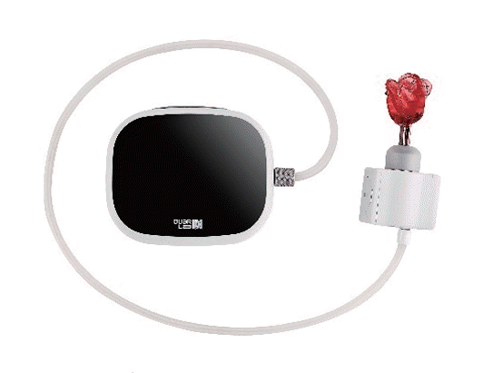
This is a prospective, single-armed, single-center trial study using the AMAD (Oxleep device) (Fig. 1). All patients who were formally diagnosed with OSA on PSG had no contraindications to the use of the device, were deemed potential candidates for custom MAD treatment, and were enrolled. Follow-up PSG with AMAD in situ was performed after 3 months of treatment. The subjects also had to complete the ESS and STOP-Bang questionnaires at this time. All adverse effects and their severity during AMAD use were assessed. In this clinical trial, subjects in the cohort served as controls for the post-intervention assessment. We used the primary and secondary outcomes obtained from subjects treated for at least 3 months. If the subjects did not complete the clinical trial protocol, they were considered withdrawn from the trial.

Primary and secondary outcome

The primary outcome of this clinical trial was to observe post-intervention improvements in respiratory parameters from PSG data, such as the AHI, supine AHI, lateral AHI, and O2 saturation (lowest or mean). Apnea was defined as a decrease in airflow of 0% to 20% lasting for more than 10 seconds. Hypopnea was defined as a decrease in airflow of at least 30% that lasted for more than 10 seconds. The secondary outcome was to observe post-intervention improvements in sleep quality parameters, including the ESS, STOP-Bang score, total sleep time, sleep efficiency, and arousal index. The arousal index is the number of cortical arousals identified via electroencephalography divided by total sleep time. Additionally, we evaluated the side effects of AMAD during the intervention as a secondary outcome.

Statistical analysis

All measured values were reported as mean (standard deviation) for continuous variables and as frequencies and percentages for categorical variables. Paired t-tests or Wilcoxon signed-rank tests were used to compare polysomnographic data between the two PSGs. All statistical analyses were performed using R version 4.0.5 (https://www.R-project.org). Two-tailed P-values of <0.05 were considered statistically significant.

RESULTS

Baseline characteristics and polysomnographic data

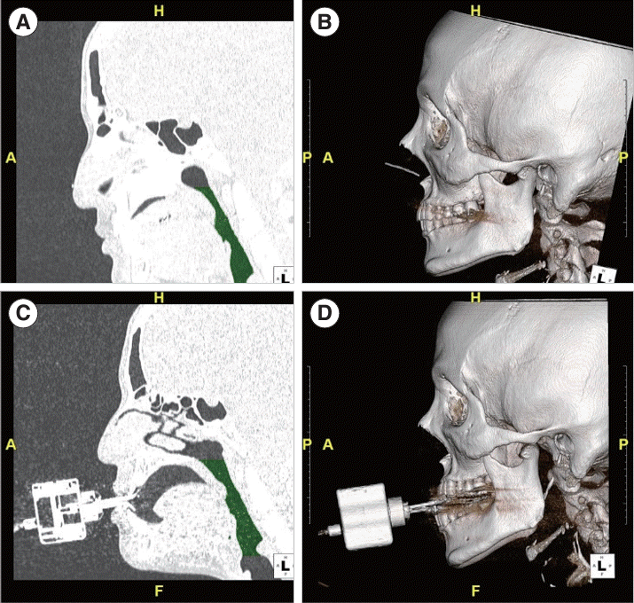
Fourteen patients with OSA (13 men and 1 woman) consented to participate in this analysis using the AMAD. The mean age of the patients was 43.0±10.5 years, and their average body mass index was 27.72±3.43 kg/m2. The baseline PSG respiratory index values for the included patients were as follows: AHI, 32.85±21.71; supine AHI, 45.91±23.58; lateral AHI, 13.94±10.95; arousal index, 33.76±21.00; lowest oxygen saturation, 79.71±6.22; mean oxygen saturation, 94.69±1.24. All descriptive data are presented in Table 1. Additionally, to visually confirm the effect of upper airway expansion, we performed three-dimensional (3D) computed tomography before and after treatment. An observed widening of the upper airway was noted when patients used the AMAD. A representative image is shown in Fig. 2.

Short-term effects of the AMAD on respiratory variables

When we assessed subjective outcomes such as ESS and STOP-Bang, there was no significant difference in the ESS scores before and after treatment (8.21±4.21 vs. 8.08±4.40, P=0.939). However, the STOP-Bang score significantly decreased from 5.00±1.00 to 3.75±1.06 (P=0.017). This reduction was noted specifically in the snoring and observed apnea items within the STOP component, but not in the Bang component. Data from the STOP-Bang risk strata indicated an overall improvement in risk assessment post-treatment (Table 2).
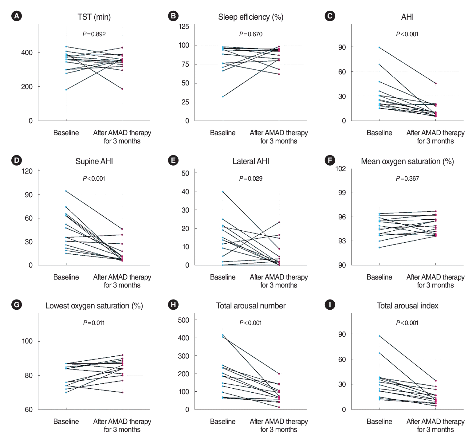
In the paired analysis of PSG data, no statistically significant differences were observed in total sleep time (P=0.892) or sleep efficiency (P=0.670). However, statistically significant improvements were noted in AHI, which decreased from 32.85±21.71 to 12.93±10.70 (P<0.001), supine AHI, which decreased from 45.91±23.58 to 15.59±12.76 (P<0.001), and lateral AHI, which decreased from 13.94±10.95 to 5.49±7.40 (P=0.029). All respiratory parameters showed improvement after the use of AMAD compared to baseline data. Additionally, significant improvements were observed in the lowest oxygen saturation, which increased from 79.71±6.22 to 84.00±5.71 (P=0.011), total arousal number, which decreased from 191.14±112.07 to 86.57±48.80 (P<0.001), and arousal index, which decreased from 33.76±21.00 to 15.05±8.42 (P<0.001). However, there was no significant change in the mean oxygen saturation (P=0.367). The paired t-test results for PSG data before and after treatment are presented in Fig. 3.
Moreover, no severe adverse effects were observed during the clinical trials. Specifically, there were no significant complaints related to pain, including tooth or jaw pain. However, some patients did report minor adverse events, such as hypersalivation and mucosal dryness (Table 3). Some patients reported mild tooth and jaw pain, which was alleviated after adjusting the device and adhering to temporomandibular joint precautions. All enrolled patients completed their treatment with AMAD during the study period.

DISCUSSION

CPAP is the preferred treatment for patients with OSA, known to enhance health-related quality of life by alleviating symptoms such as apnea and excessive daytime sleepiness, and potentially reducing the risk of cardiovascular diseases through the lowering of blood pressure and angina. However, due to its greater convenience and compliance, MAD treatment has become an increasingly popular alternative [15]. The American Academy of Sleep Medicine and the American Academy of Dental Sleep Medicine have also endorsed MAD as an alternative therapy for OSA patients who cannot tolerate CPAP therapy [9]. Recently, OUaR LaB Inc. (Seoul, Korea) developed a new type of MAD, which received approval from the Korea Food and Drug Administration. This device underwent GMP inspections for marketing authorization of advanced therapy medicinal products, adhering to relevant regulations and guidelines. It features a unique mechanism that automatically adjusts the mandibular traction distance based on the patient’s sleep position, leading us to name it the AMAD. These features not only provide therapeutic effects on OSA but also minimize the potential side effects associated with static jaw traction during overnight sleep. Thus, we conducted a clinical trial to assess its therapeutic efficacy and to determine if it could mitigate the common side effects associated with short-term MAD use. Interestingly, the trial revealed that AMAD not only significantly improved respiratory parameters such as AHI and lowest oxygen saturation but also resulted in no severe side effects in OSA patients after short-term use.
The primary mechanism of action for MADs involves enhancing the dimensions of the upper airway by protruding the mandible. There is a wide range of MADs available on the market, each featuring different design elements [16-21]. Customized biblock MADs are generally preferred as they allow precise adjustments in mandibular advancement, given that their upper and lower parts are separate yet interconnected [9]. These devices are often referred to as titratable MADs, which are engineered to incrementally advance the mandible using a straightforward mechanical mechanism until an effective protrusive position for alleviating sleep apnea symptoms is reached. Customized bi-block titratable MADs are available in two main types, differentiated by the degree of mandibular vertical opening they permit. However, findings from one randomized controlled trial indicate that MADs with either limited or free vertical opening produce similar outcomes in terms of respiratory parameters and upper airway dimensions in patients with mild to moderate OSA [22]. To date, various titration protocols have been explored to identify the optimal protrusive position. Nonetheless, no gold standard for titration has been established, making the adjustment of MADs a “trial and error” process in everyday clinical practice.
The AMAD is a state-of-the-art MAD that automatically increases the forward distance when the patient is in the supine position and decreases it when the patient is lying in the lateral position. The AMAD (Oxleep) introduces a novel concept in titration protocols compared to other state-of-the-art MADs. Traditional titratable MADs determine the degree of mandibular advancement by measuring how far a patient can protrude their mandible while awake. In contrast, the AMAD (Oxleep) detects changes in the patient’s sleeping position and automatically adjusts the mandibular advancement within the preset upper and lower limits. This dynamic titration process increases mandibular protrusion in the supine position and decreases it in the lateral position during sleep, allowing the AMAD to achieve a therapeutic effect similar to that of auto-PAP in patients with OSA. The basic operating principle of the AMAD is as follows. The device includes a 3D accelerometer that identifies whether the patient’s position is supine or lateral. Medical professionals set the mandibular advancement distance based on the patient’s AHI and sleep position to optimize airway opening. The device then adjusts the mandible forward or backward according to the preconfigured advancement settings. For example, if the advancement setting includes a base advancement of 4 mm with an additional 2 mm for supine sleeping, the device will advance the mandible by the base distance (4 mm) when the patient is in a lateral position. If the patient shifts to a supine position, the device will further advance the mandible by an additional 2 mm. Should the patient return to a lateral position, the device will retract by 2 mm. Moreover, if the mandibular advancement is maintained for an extended period, the device can enter a rest mode, allowing the jaw to relax for a set duration. These features of differential adjustment and rest mode contribute to effective sleep apnea management while enhancing patient comfort.
In this study, we confirmed that changes in the degree of mandibular protrusion during sleep did not interfere with sleep quality. We observed no difference in total sleep time or sleep efficiency, and notably, the arousal index decreased with the use of AMAD. Additionally, AMAD dynamic titration reduced the patients’ exposure time to the maximum duration of mandible protrusion, significantly lowering the incidence of common MAD-related complications, particularly those related to pain. These unique advantages are anticipated to address changes in dental occlusion, such as overjet and overbite, following long-term MAD use, a hypothesis that future long-term studies of AMAD will need to confirm [23,24]. Furthermore, a recent study indicated that sleep stage transitions from N3 and REM sleep to wakefulness were significantly more frequent during changes in body position [25]. This finding suggests that leveraging the influence of sleeping positions could enhance sleep structure and quality in patients with OSA.
Although the benefits of MAD therapy include significant improvements in the quality of life and sleep quality for both the patient and their bed partner, several studies have shown that MAD is generally considered less effective than CPAP [26-29]. However, a previous randomized controlled trial demonstrated that the key health outcomes were similar after 1 month of optimal treatment with both MAD and CPAP in patients with moderate to severe OSA [30]. It also showed that CPAP therapy was more effective in reducing AHI than MAD, but MAD had higher compliance than CPAP. Additionally, another long-term study indicated no significant difference in the success rates of treating mild to moderate OSA between MAD therapy and CPAP over a 2-year follow-up period [24]. Interestingly, when evaluating subjective outcomes such as ESS and STOP-Bang, there was no significant change in ESS scores before and after treatment. However, the STOP-Bang score significantly decreased from 5.00±1.00 to 3.75±1.06, suggesting an overall improvement in risk assessment according to the STOP-Bang risk stratum data. The considerable standard deviation in ESS scores between the two groups likely affected the achievement of statistical significance. It is anticipated that with more participants in future studies, a statistically significant difference in ESS scores might be observed. In the current clinical trial, we observed a significant reduction in the mean AHI from 32.85±21.71 to 12.93±10.70. Furthermore, both the supine and lateral AHI showed statistically significant improvements in the paired analysis. Consistent with previous findings [31,32], AMAD reduced AHI by at least 50%. We noted that AMAD treatment led to a statistically significant reduction in AHI of approximately 57%. Additionally, significant improvements were seen in the lowest oxygen saturation, arousal index, and overall risk assessment based on the STOP-Bang questionnaire after 3 months of AMAD use in patients with OSA. This suggests that automatic titration during sleep does not interfere with improvements in sleep quality and quality of life. Specifically, only one participant showed no improvement in AHI and lowest O2 saturation after using the AMAD. This case occurred early in the study when the advancement distance setting of the AMAD was not effectively or sufficiently adjusted. As the pilot study progressed, our clinical experience with setting the advancement distance improved, preventing the recurrence of such errors.
Our study had some limitations. First, it had a short follow-up time, a small sample size, and lacked a control group (no treatment/other MAD). These limitations may be overcome by recruiting a larger sample, extending the therapeutic monitoring time, and controlling the randomized selection. Second, this was a simple short-term study that did not evaluate long-term effects. It remains to be determined whether AMAD can reduce the risk of cardiovascular diseases and related complications. Future studies should also evaluate long-term side effects, adherence rates, and changes in dental occlusion. Consequently, well-controlled, large-scale, long-term clinical trials are necessary to verify the clinical efficacy of AMAD. Finally, our titration protocol was solely based on sleep position and did not take into account oxygen saturation. While lateral AHI showed improvement with AMAD in situ, incorporating oxygen saturation status into the titration protocol could potentially optimize mandible protrusion, thereby enhancing the treatment’s success rate.
In conclusion, our clinical trial suggests that AMAD could serve as an effective alternative for treating OSAS, significantly improving several PSG parameters, including AHI, lowest oxygen saturation, and arousal index. Additionally, we observed no severe common side effects such as hypersalivation, tooth pain, mucosal dryness, or jaw pain during the short-term use of AMAD. While the primary and secondary outcomes were promising, further research is necessary to develop standardized titration protocols for AMAD.

HIGHLIGHTS

▪ We conducted a clinical trial to assess the efficacy of an autotitrating mandibular advancement device (AMAD) in treating obstructive sleep apnea (OSA).
▪ After short-term AMAD therapy, OSA patients showed an improved apnea-hypopnea index, lower oxygen saturation, and arousal index, whereas there were no significant troublesome complaints regarding side effects.
▪ AMAD may be effective for treating OSA while minimizing side effects.

Notes

DKK serves on the Board of Directors for OUaR LaB Inc. and owns OUaR LaB Stock, which is subject to certain restrictions under university policy. No other potential conflicts of interest relevant to this article were reported.

AUTHOR CONTRIBUTIONS

Conceptualization: SWO, SHB, SJH. Data curation: DKK. Formal analysis: SWO, SHB, MHL, JHL, SJH. Funding acquisition: SJH, SHB. Methodology: DKK. Project administration: SHB, SJH. Visualization: SJH. Writing–original draft: DKK, SWO, SJH. Writing–review & editing: DKK, KCL, SJH. All authors read and agreed to the published version of the manuscript.

ACKNOWLEDGMENTS

This research was supported by a grant from the Korea Health Technology R&D Project through the Korea Health Industry Development Institute (KHIDI), funded by the Ministry of Health and Welfare, Republic of Korea (grant no. HI20C2114). This work was also partly supported by National IT Industry Promotion Agency (NIPA) grant funded by the Korea government (MSIT) (S1402-23-1001, AI Diagnostic Assisted Virtual Surgery and Digital Surgical Guide for Dental Implant Treatment in the Post-Aged Society: A Multicenter Clinical Demonstration).

REFERENCES

1. Sankri-Tarbichi AG. Obstructive sleep apnea-hypopnea syndrome: etiology and diagnosis. Avicenna J Med. 2012; Jan. 2(1):3–8.
crossref
2. Correction to: obstructive sleep apnea and cardiovascular disease: a scientific statement from the American Heart Association. Circulation. 2022; Mar. 145(12):e775.
3. Yeghiazarians Y, Jneid H, Tietjens JR, Redline S, Brown DL, El-Sherif N, et al. Obstructive sleep apnea and cardiovascular disease: a scientific statement from the American Heart Association. Circulation. 2021; Jul. 144(3):e56–67.
crossref
4. Lin WC, Winkelman JW. Obstructive sleep apnea and severe mental illness: evolution and consequences. Curr Psychiatry Rep. 2012; Oct. 14(5):503–10.
crossref
5. Patil SP, Ayappa IA, Caples SM, Kimoff RJ, Patel SR, Harrod CG. Treatment of adult obstructive sleep apnea with positive airway pressure: an American Academy of Sleep Medicine systematic review, meta-analysis, and GRADE assessment. J Clin Sleep Med. 2019; Feb. 15(2):301–34.
crossref
6. Xanthopoulos MS, Kim JY, Blechner M, Chang MY, Menello MK, Brown C, et al. Self-efficacy and short-term adherence to continuous positive airway pressure treatment in children. Sleep. 2017; Jul. 40(7):zsx096.
crossref
7. Stepnowsky CJ Jr, Marler MR, Ancoli-Israel S. Determinants of nasal CPAP compliance. Sleep Med. 2002; May. 3(3):239–47.
crossref
8. Weaver TE, Grunstein RR. Adherence to continuous positive airway pressure therapy: the challenge to effective treatment. Proc Am Thorac Soc. 2008; Feb. 5(2):173–8.
crossref
9. Ramar K, Dort LC, Katz SG, Lettieri CJ, Harrod CG, Thomas SM, et al. Clinical practice guideline for the treatment of obstructive sleep apnea and snoring with oral appliance therapy: an update for 2015. J Clin Sleep Med. 2015; Jul. 11(7):773–827.
crossref
10. Jayesh SR, Bhat WM. Mandibular advancement device for obstructive sleep apnea: an overview. J Pharm Bioallied Sci. 2015; Apr. 7(Suppl 1):S223–5.
11. Ferguson KA, Ono T, Lowe AA, al-Majed S, Love LL, Fleetham JA. A short-term controlled trial of an adjustable oral appliance for the treatment of mild to moderate obstructive sleep apnoea. Thorax. 1997; Apr. 52(4):362–8.
crossref
12. Fritsch KM, Iseli A, Russi EW, Bloch KE. Side effects of mandibular advancement devices for sleep apnea treatment. Am J Respir Crit Care Med. 2001; Sep. 164(5):813–8.
crossref
13. Ishida E, Kunimatsu R, Medina CC, Iwai K, Miura S, Tsuka Y, et al. Dental and occlusal changes during mandibular advancement device therapy in Japanese patients with obstructive sleep apnea: four years follow-up. J Clin Med. 2022; Dec. 11(24):7539.
crossref
14. Jo SY, Lee SM, Lee KH, Kim DK. Effect of long-term oral appliance therapy on obstruction pattern in patients with obstructive sleep apnea. Eur Arch Otorhinolaryngol. 2018; May. 275(5):1327–33.
crossref
15. Zhu Y, Long H, Jian F, Lin J, Zhu J, Gao M, et al. The effectiveness of oral appliances for obstructive sleep apnea syndrome: a meta-analysis. J Dent. 2015; Dec. 43(12):1394–402.
crossref
16. Ghazal A, Sorichter S, Jonas I, Rose EC. A randomized prospective long-term study of two oral appliances for sleep apnoea treatment. J Sleep Res. 2009; Sep. 18(3):321–8.
crossref
17. Zhou J, Liu YH. A randomised titrated crossover study comparing two oral appliances in the treatment for mild to moderate obstructive sleep apnoea/hypopnoea syndrome. J Oral Rehabil. 2012; Dec. 39(12):914–22.
crossref
18. Umemoto G, Toyoshima H, Yamaguchi Y, Aoyagi N, Yoshimura C, Funakoshi K. Therapeutic efficacy of twin-block and fixed oral appliances in patients with obstructive sleep apnea syndrome. J Prosthodont. 2019; Feb. 28(2):e830–6.
crossref
19. Bloch KE, Iseli A, Zhang JN, Xie X, Kaplan V, Stoeckli PW, et al. A randomized, controlled crossover trial of two oral appliances for sleep apnea treatment. Am J Respir Crit Care Med. 2000; Jul. 162(1):246–51.
crossref
20. Segu M, Campagnoli G, Di Blasio M, Santagostini A, Pollis M, Levrini L. Pilot study of a new mandibular advancement device. Dent J (Basel). 2022; Jun. 10(6):99.
crossref
21. El Ibrahimi M, Laabouri M. Pilot study of a new adjustable thermoplastic mandibular advancement device for the management of obstructive sleep apnoea-hypopnoea syndrome: a brief research letter. Open Respir Med J. 2016; Jul. 10:46–50.
crossref
22. Shi X, Lobbezoo F, Chen H, Rosenmoller BR, Berkhout E, de Lange J, et al. Comparisons of the effects of two types of titratable mandibular advancement devices on respiratory parameters and upper airway dimensions in patients with obstructive sleep apnea: a randomized controlled trial. Clin Oral Investig. 2023; May. 27(5):2013–25.
crossref
23. Martins OFM, Chaves Junior CM, Rossi RRP, Cunali PA, Dal-Fabbro C, Bittencourt L. Side effects of mandibular advancement splints for the treatment of snoring and obstructive sleep apnea: a systematic review. Dental Press J Orthod. 2018; Aug. 23(4):45–54.
crossref
24. Doff MH, Hoekema A, Wijkstra PJ, van der Hoeven JH, Huddleston Slater JJ, de Bont LG, et al. Oral appliance versus continuous positive airway pressure in obstructive sleep apnea syndrome: a 2-year follow-up. Sleep. 2013; Sep. 36(9):1289–96.
crossref
25. Ahn HK, Kang YJ, Yoon W, Shin HW. Analysing the impact of body position shift on sleep architecture and stage transition: a comprehensive multidimensional study using event-synchronised polysomnography data. J Sleep Res. 2024; Aug. 33(4):e14115.
crossref
26. Sharples LD, Jackson C, Wheaton E, Griffith G, Annema JT, Dooms C, et al. Clinical effectiveness and cost-effectiveness of endobronchial and endoscopic ultrasound relative to surgical staging in potentially resectable lung cancer: results from the ASTER randomised controlled trial. Health Technol Assess. 2012; 16(18):1–75.
crossref
27. Sharples LD, Clutterbuck-James AL, Glover MJ, Bennett MS, Chadwick R, Pittman MA, et al. Meta-analysis of randomised controlled trials of oral mandibular advancement devices and continuous positive airway pressure for obstructive sleep apnoea-hypopnoea. Sleep Med Rev. 2016; Jun. 27:108–24.
28. Marklund M. Update on oral appliance therapy for OSA. Curr Sleep Med Rep. 2017; 3(3):143–51.
crossref
29. Schwartz M, Acosta L, Hung YL, Padilla M, Enciso R. Effects of CPAP and mandibular advancement device treatment in obstructive sleep apnea patients: a systematic review and meta-analysis. Sleep Breath. 2018; Sep. 22(3):555–68.
crossref
30. Phillips CL, Grunstein RR, Darendeliler MA, Mihailidou AS, Srinivasan VK, Yee BJ, et al. Health outcomes of continuous positive airway pressure versus oral appliance treatment for obstructive sleep apnea: a randomized controlled trial. Am J Respir Crit Care Med. 2013; Apr. 187(8):879–87.
crossref
31. Marklund M, Verbraecken J, Randerath W. Non-CPAP therapies in obstructive sleep apnoea: mandibular advancement device therapy. Eur Respir J. 2012; May. 39(5):1241–7.
crossref
32. Ferguson KA, Cartwright R, Rogers R, Schmidt-Nowara W. Oral appliances for snoring and obstructive sleep apnea: a review. Sleep. 2006; Feb. 29(2):244–62.
crossref

Fig. 1.
Appearance of the auto-titrating mandibular advancement device (Oxleep).
ceo-2024-00124f1.tif
Fig. 2.
Pre-treatment and post-treatment three-dimensional computed tomography sagittal views of the upper respiratory tract. (A) Pretreatment sagittal view of computed tomography showing the upper respiratory tract. (B) Pre-treatment sagittal view of the three-dimensional reconstructed image. (C) Post-treatment sagittal view of computed tomography showing the upper respiratory tract. (D) Post-treatment sagittal view of the three-dimensional reconstructed image. The green area represents the upper airway cross-sectional area in the pre-treatment (A) and post-treatment (C) images.
ceo-2024-00124f2.tif
Fig. 3.
Short-term effects of the auto-titrating mandibular advancement device (AMAD) on respiratory variables. (A) Total sleep time (TST). (B) Sleep efficiency. (C) Apnea-hypopnea index (AHI). (D) Supine AHI. (E) Lateral AHI. (F) Mean oxygen saturation. (G) Lowest oxygen saturation. (H) Total arousal number. (I) Total arousal index.
ceo-2024-00124f3.tif
Table 1.
Demographic data and sleep parameters for the enrolled population
Variable Patients with AMAD (n=14)
Subjects characteristics
 Age (yr) 43.0±10.5
 Sex (male) 13 (92.86)
 Body mass index (kg/m2) 27.72±3.43
Polysomnographic data
 Total sleep time (min) 345.65±63.78
 Sleep efficiency (%) 84.10±18.53
 AHI (events/hr) 32.85±21.71
 Supine AHI (events/hr) 45.91±23.58
 Left AHI (events/hr) 7.28±10.88
 Right AHI (events/hr) 6.66±6.88
 Lateral AHI (events/hr) 3.94±10.95
 Arousal index (events/hr) 33.76±21.00
 Arousal number 191.14±112.07
 Lowest O2 saturation (%) 79.71±6.22
 Mean O2 saturation (%) 94.69±1.24

Values are presented as mean±standard deviation or number (%).

AMAD, auto-titrating mandibular advancement device; AHI, apnea-hypopnea index.

Table 2.
Comparison of STOP-Bang scores before and after using the auto-titrating mandibular advancement device
Risk assessment (n=14) Baseline After AMAD therapy
Low risk 0 1 (7.1)
Intermediate risk 4 (28.6) 11 (78.6)
High risk 10 (71.4) 2 (14.3)

Values are presented as number (%).

AMAD, auto-titrating mandibular advancement device.

Table 3.
The intensity of the most commonly reported subjective side effects
Variable Patients with AMAD (n=14)
Hypersalivation
 None 5 (35.7)
 Mild 6 (42.9)
 Moderate 3 (21.4)
 Severe 0
Tooth pain
 None 8 (57.1)
 Mild 6 (42.9)
 Moderate 0
 Severe 0
Mucosal dryness
 None 7 (50.0)
 Mild 4 (28.6)
 Moderate 3 (21.4)
 Severe 0
Jaw pain
 None 15 (71.4)
 Mild 4 (28.6)
 Moderate 0
 Severe 0

Values are presented as number (%).

AMAD, auto-titrating mandibular advancement device.

TOOLS
Similar articles