This article has been
cited by other articles in ScienceCentral.
Abstract
IgG4-related disease (IgG4-RD) is a systemic inflammatory condition characterized by tissue infiltration with IgG4-positive plasma cells and a tendency to form mass-like lesions in various organs. IgG4-related sinusitis, although a relatively rare manifestation of IgG4-RD, significantly impacts the paranasal sinuses. A 52-year-old man presented with persistent rhinorrhea, nasal obstruction, and headaches. He was diagnosed with IgG4-RD involving the bilateral nasal cavity, paranasal sinuses, submandibular glands, lacrimal glands, and parotid glands. We recently managed a case of IgG4-related sinusitis, which was successfully diagnosed and treated. This condition represents a distinct subset of chronic rhinosinusitis, with unique pathophysiological and clinical features. Accurate diagnosis and effective management of IgG4-related sinusitis require a high index of suspicion and a multidisciplinary approach.
Go to :

Keywords: IgG4-related disease, Sinusitis, Headache
INTRODUCTION
IgG4-related disease (IgG4-RD) is a systemic inflammatory condition characterized by the infiltration of tissues with IgG4-positive plasma cells and a propensity to form mass-like lesions in various organs. Among its many manifestations, IgG4-related sinusitis is a relatively rare yet significant condition that affects the paranasal sinuses [
1,
2].
IgG4-related sinusitis is part of the broader spectrum of IgG4-RD, which encompasses conditions such as autoimmune pancreatitis, sclerosing cholangitis, and retroperitoneal fibrosis [
1].
This condition typically presents with chronic sinus symptoms, including nasal obstruction, rhinorrhea, facial pain, and occasionally anosmia [
3].
Unlike typical chronic rhinosinusitis, IgG4-related sinusitis frequently exhibits a unique histopathological pattern. This pattern is characterized by a dense lymphoplasmacytic infiltrate, storiform fibrosis, and obliterative phlebitis, accompanied by an elevated number of IgG4-positive plasma cells [
4].
The etiology of IgG4-related sinusitis remains unclear, but it is believed to involve an aberrant immune response. This condition often requires differentiation from other types of chronic rhinosinusitis, as well as from malignancies and other systemic inflammatory diseases, making accurate diagnosis crucial. Diagnostic criteria typically include clinical presentation, elevated serum IgG4 levels, radiological findings, and histopathological confirmation through biopsy [
5,
6].
The management of IgG4-related sinusitis typically involves corticosteroids as the first-line treatment, which can lead to significant improvement in symptoms and reduction in tissue infiltration [
3]. However, relapses are common, and long-term immunosuppressive therapy may be necessary for some patients [
7].
Early recognition and appropriate management of this condition are essential to prevent complications and improve the quality of life for affected individuals [
8].
Go to :

CASE REPORT
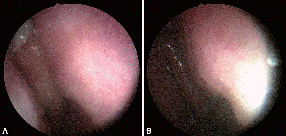
A 52-year-old man presented with persistent rhinorrhea, nasal obstruction, and headaches. These symptoms persisted for one year, and although periodic medication provided temporary relief, it did not prevent their recurrence. His medical history included pulmonary sarcoidosis, for which he had received steroid treatment. Upon examination, the patient exhibited chronic fatigue, bilateral submandibular gland swelling, and bilateral exophthalmos. Nasal endoscopy revealed polyps and purulent discharge in both nasal cavities, as well as mucosal swelling (
Fig. 1).
 | Fig. 1.Preoperative endoscopic findings show generalized mucosal swelling with thick mucoid rhinorrhea. A: Right. B: Left. 
|
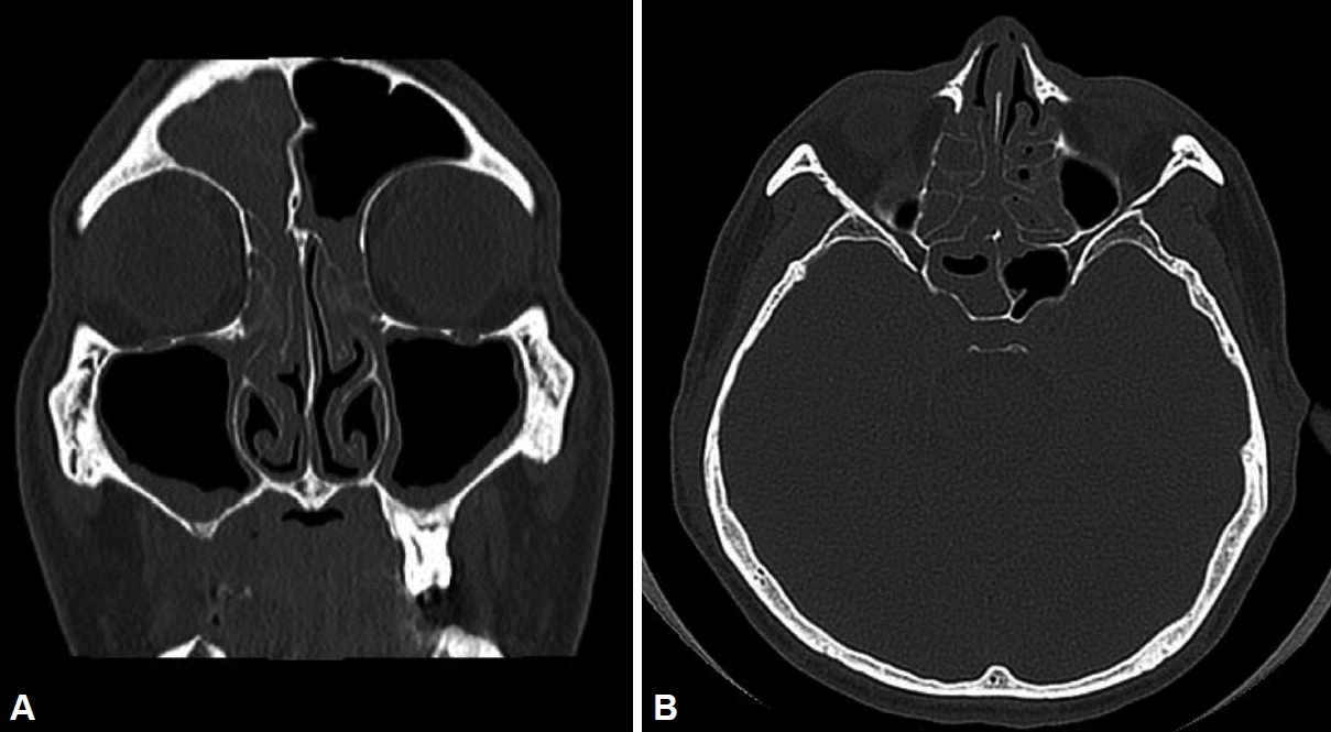
Head and neck computed tomography (CT) revealed generalized enlargement of the bilateral parotid and submandibular glands, enlargement of the bilateral lacrimal glands, and enlarged lymph nodes at levels 1–2 on both sides of the neck (
Fig. 2). CT of the paranasal sinuses showed signs of sinusitis in the frontal, ethmoid, maxillary, and sphenoid sinuses, along with polypoid lesions in the nasal cavity. The chest X-ray appeared normal. Blood tests indicated an elevated erythrocyte sedimentation rate of 42 mm/h, while the complete blood cell count, electrolyte panel, serology tests, urinalysis, complement C3, C4, fluorescent antinuclear antibody, anti-DNA antibody, anti-Sjögren syndrome-A & B antibodies, and coagulation tests were all within normal limits. Anosmia was noted during olfactory testing.
 | Fig. 2.Preoperative computed tomography findings demonstrate right frontal, ethmoid, and sphenoid sinusitis, as well as left ethmoid sinusitis. There is mucosal thickening of the bilateral maxillary sinuses. A: Coronal. B: Axial. 
|
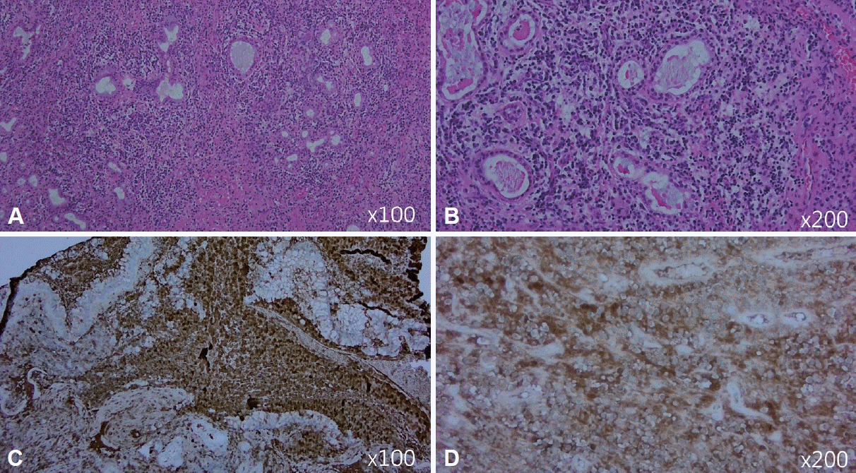
The patient expressed a desire for surgical intervention to address his sinusitis, leading to the performance of bilateral endoscopic sinus surgery. Immunohistochemical analysis revealed dense lymphoplasmacytic and eosinophilic infiltration, with an IgG4/IgG positive plasma cell ratio exceeding 40%, indicative of IgG4-RD (
Fig. 3). A subsequent ultrasound-guided biopsy of the submandibular glands showed similar dense lymphoplasmacytic infiltration and stromal fibrosis, with 50–78 IgG4-positive plasma cells per high-power field.
 | Fig. 3.Histopathologic findings of IgG4-related sinusitis. A and B: Infiltration by inflammatory cells, consisting of lymphocytes and plasma cells (hematoxylin and eosin stain). C and D: Prominently increased IgG4 plasma cells with inflammation (IgG4 special stain). 
|
The patient was ultimately diagnosed with IgG4-RD, which affected the bilateral nasal cavity, paranasal sinuses, submandibular glands, lacrimal glands, and parotid glands. He was subsequently referred to the rheumatology department for further evaluation. Tests revealed an IgG level of 1,956 mg/dL, which is above the normal range of 700–1,600 mg/dL, and an IgG4 subclass level of 1,340 mg/dL, within the reference range of 30-2,010 mg/L. Treatment commenced with oral corticosteroids (prednisolone 30 mg/day) and methotrexate. The patient experienced symptom improvement, and the medications were gradually tapered off until treatment was discontinued. Postoperatively, the wounds in the nasal cavity and sinuses healed well, showing no signs of recurrence.
Go to :

DISCUSSION
IgG4-related sinusitis, a manifestation of the systemic IgG4-RD, is a rare yet increasingly recognized condition that presents diagnostic and therapeutic challenges. It is crucial to understand its pathophysiology, clinical presentation, and management to provide effective patient care [
1,
5]. Involvement of the nasal cavity and paranasal sinuses is rare worldwide, with only seven cases reported in South Korea to date (
Table 1) [
9-
13].
Table 1.
Overview of the reviewed cases of IgG4-related disease with sinusitis
|
Case |
Age (yr) |
Sex |
Underlying diseases |
Presenting symptoms |
Involvement sites
|
Diagnostic findings
|
Treatment |
Progress |
|
Intranasal |
Extranasal |
Serum IgG4 (reference range; 30–2,010 mg/L) |
Pathology |
|
Lee et al. [9] |
54 |
F |
None |
NO, Rt |
IT, Rt |
Pituitary gland |
550 mg/L |
IgG4(+) plasma cells (>106/HPF) |
- Diagnostic endoscopic IT mass excision, Rt |
- Recurred 10 months after surgery, prednisolone 60 mg/day for 3 weeks and then tapering for 10 months |
|
R, Rt |
- Endoscopic TSA for pituitary lesion |
- No symptoms or recurrence for 4 years thereafter |
|
Lee et al. [9] |
26 |
F |
None |
NO, both |
Septum |
None |
Unknown |
IgG4(+) plasma cells (>30/HPF) |
- Methylprednisolone 32 mg/day for 4 weeks and taper |
- No symptoms or recurrence for 2 months after treatment and follow-up loss |
|
R, both |
Cribriform plate |
|
PND |
Inflammatory myofibroblastic tumor |
|
Sneezing |
|
Lee et al. [9] |
20 |
M |
None |
NO, both |
MS, both |
None |
530 mg/L |
Infiltration of lymphoplasma cells and fibrosis |
- Diagnostic ESS, both |
- No symptoms or recurrence for 2 months after treatment and follow-up loss |
|
ES, both |
- Postoperative methylprednisolone 16 mg/day for 6 weeks |
|
FS, Lt |
IgG4(+) plasma cells |
|
Chung and Lee [10] |
43 |
F |
None |
Facial pain, Lt |
MS, Lt |
None |
3,000 mg/L |
Infiltration of lymphoplasma cells and fibrosclerosis |
- Diagnostic ESS, Lt |
- No symptoms or recurrence for 6 months after treatment |
|
NO, Lt |
ES, Lt |
- Postoperative prednisolone 0.6 mg/kg for 4 weeks and 10 mg/day for 2 months in sequence |
|
Headache |
FS, Lt |
IgG4(+) plasma cells (>60/HPF) |
|
LP, Lt |
|
Ko et al. [11] |
40 |
F |
Unknown |
Saddle nose |
MS, Lt |
None |
37.5 mg/L |
Infiltration of lymphoplasma cells and fibrosclerosis |
- Prednisolone 60 mg/day for 5 weeks |
- Still no improvement, stopping MTX and changing to AZA 100 mg/day, showing response |
|
Epistaxis |
LNW, Lt |
- Steroid-only therapy showed no improvement and was changed to combined therapy with MTX 7.5 mg/ day for 4 weeks |
|
Crust |
Septum |
IgG4(+) plasma cells (>35/HPF) |
- No symptoms or recurrence for 2 months after low-dose steroid and AZA combination therapy |
|
UP, both |
IgG4/IgG ratio 40% |
|
Mun et al. [12] |
69 |
F |
HTN |
Epistaxis |
MS, both |
None |
1,028 mg/L |
Infiltration of lymphoplasma cells |
- Diagnostic ESS, both |
- Symptoms and endoscopic findings improved for 6 months after treatment |
|
CKD |
Mucopurulent |
ES, both |
IgG4(+) plasma cells (>90/HPF) |
- Postoperative prednisolone 40 mg/day for 1 week, 20 mg/day for 2 weeks, and 10 mg/day for 6 months in sequence |
- Intermittent use of steroids as needed |
|
Pulmonary Tb |
R, both |
Septum |
IgG4/IgG ratio 70% |
- Acute cerebral infarction in the left pons 3 months after ESS |
|
Corneal ulcer |
IT, both |
- Died from complications of cerebral infarction 1 year after onset |
|
Han et al. [13] |
70 |
M |
HTN |
NO, both |
MS, Rt |
Lung, both |
4,960 mg/L |
Infiltration of lymphoplasma cells and storiform fibrosis |
- Prednisolone 40 mg/day for 4 weeks, tapering for 6 months |
- No symptoms or recurrence for 6 months after treatment |
|
CRS |
PND |
ES, Rt |
- Curative ESS, both |
- Prednisolone 5 mg/day maintenance therapy for residual lung lesions |
|
Dry nose |
SS, Rt |
IgG4(+) plasma cells (>100/HPF) |
|
Crust |
|
Hyposmia |
IgG4/IgG ratio 30% |
|
Our case |
52 |
M |
None |
Rhinorrhea, Nasal obstruction, headache, Anosmia |
MS, both |
Lacrimal gland, Parotid gland, Submandibular gland |
1,956 mg/L |
Infiltration of lymphoplasma cells and stromal fibrosis |
- Prednisolone 0 mg/day for 2 weeks, tapering for 6 months |
- No symptoms or recurrence for 6 months after treatment |
|
ES, both |
IgG4(+) plasma cells (50–78/HPF) |
- Curative ESS, both |
|
FS, both |
|
SS, both |
IgG4/IgG ratio >40% |

Pathophysiology and clinical presentation
IgG4-related sinusitis is characterized by chronic inflammation and tissue infiltration by IgG4-positive plasma cells. The exact etiology of this immune-mediated condition remains unclear; however, it is believed to involve a dysregulated immune response. Unlike typical chronic rhinosinusitis, IgG4-related sinusitis is distinguished by a unique histopathological triad: dense lymphoplasmacytic infiltrate, storiform fibrosis, and obliterative phlebitis. Clinically, patients may experience nasal obstruction, rhinorrhea, facial pain, and sometimes anosmia. Although these symptoms resemble those of chronic rhinosinusitis, the underlying pathology is markedly different [
14].
Diagnostic challenges
Diagnosing IgG4-related sinusitis can be challenging due to its nonspecific clinical symptoms and the necessity to differentiate it from other forms of chronic rhinosinusitis, malignancies, and systemic inflammatory diseases. Key diagnostic criteria include elevated serum IgG4 levels, characteristic radiological findings, and histopathological confirmation via biopsy. Imaging studies frequently show diffuse thickening of the sinus mucosa and occasionally mass-like lesions. A definitive diagnosis is confirmed through biopsy, which reveals the hallmark features of IgG4-RD and a significant presence of IgG4-positive plasma cells [
1,
15].
Management strategies
The cornerstone of treatment for IgG4-related sinusitis is corticosteroid therapy, which typically leads to a marked improvement in symptoms and a reduction in inflammatory tissue infiltration. The standard initial dose of prednisone or an equivalent corticosteroid is often effective; however, the dosage must be carefully tapered to prevent relapse. Despite initial responsiveness to steroids, relapses are common, necessitating long-term management strategies. In cases of recurrent or refractory disease, immunosuppressive agents such as azathioprine, mycophenolate mofetil, or rituximab may be considered [
4-
6].
Long-term outcomes and follow-up
The long-term prognosis for patients with IgG4-related sinusitis varies. Although many respond favorably to corticosteroids, the disease’s chronic and relapsing nature can significantly affect their quality of life. Regular follow-up is crucial to monitor disease recurrence and manage any long-term treatment effects. A multidisciplinary approach involving otolaryngologists, rheumatologists, and immunologists is essential for comprehensive management and improved patient outcomes [
1,
8,
14].
Future directions
Further research is needed to elucidate the pathogenesis of IgG4-related sinusitis and to develop targeted therapies. Exploring the genetic and environmental factors that contribute to this condition could lead to more effective treatments and potential preventive strategies. Additionally, studies focusing on long-term outcomes and optimal management protocols are crucial for enhancing patient care and quality of life [
2].
Conclusion
IgG4-related sinusitis is a distinct subset of chronic rhinosinusitis, characterized by unique pathophysiological and clinical features. Accurate diagnosis and effective management of this condition necessitate a high index of suspicion and a multidisciplinary approach. Although corticosteroids are the primary treatment, ongoing research and clinical vigilance are essential to optimize patient outcomes for this challenging condition.
Go to :


 PDF
PDF Citation
 Print
Print


 XML Download