Journal List > J Pathol Transl Med > v.58(6) > 1516088934

Park, Shin, Kim, Oh, and Cho: Histopathologic classification and immunohistochemical features of papillary renal neoplasm with potential therapeutic targets

Abstract

Background

Papillary renal cell carcinoma (pRCC) is the second most common histological subtype of renal cell carcinoma and is considered a morphologically and molecularly heterogeneous tumor. Accurate classification and assessment of the immunohistochemical features of possible therapeutic targets are needed for precise patient care. We aimed to evaluate immunohistochemical features and possible therapeutic targets of papillary renal neoplasms

Methods

We collected 140 papillary renal neoplasms from three different hospitals and conducted immunohistochemical studies on tissue microarray slides. We performed succinate dehydrogenase B, fumarate hydratase, and transcription factor E3 immunohistochemical studies for differential diagnosis and re-classified five cases (3.6%) of papillary renal neoplasms. In addition, we conducted c-MET, p16, c-Myc, Ki-67, p53, and stimulator of interferon genes (STING) immunohistochemical studies to evaluate their pathogenesis and value for therapeutic targets.

Results

We found that c-MET expression was more common in pRCC (classic) (p = .021) among papillary renal neoplasms and Ki-67 proliferation index was higher in pRCC (not otherwise specified, NOS) compared to that of pRCC (classic) and papillary neoplasm with reverse polarity (marginal significance, p = .080). Small subsets of cases with p16 block positivity (4.5%) (pRCC [NOS] only) and c-Myc expression (7.1%) (pRCC [classic] only) were found. Also, there were some cases showing STING expression and those cases were associated with increased Ki-67 proliferation index (marginal significance, p = .063).

Conclusions

Our findings suggested that there are subsets of pRCC with c-MET, p16, c-MYC, and STING expression and those cases could be potential candidates for targeted therapy.

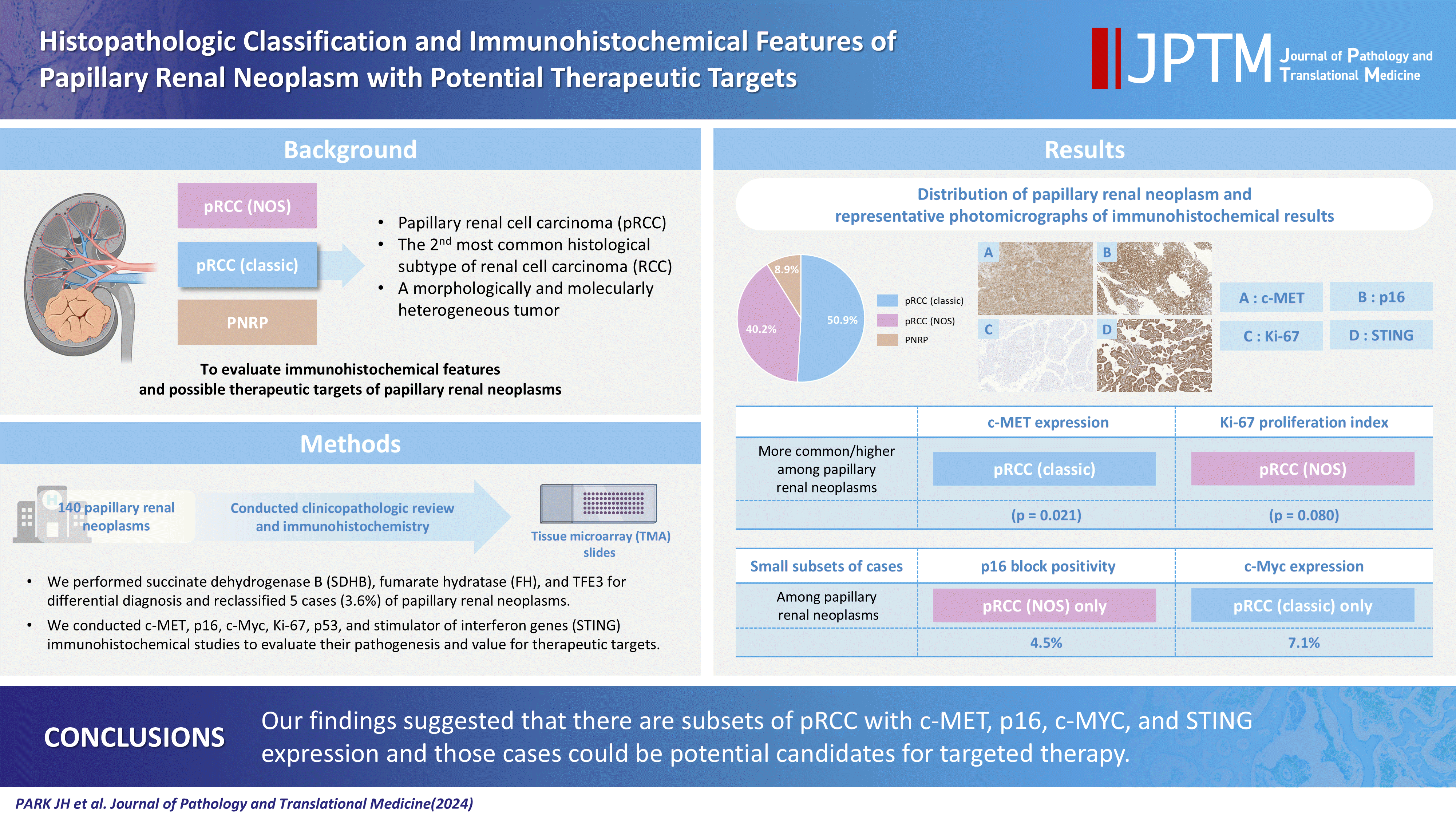
GRAPHICAL ABSTRACT

Kidney cancer is one of the most lethal genitourinary tumors worldwide with an incidence and mortality rate of approximately 2% among all cancers [1]. Kidney cancer is the seventh and tenth most common cancer in men and women, respectively [2], and accounts for the 16th most common cause of cancer-related deaths worldwide [3]. The 5-year survival rate of patients with kidney cancer is approximately 70% for stage I–II, 30%–55% for stage III, and 5% for stage IV [4]. Kidney cancer can spread to various organs including the lungs, lymph nodes, and bones [5]. Histologically, kidney cancer is classified into approximately 20 histologic subtypes, including clear cell renal cell carcinoma (ccRCC), papillary renal cell carcinoma (pRCC), and chromophobe renal cell carcinoma, which account for 90% of renal cell carcinoma (RCC) cases [6].
pRCC is the second most common histological subtype of RCC and occupies approximately 13%–20% of all RCC cases [6,7]. pRCC is defined as a malignant neoplasm characterized by papillary or tubulopapillary architecture [6]. pRCC may show multifocal occurrence or bilaterality. pRCC is considered a molecularly heterogeneous group with chromosomal changes (gains of chromosomes 7 and 17, loss of the Y chromosome) [8,9], MET alterations [9], CDKN2A [9], MYC [10], NRF2/ARE [11], and chromatin modification [9] pathways are thought to be pathogenic alterations. The prognosis of pRCC is more favorable than that of ccRCC or unclassified RCC. American Joint Committee on Cancer (AJCC) staging and World Health Organization/International Society of Urological Pathology (WHO/ISUP) grading are the best prognostic indicators of pRCC [6].
Until 2016, pRCC was classified as type 1 and type 2, based on the growth pattern and nuclear features of the tumors. However, a molecular study revealed new entities of RCC, pRCC type 2 was considered to include different RCC entities such as fumarate hydratase (FH)–deficient RCC and translocation RCC [6]. Type 1 pRCC is considered a classic pattern of pRCC with papillary and tubular architecture and a lining of cuboidal cells with scant or light basophilic cytoplasm [6]. Other morphological patterns include predominantly solid growth, predominantly vacuolated cells, biphasic patterns with squamoid cells with glandular lumina, brisk inflammation, and low-grade oncocytic tumors with reverse polarity [6]. As papillary renal neoplasms have been subdivided histologically, and molecular alterations have been identified, subclassification of papillary renal neoplasms and evaluation of immunohistochemical features for the classification and assessment of pathogenesis are needed.
In this study, we evaluated the histopathological features and immunohistochemical features to further classify the papillary renal neoplasms. Additionally, we performed immunohistochemical studies on cancer treatment and pathogenic pathways. Finally, we assessed the clinicopathological correlation between papillary renal neoplasm subtypes and immunohistochemical findings.

MATERIALS AND METHODS

Case collection and clinicopathologic review

We collected cases of papillary renal neoplasm based on the 4th edition of the World Health Organization (WHO) classification of Urogenital Tumors [12]. We conducted a multicenter papillary renal neoplasm study and analyzed 140 cases from three different hospitals. Cases were collected from the Asan Medical Center (AMC), Seoul Metropolitan Government-Seoul National University (SMG-SNU) Boramae Medical Center (BMC), and Paik Hospital by aided Korean Society of Urogenital Pathologists. A total of 140 cases were included in this study (94 cases from AMC, 34 cases from BMC, and 12 cases from Paik Hospital). Cases from AMC were retrieved from January 2009 to December 2011 and reviewed by Y.M.C. Cases from BMC included the cases from January 2009 to December 2020 and were reviewed by J.H.P. Cases from Paik Hospital between January 2004 and February 2013 were retrieved and reviewed by H.J.K. All cases included in this study were independently reviewed by J.H.P based of 5th edition of WHO classification of urinary and male genital tumors [6]. Histologically, papillary renal neoplasms were classified as pRCC (classic pattern), pRCC (not otherwise specified, NOS), papillary neoplasm with reverse polarity (PNRP), and others. We classified pRCC (classic pattern) and pRCC (NOS) based on WHO classification, further classifying pRCC (classic pattern) as pRCC showing mainly papillary or tubular architecture and tumor cells lined by cuboidal cells with scant or light basophilic cytoplasm and pRCC (NOS) as pRCC showing solid or diverse growth pattern or pseudostratification and tumor cells showing abundant eosinophilic or basophilic, squamoid, or vacuolated cells with variable inflammatory cells infiltration [6,13]. If there was a disagreement on the pathologic diagnosis, the immunohistochemical results from the original hospital were revisited and discussed with a pathologist from the original hospital for diagnostic consensus. Clinicopathological data included patient’s age, sex, original pathological diagnosis, WHO/ISUP grade, and presence of hemorrhage, necrosis, and sarcomatoid change. For assessing pathologic features, we reviewed original pathology report and whole pathology slides.

Tissue microarray

For immunohistochemical study, representative areas were used for tissue microarray (TMA). In cases of AMC, three cores of 1 mm diameter from representative areas were taken from the widest region and those with the worst (most advanced) histology. In the cases of BMC and Paik Hospital, two cores of 2 mm diameter with representative areas were used for TMA. Tissue areas with necrosis or hemorrhage were excluded. In the TMA of AMC, normal tissue was also included for comparison with cancer tissue.

Immunohistochemistry

We conducted an immunohistochemical analysis of the TMA slides using nine antibodies. These included succinate dehydrogenase B (SDHB; 1:100, 21A11AE7, Abcam, Fremont, CA, USA), FH (1:200, sc-100743 (J-13), Santa Cruz Biotechnology, Santa Cruz, Dallas, TX, USA), transcription factor E3 (TFE3; MRQ-37, ready-to-use (RTU), Roche, Tucson, AZ, USA), c-MET (SP44, RTU, Roche), p16 (E6H4, RTU, Roche), c-Myc (EP121, Cell Marque, Rocklin, CA, USA), Ki-67 (1:300, MRQ-64, Cell Marque), p53 (DO-7, 1:1, Roche), and stimulator of interferon genes (STING; 1:4,000, EPR13130-55, Abcam). Immunohistochemical studies for SDHB, FH, and TFE3 were conducted for the differential diagnosis of papillary renal neoplasms with SDH-deficient RCC, FH-deficient RCC, and translocation RCC, respectively, and the results were interpreted according to the manufacturer’s recommendations. c-MET, p16, c-Myc, and STING immunoreactivity were evaluated for pathogenic aspects and because those markers were thought to be possible therapeutic targets. The interpretation of c-MET expression was based on the intensity, proportion, and location of immunopositivity. Immunopositivity for p16 was assessed as negative with a few positive cells, patchy positivity, and block positivity. c-Myc immunostaining was performed based on the proportion of nuclear staining. STING immunostaining was divided into negative and positive. We analyzed p53 immunohistochemistry because p53 is one of the most frequently altered genes in cancer and can be a therapeutic target [14]. The p53 immunoreactivity was classified as normal pattern (a few positive cells) and abnormal pattern (complete loss or diffuse positive). Also, we analyzed Ki-67 immunohistochemistry because Ki-67 is a general marker for assessing proliferation of tumor cells and can be used for tumor grading. Immunohistochemistry was conducted based on the avidin-biotin-peroxidase detection system using Ventana BenchMark Ultra (Roche Diagostics, Basel, Switzerland) for SDHB, FH, TFE3, c-MET, p16, Ki-67, and STING, and Dako Omnis (Agilent, Santa Clara, CA, USA) for c-Myc and p53, according to the manufacturer’s recommendations.

Statistical analysis

We assessed the clinicopathological correlations between the subtypes of papillary renal neoplasm (pRCC [classic], pRCC [NOS], PNRP, and others) and immunohistochemical results. For baseline characteristics, one-way analysis of variance (ANOVA) was used for continuous variables. Categorical variables were analyzed by Pearson’s chi-square test and Fisher exact test. A 2-tailed p<.05 was regarded as statistically significant in all statistical analyses. For multiple comparisons, Bonferroni correction was applied. Statistical analyses were performed using the IBM SPSS Statistics ver. 26 (IBM SPSS Statistics, Armonk, NY, USA).

RESULTS

Histologic classification of papillary renal neoplasm

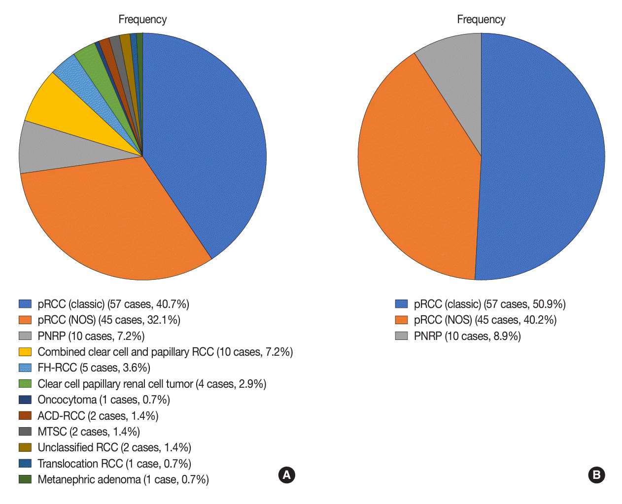
A total of 140 papillary renal neoplasms from three different hospitals were evaluated and re-classified based on the WHO classification of urinary and male genital tumors 5th edition (Fig. 1) [6]. Specifically, the cases from AMC included 40 cases of pRCC (classic); 22 cases of pRCC (NOS); eight cases of PNRP; seven cases of combined clear cell and papillary RCC; five cases of FH-deficient RCC; three cases of clear cell papillary renal neoplasm; two cases of unclassified RCC, mucinous tubular spindle cell carcinoma, and acquired cystic disease-associated RCC; and one case of TFE3-rearranged RCC, oncocytoma, and metanephric adenoma. The cases of BMC included 15 cases of pRCC (classic), 18 cases of pRCC (NOS), and one case of PNRP. Cases from Paik Hospital comprised of two cases of pRCC (classic), five cases of pRCC (NOS), three cases of combined clear cell and papillary RCC, and one case each of clear cell papillary renal neoplasm and PNRP. Among papillary renal neoplasms, pRCC (classic) accounted for 57 cases (50.9%), pRCC (NOS) for 45 cases (40.2%), and PNRP for 10 cases (8.9%).

Clinical and histopathologic features of papillary renal neoplasm

We evaluated the clinical and histopathological features of papillary neoplasms after re-classifying the subtypes (Table 1, Fig. 2). The mean ages at the diagnosis were 59.3 years for pRCC (classic), 61.3 years for pRCC (NOS), and 55.9 years for PNRP. Although the mean age was higher in the pRCC (NOS) group and lower in the PNRP group, the difference was not statistically significant (p=.374). All three groups showed a male predilection with a 3.07–4.0:1 male-to-female ratio (p>.99). WHO/ISUP grade revealed that there were more patients with high WHO/ISUP grade in pRCC (NOS) among papillary renal neoplasm (p<.001). We further performed post-hoc Bonferroni analysis and showed patients with high WHO/ISUP grade were more common in pRCC (NOS) compared to pRCC (classic) (corrected p<.001) and PNRP (corrected p=.009), respectively. The presence of hemorrhage was found in 28.1% (pRCC [classic]), 31.1% (pRCC [NOS]), and 20.0% (PNRP). Also, we could find necrosis in 22.8% (pRCC [classic]), 22.2% (pRCC [NOS]), and 0.0% (PNRP). The presence of sarcomatoid change were rare and identified in 1.8% (pRCC [classic]), 6.7% (pRCC [NOS]), and 0.0% (PNRP). There was no statistical significance in presence of hemorrhage, necrosis, and sarcomatoid change. In the BMC cases, we identified multifocality and bilaterality. Among the 31 patients, two (one male and one female) had multiple pRCC, and both cases were histologically pRCC (NOS).

Immunohistochemical results for differential diagnosis of papillary renal neoplasm

We performed immunohistochemical analysis for the differential diagnosis of papillary renal neoplasms. Based on the TFE3 and FH immunohistochemical studies, we identified one case of TFE3-rearranged RCC and five cases of FH-deficient RCC. Originally, TFE3-rearranged RCC was classified as TFE3-rearranged RCC. However, FH-deficient RCCs were originally diagnosed as pRCC type 1 (classic pRCC in this study), pRCC type 2 (pRCC NOS in this study), or unclassified RCC. There were no cases of SDHB loss on immunohistochemistry.

Immunohistochemical features of papillary renal neoplasm for pathogenic pathways and targets

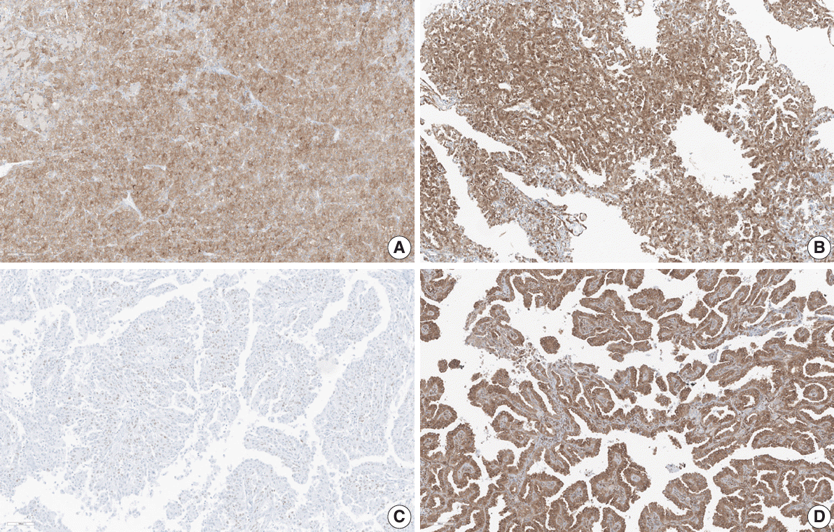
c-MET, p16, c-Myc, Ki-67, and STING were used to assess the pathogenic pathways and targets of papillary renal neoplasms (Table 2, Fig. 3). In the c-MET immunohistochemical study, 22 cases (39.3%) of pRCC (classic) and seven cases (15.9%) of pRCC (NOS) showed focal or diffuse moderate-to-strong membranous staining. There were no cases of such staining in PNRP. We found that c-MET expression was more common in pRCC (classic) than in other papillary renal neoplasm subtypes (p=.021). We conducted post-hoc Bonferroni analysis and revealed that there was marginal significance between pRCC (classic) and pRCC (NOS) (corrected p=.097) and between pRCC (classic) and PNRP (corrected p=.081). In the p16 immunohistochemical study, only two cases of pRCC (NOS) (4.5%) revealed block positivity and there were no cases with block positivity in pRCC (classic) and PNRP (p=.328). In the c-Myc immunohistochemical study, only a small subset of pRCC (classic) cases (4 cases, 7.1%) showed immunoreactivity and cases of pRCC (NOS) and PNRP did not show immunoreactivity (p=.199). Proliferative activity was assessed using Ki-67 immunohistochemistry. Cases with pRCC (NOS) showed increased proliferative activity other than papillary renal neoplasms (marginal significance, p=.081 (continuous variables); p=.080 (categorical variables). When comparing pRCC (classic) and pRCC (NOS), five cases (8.8%) of pRCC (classic) and nine cases (20.5%) of pRCC (NOS) showed increased proliferative activity (≥1%). There was no case with abnormal p53 immunohistochemical pattern. We identified subsets of each papillary renal neoplasm with STING positivity. There were 14 cases (24.6%), 12 cases (26.7%), and seven cases (70.0%) from pRCC (classic), pRCC (NOS), and PNRP, respectively, with STING immunoreactivity (p=.020). Posthoc Bonferroni analysis showed that there was statistical significance between pRCC (classic) and PNRP (corrected p=.024) and marginal significance between pRCC (NOS) and PNRP (corrected p=.068). Additionally, we assessed the correlation between immunohistochemical findings. We found the tendency that increased proliferative activity (high Ki-67 proliferation index) was associated with STING positivity (marginal significance, p=.063) (Table 3). Although, the number of cases were small, there were less than 30% of STING-positive cases with low Ki-67 proliferation index (≤3%) (29 cases out of 106 cases) compared to 80.0% STING STING-positive cases with high Ki-67 proliferation index (>3%) (4 cases out of 5 cases).

DISCUSSION

According to the 5th edition of WHO classification of urinary and male genital tumors, pRCC is defined as a malignant neoplasm characterized by papillary or tubulopapillary architecture with no specific features of RCC with papillary architecture [6]. pRCC is considered a morphologically and molecularly heterogeneous group and can be classified into the classic (previously type 1), PRNP, and NOS types (majority cases of previously type 2 pRCC). As the morphological and molecular features of pRCC have been assessed, new entities with similar morphological features have been identified. Currently, FH-deficient RCC, eosinophilic solid and cystic RCC, tubulocystic RCC, collecting duct RCC, or MiT family gene–rearranged RCCs are considered major differential diagnoses for pRCC (NOS), and appropriate immunohistochemical and molecular studies are needed for accurate diagnosis [6]. In this study, we conducted immunohistochemical analysis and re-classified papillary renal neoplasms based on 5th edition of the WHO classification. By doing this, we could re-classify five cases (3.6%) of papillary renal neoplasm based on histologic and immunohistochemical results (four cases of pRCC and one case of unclassified RCC from original diagnosis were re-classify as FH-deficient RCC).
Molecular studies on pRCC have identified several pathogenic molecular features along with previous cytogenetic findings. Along with chromosomal gains (chromosomes 7 and 17) and loss (chromosome Y) [8,9], alterations in MET, CDKN2A genes [9], and genes related to MYC [10], NRF2/ARE [11], and chromatin modifier [9] pathways have been implicated in the pathogenesis of pRCC. In previous studies, MET alterations were more common in low-grade pRCC [9], while alterations in the MYC [10], NRF2/ARE [11], and chromatin modifier [9] pathways were found in high-grade pRCC. To assess the aforementioned genetic and pathway alterations, we performed immunohistochemical analyses of c-MET, p16, and c-Myc. Additionally, we conducted a STING immunohistochemical study since there are reports that pRCC has compromised oxidative phosphorylation [15], and STING activation could be a therapeutic target for pRCC [16].
In pRCC, MET alterations are found in approximately 15% of patients with pRCC [17,18]. The importance of MET mutations was suggested by the finding that hereditary pRCC activates germline MET mutations [19]. In previous studies, MET alterations were mainly found in type 1 pRCC (pRCC (classic) in this study), which was consistent with our finding that c-MET immunoreactivity is more common in pRCC (classic). c-MET belongs to the MET family and is a receptor tyrosine kinase activated by its ligand, hepatocyte growth factor [20]. Activation of c-MET leads to various cellular signaling associated with proliferation, invasion, and angiogenesis [21]. There are several FDA-approved MET inhibitors for selected cancer types, such as non-small cell lung cancer and RCC [22]. As, MET alteration is one of the important genetic alterations in pRCC, there have been conducted several clinical trials targeting MET in pRCC using i.e., savolitinib or crizotinib [23]. Growing results have been revealed that targeting MET pathway may beneficial for patient outcome of pRCC and may result favorable effect when combining with immune checkpoint inhibitors [24]. As c-MET immunoreactivity was found in a substantial number of cases (39.3% of pRCC [classic] and 15.9% of pRCC [NOS] in this study) and could serve as a therapeutic target, c-MET immunohistochemical studies would be beneficial not only for the differential diagnosis between pRCC (classic) and pRCC (NOS) but also for patient treatment.
In contrast to MET alterations, aberrations in CDKN2A and MYC are more common in type 2 pRCC [9,10]. The CKDN2A gene alterations were found in approximately 25% of patients with type 2 pRCC. This gene encodes p16 and p14 and functions as a tumor suppressor gene [25]. Loss-of-function mutations in CDKN2A lead to cell cycle progression [26]. There are some FDA-approved CDK4/6 inhibitors for breast cancer [27], and there were some preclinical studies for RCC [28]. The preclinical studies suggested that CDK4/6 inhibitor in kidney cancer might be beneficial when it treated alone or in combination with mammalian target of the rapamycin (mTOR) or immune checkpoint inhibitors [28]. In the case of MYC alterations, MYC activation has been observed in 67% of high-grade pRCC type 2 [10]. The protein of MYC is a transcription factor with an oncogenic function [29]. The C-MYC is a member of the MYC oncogene family that encodes c-Myc [30]. MYC activation leads to cell growth, proliferation, metabolism, and signal transduction [29,30]. There have been many studies to find MYC inhibitor, however, currently there are no FDA-approved drugs. However, some drugs have shown promising results [31] and there have been studied effective strategy to inhibit oncogenic function of MYC [32]. Patients with MYC overexpression may be beneficial if the new drug or approach for MYC inhibition is developed. Based on our immunohistochemical results, we could find that 4.5% of patients with pRCC (NOS) showed block positivity on p16 immunohistochemistry. Furthermore, c-Myc immunoreactivity was found in 7.1% of pRCC (classic) only and there was no case with c-Myc immunoreactivity in pRCC (NOS) and PNRP. Although we could not evaluate genetic alterations in both genes or genetic-proteomic differences, there were small subsets that could be helpful for pRCC treatment when using CDK4/6 or MYC inhibition.
We also evaluated STING immunohistochemical results. Although the correlation between hypoxia and cancer is not as evident compared to ccRCC [9,33], pRCC shows impaired oxidative phosphorylation [15], and STING immunoreactivity could be a marker for STING activation. [16]. The STING pathway is important for innate immunity, and recently, activation of the pathway has been thought to be associated with adaptive anticancer immune responses and angiogenesis [16,34]. The expression of STING in various cancers [35-37] has been studied and STING is considered a novel therapeutic target for cancer treatment [38]. The pathogenic role of STING expression is thought to be exerted by cyclic GMP-AMP synthase (cGAS)/STING pathway [38]. cGAS/STING pathway showed dichotomous role in tumor and possible mechanism related to anti-tumor or tumor-promoting role has been elucidated. Meta-analysis for prognostic significance of STING expression in various tumors revealed mainly anti-tumor effect of STING expression, however, in kidney canner, STING expression was associated with unfavorable prognosis [39,40]. Interestingly, STING expression is associated with chromosomal instability (CIN), such as pRCC and survival of tumor cells with CIN, which implies possible tumor-promoting role of cGAS/STING pathway and STING as a potential therapeutic target [6,8,41]. Also, there have been reported that STING expression is associated with drug resistance in colorectal cancer [42] and normalization of tumor vasculature and further possible synergistic effect with antiangiogenic therapy [16].
In this study, we found that approximately 25% of pRCC showed immunoreactivity for STING, and the STING expression was marginally correlated with the Ki-67 proliferation index. This finding was of interest because there was evidence that upregulation of STING expression could lead tumor cell proliferation by regulating adenosine 5'-monophosphate (AMP)-activated protein kinase–mTOR pathway in colorectal cancer [42]. Further studies using cell lines or larger samples are required to evaluate the beneficial effects of STING-targeted therapy.
This study had several limitations. First, the enrolled papillary renal neoplasm cases were determined solely based on the experience of the pathologist. One pathologist selected cases of papillary renal neoplasm in which papillary features were dominant and pRCC could be a differential diagnosis. Another pathologist chose papillary renal neoplasms as cases diagnosed with pRCC in the original report based on previous WHO classification. Consequently, this may cause selection bias and the real-world incidence of papillary renal neoplasms based on the histological subtypes could not be assessed. Second, we collected papillary renal neoplasm cases from three different hospitals; however, there was no detailed clinicopathological information on papillary renal neoplasms. More detailed clinicopathological features could be collected in the following studies such as multiplicity, bilaterality, presence of end-stage renal disease, disease progression status, metastasis status, disease-free survival, cancer-specific survival, and more detailed clinicopathological features of papillary renal neoplasms could be evaluated. Third, we assessed histopathological and immunohistochemical features using TMA slides and classified papillary renal neoplasms into pRCC (classic), pRCC (NOS), and PNRP. It would be helpful investigating whether there are any histopathologic features to distinguish those subtypes; however, we could not evaluate whole histopathological features because we reviewed TMA slides. Fourth, we should address that there would be interobserver variability for diagnosing papillary renal neoplasms. Further study such as reader study of papillary renal neoplasm with equivocal histopathological or immunohistochemical features would be interesting and helpful for evaluating clinical usefulness of our classification. Fifth, we could not identify or perform molecular studies such as next-generation sequencing. Further studies evaluating the molecular features of papillary renal neoplasms will be useful for further classifying and assessing the possible pathogenic pathways and targets.
In summary, we collected 140 cases of papillary renal neoplasms from three hospitals. We conducted immunohistochemical study for differential diagnosis and assessment of pathogenesis and therapeutic targets. After re-classifying the papillary renal neoplasms, we analyzed pRCC (classic), pRCC (NOS), and PNRP. In immunohistochemical study, we found that c-MET and STING expression were significantly different among papillary renal neoplasms. Additionally, we identified small subsets positive for p16 or c-Myc that could be beneficial as CDK4/6 or MYC inhibitors. Our study suggests that pRCC is a heterogeneous tumor, and immunohistochemical studies would be helpful in assessing possible therapeutic targets. Further studies using artificial intelligence for classification and molecular studies, along with clinicopathological features, would be helpful in understanding the pathogenesis and patient care.

Notes

Ethics Statement

This study was approved by the regional Institutional Review Board of the three hospitals (IRB No. 2012-0788 (AMC), 10-2021-74 (SMG-SNU BMC), SSPAIK2024-04-011 (Paik Hospital)). Formal written informed consent was not required with a waiver by the appropriate IRB and/or national research ethics committee.

Availability of Data and Material

The datasets generated or analyzed during the study are available from the corresponding author on reasonable request.

Code Availability

Not applicable.

Author Contributions

Conceptualization: JHP, YMC. Data curation: JHP, SJS, HJK, YMC. Methodology: JHP, SJS, HJK, SO, YMC. Project administration: JHP, YMC. Resources: JHP, SJS, HJK, YMC. Supervision: JHP, YMC. Validation: JHP, YMC. Visualization: JHP. Writing—original draft: JHP. Writing—review & editing: all authors. Approval of the final manuscript: all authors.

Conflicts of Interest

J.H.P., a contributing editor of the Journal of Pathology and Translational Medicine, was not involved in the editorial evaluation or decision to publish this article. All remaining authors have declared no conflicts of interest.

Funding Statement

This research was supported by the Korean Society of Pathologists Grant No. KSPG2020-02.

ACKNOWLEDGMENTS

We appreciate the cooperation and discussion among the members of the Korean Society of Uropathologists. We are also grateful to Hye Young Kim for her assistance.

References

1. Padala SA, Barsouk A, Thandra KC, et al. Epidemiology of renal cell carcinoma. World J Oncol. 2020; 11:79–87.
crossref
2. Sung WW, Wang SC, Hsieh TY, et al. Favorable mortality-to-incidence ratios of kidney cancer are associated with advanced health care systems. BMC Cancer. 2018; 18:792.
crossref
3. Bai X, Yi M, Dong B, Zheng X, Wu K. The global, regional, and national burden of kidney cancer and attributable risk factor analysis from 1990 to 2017. Exp Hematol Oncol. 2020; 9:27.
crossref
4. SEER training modules, module name [Internet]. Bethesda: National Cancer Institute;2023. [cited 2024 Jan 26]. Available from: https://training.seer.cancer.gov/.
5. Dudani S, de Velasco G, Wells JC, et al. Evaluation of clear cell, papillary, and chromophobe renal cell carcinoma metastasis sites and association with survival. JAMA Netw Open. 2021; 4:e2021869.
crossref
6. WHO Classification of Tumours Editorial Board. Urinary and male genital tumours. WHO classification of tumours series, 5th ed., Vol. 8 [Internet]. Lyon: International Agency for Research on Cancer;2022. [cited 2023 Jul 22]. Available from: https://tumourclassification.iarc.who.int/chapters/36.
7. Courthod G, Tucci M, Di Maio M, Scagliotti GV. Papillary renal cell carcinoma: a review of the current therapeutic landscape. Crit Rev Oncol Hematol. 2015; 96:100–12.
crossref
8. Pitra T, Pivovarcikova K, Alaghehbandan R, Hes O. Chromosomal numerical aberration pattern in papillary renal cell carcinoma: review article. Ann Diagn Pathol. 2019; 40:189–99.
crossref
9. Cancer Genome Atlas Research Network, Linehan WM, Spellman PT, et al. Comprehensive molecular characterization of papillary renal-cell carcinoma. N Engl J Med. 2016; 374:135–45.
crossref
10. Furge KA, Chen J, Koeman J, et al. Detection of DNA copy number changes and oncogenic signaling abnormalities from gene expression data reveals MYC activation in high-grade papillary renal cell carcinoma. Cancer Res. 2007; 67:3171–6.
11. Ooi A, Dykema K, Ansari A, et al. CUL3 and NRF2 mutations confer an NRF2 activation phenotype in a sporadic form of papillary renal cell carcinoma. Cancer Res. 2013; 73:2044–51.
12. Moch H, Humphrey PA, Ulbright TM, Reuter VE. WHO classification of tumours. 4th ed. Vol. 8. WHO classification of tumours of the urinary system and male genital organs. Geneva: World Health Organization;2016.
13. Chevarie-Davis M, Riazalhosseini Y, Arseneault M, et al. The morphologic and immunohistochemical spectrum of papillary renal cell carcinoma: study including 132 cases with pure type 1 and type 2 morphology as well as tumors with overlapping features. Am J Surg Pathol. 2014; 38:887–94.
14. Chen X, Zhang T, Su W, et al. Mutant p53 in cancer: from molecular mechanism to therapeutic modulation. Cell Death Dis. 2022; 13:974.
crossref
15. Al Ahmad A, Paffrath V, Clima R, et al. Papillary renal cell carcinomas rewire glutathione metabolism and are deficient in both anabolic glucose synthesis and oxidative phosphorylation. Cancers (Basel). 2019; 11:1298.
crossref
16. Yang H, Lee WS, Kong SJ, et al. STING activation reprograms tumor vasculatures and synergizes with VEGFR2 blockade. J Clin Invest. 2019; 129:4350–64.
crossref
17. Schmidt L, Junker K, Nakaigawa N, et al. Novel mutations of the MET proto-oncogene in papillary renal carcinomas. Oncogene. 1999; 18:2343–50.
crossref
18. Durinck S, Stawiski EW, Pavia-Jimenez A, et al. Spectrum of diverse genomic alterations define non-clear cell renal carcinoma subtypes. Nat Genet. 2015; 47:13–21.
crossref
19. Schmidt L, Duh FM, Chen F, et al. Germline and somatic mutations in the tyrosine kinase domain of the MET proto-oncogene in papillary renal carcinomas. Nat Genet. 1997; 16:68–73.
crossref
20. Zhang Y, Xia M, Jin K, et al. Function of the c-Met receptor tyrosine kinase in carcinogenesis and associated therapeutic opportunities. Mol Cancer. 2018; 17:45.
crossref
21. Sierra JR, Tsao MS. c-MET as a potential therapeutic target and biomarker in cancer. Ther Adv Med Oncol. 2011; 3(1 Suppl):S21–35.
crossref
22. Puccini A, Marin-Ramos NI, Bergamo F, et al. Safety and tolerability of c-MET inhibitors in cancer. Drug Saf. 2019; 42:211–33.
crossref
23. Choueiri TK, Heng DYC, Lee JL, et al. Efficacy of savolitinib vs sunitinib in patients with MET-driven papillary renal cell carcinoma: the SAVOIR phase 3 randomized clinical trial. JAMA Oncol. 2020; 6:1247–55.
crossref
24. Rhoades Smith KE, Bilen MA. A review of papillary renal cell carcinoma and MET inhibitors. Kidney Cancer. 2019; 3:151–61.
crossref
25. Foulkes WD, Flanders TY, Pollock PM, Hayward NK. The CDKN2A (p16) gene and human cancer. Mol Med. 1997; 3:5–20.
crossref
26. Zhao R, Choi BY, Lee MH, Bode AM, Dong Z. Implications of genetic and epigenetic alterations of CDKN2A (p16(INK4a)) in Cancer. EBioMedicine. 2016; 8:30–9.
crossref
27. Cicenas J, Simkus J. CDK inhibitors and FDA: approved and orphan. Cancers (Basel). 2024; 16:1555.
crossref
28. Sager RA, Backe SJ, Ahanin E, et al. Therapeutic potential of CDK4/6 inhibitors in renal cell carcinoma. Nat Rev Urol. 2022; 19:305–20.
crossref
29. Dang CV. MYC on the path to cancer. Cell. 2012; 149:22–35.
crossref
30. Chen H, Liu H, Qing G. Targeting oncogenic Myc as a strategy for cancer treatment. Signal Transduct Target Ther. 2018; 3:5.
crossref
31. Garralda E, Beaulieu ME, Moreno V, et al. MYC targeting by OMO103 in solid tumors: a phase 1 trial. Nat Med. 2024; 30:762–71.
crossref
32. Llombart V, Mansour MR. Therapeutic targeting of “undruggable” MYC. EBioMedicine. 2022; 75:103756.
crossref
33. Wohlrab C, Vissers MC, Phillips E, Morrin H, Robinson BA, Dachs GU. The association between ascorbate and the hypoxia-inducible factors in human renal cell carcinoma requires a functional von Hippel-Lindau protein. Front Oncol. 2018; 8:574.
crossref
34. Zhu Y, An X, Zhang X, Qiao Y, Zheng T, Li X. STING: a master regulator in the cancer-immunity cycle. Mol Cancer. 2019; 18:152.
crossref
35. Lin Z, Liu Y, Lin P, Li J, Gan J. Clinical significance of STING expression and methylation in lung adenocarcinoma based on bioinformatics analysis. Sci Rep. 2022; 12:13951.
crossref
36. Xia T, Konno H, Ahn J, Barber GN. Deregulation of STING signaling in colorectal carcinoma constrains DNA damage responses and correlates with tumorigenesis. Cell Rep. 2016; 14:282–97.
crossref
37. Parkes EE, Humphries MP, Gilmore E, et al. The clinical and molecular significance associated with STING signaling in breast cancer. NPJ Breast Cancer. 2021; 7:81.
crossref
38. Gan Y, Li X, Han S, et al. The cGAS/STING pathway: a novel target for cancer therapy. Front Immunol. 2021; 12:795401.
39. Kim Y, Cho NY, Jin L, Jin HY, Kang GH. Prognostic significance of STING expression in solid tumor: a systematic review and meta-analysis. Front Oncol. 2023; 13:1244962.
crossref
40. Marletta S, Calio A, Bogina G, et al. STING is a prognostic factor related to tumor necrosis, sarcomatoid dedifferentiation, and distant metastasis in clear cell renal cell carcinoma. Virchows Arch. 2023; 483:87–96.
crossref
41. Yan H, Lu W, Wang F. The cGAS-STING pathway: a therapeutic target in chromosomally unstable cancers. Signal Transduct Target Ther. 2023; 8:45.
crossref
42. Yao H, Wang S, Zhou X, et al. STING promotes proliferation and induces drug resistance in colorectal cancer by regulating the AMPK-mTOR pathway. J Gastrointest Oncol. 2022; 13:2458–71.
crossref

Fig. 1.
Histologic subtypes of papillary renal neoplasms evaluated in this study. (A) Distribution of renal cell neoplasm with papillary feature. (B) Distribution of papillary renal neoplasm. ACD-RCC; acquired cystic disease-associated renal cell carcinoma; FH-RCC, fumarate hydratase-deficient renal cell carcinoma; MTSC, mucinous tubular and spindle cell carcinoma; NOS, not otherwise specified; PNRP, papillary neoplasm with reverse polarity; pRCC, papillary renal cell carcinoma; RCC, renal cell carcinoma.
jptm-2024-07-31f1.tif
Fig. 2.
Representative photomicrographs of papillary renal neoplasms. (A) Papillary renal cell carcinoma (pRCC), classic. (B) pRCC, not otherwise specified. (C) Papillary neoplasm with reverse polarity.
jptm-2024-07-31f2.tif
Fig. 3.
Representative photomicrographs of immunohistochemical results of c-MET (A), p16 (B), Ki-67 (C), and stimulator of interferon genes (D).
jptm-2024-07-31f3.tif
jptm-2024-07-31f4.tif
Table 1.
Clinicopathologic features of papillary renal neoplasm
pRCC (classic) (n = 57) pRCC (NOS) (n = 45) PNRP (n = 10) p-value
Age (yr) 59.3 ± 12.2 61.3 ± 11.5 55.9 ± 9.6 .374
Sex > .99
 Male 43 (75.4) 34 (75.6) 8 (80.0)
 Female 14 (24.6) 11 (24.4) 2 (20.0)
WHO/ISUP grade < .001a
 1/2 50 (87.7) 22 (48.9) 10 (100)
 3/4 7 (12.3) 23 (51.1) 0
Hemorrhage .820
 Absent 41 (71.9) 31 (68.9) 8 (80.0)
 Present 16 (28.1) 14 (31.1) 2 (20.0)
Necrosis .285
 Absent 44 (77.2) 35 (77.8) 10 (100)
 Present 13 (22.8) 10 (22.2) 0
Sarcomatoid change .338
 Absent 56 (98.2) 42 (93.3) 10 (100)
 Present 1 (1.8) 3 (6.7) 0

Values are presented as mean ± SD or number (%).

pRCC, papillary renal cell carcinoma; NOS, not otherwise specified; PNRP, papillary neoplasm with reverse polarity; WHO/ISUP, World Health Organization/International Society of Urological Pathology; SD, standard deviation.

a Post-hoc Bonferroni analysis revealed statistical significance between two groups.

Table 2.
Immunohistochemical features of papillary renal neoplasm
pRCC (classic) (n = 57) pRCC (NOS) (n = 45) PNRP (n = 10) p-value
c-MET 0.021
 Negative 18 (32.1) 24 (54.5) 8 (80.0)
 Faint to weak membranous staining 16 (28.6) 13 (29.5) 2 (20.0)
 Focal or diffuse moderate membranous staining 19 (33.9) 5 (11.4) 0
 Diffuse strong membranous staining 3 (5.4) 2 (4.6) 0
p16 0.328
 Negative/patchy positivity 57 (100) 42 (95.5) 10 (100)
 Block positivity 0 2 (4.5) 0
c-Myc 0.199
 Negative 52 (92.9) 44 (100) 10 (100)
 Postive 4 (7.1) 0 0
Ki-67 proliferation index (continuous) (mean ± SD) 0.3 ± 0.5 0.8 ± 2.0 0.2 ± 0.3 0.081
Ki-67 proliferation index (categorized) (%) 0.080
 < 1 52 (91.2) 35 (79.5) 10 (100)
 1–3 5 (8.8) 4 (9.1) 0
 > 3 0 5 (11.4) 0
p53 NA
 Normal pattern 56 (100) 45 (100) 10 (100)
 Abnormal pattern 0 0 0
STING 0.020a
 Negative 43 (75.4) 33 (73.3) 3 (30.0)
 Positive 14 (24.6) 12 (26.7) 7 (70.0)

Values are presented as number (%) unless otherwise indicated.

pRCC, papillary renal cell carcinoma; NOS, not otherwise specified; PNRP, papillary neoplasm with reverse polarity; SD, standard deviation; NA, not available; STING, stimulator of interferon genes.

a Post-hoc Bonferroni analysis revealed statistical significance between two groups.

Table 3.
Correlation between Ki-67 proliferation index and STING immunoreactivity of papillary renal neoplasm
STING Ki-67 proliferation index
p-value
<1% (n = 97) 1%–3% (n = 9) > 3% (n = 5)
Negative (n = 78) 70 (72.2) 7 (77.8) 1 (20.0) .063
Positive (n = 33) 27 (27.8) 2 (22.2) 4 (80.0)

Values are presented as number (%).

STING, stimulator of interferon genes.

TOOLS
Similar articles