Abstract
Peripheral ossifying fibroma (POF) is a benign tumor characterized by dystrophic calcification or ossification within the gingiva, primarily affecting the anterior maxilla of females and young adults. Its pathogenesis is unclear but linked to local irritants such as trauma, biofilm, dental calculus, and poorly fitting prostheses. In this study, a 63-year-old male presented at Dankook University Dental Hospital with a large nodular lesion on the left maxillary bucco-palatal gingiva. Preoperative imaging, including panoramic radiography and cone-beam computed tomography, was performed. Surgical excision and histological examination confirmed POF with specific morphological characteristics, including mineralized tissue with varied deposition patterns, mature and immature bone, cementum-like tissue, and dystrophic calcification. In conclusion, POF is a rare oral tumor, more common in younger females, typically presenting asymptomatically on the anterior maxilla. Histopathological analysis is crucial for diagnosis. Standard treatment involves conservative local resection, but recurrence rates range from 8% to 20%, necessitating continuous follow-up. This report aims to enhance understanding of POF by presenting a rare case of a large POF in the maxillary posterior bucco-palatal gingiva of an elderly male.
Peripheral ossifying fibroma (POF) is a clinically rare benign tumor that commonly occurs in periodontal tissues1. This lesion was first described by Shepherd2 in 1843 as alveolar exostosis; later, Eversole and Robin et al.3 introduced the term “peripheral ossifying fibroma.” This type of lesion is also known by various other names, including peripheral cemento-ossifying fibroma, ossifying fibro-epithelia polyp, peripheral fibroma with osteogenesis, peripheral fibroma with cementogeneis, peripheral fibroma with calcification, calcifying or ossifying fibroma epulis, and calcifying fibroblastic granuloma2,4.
POF typically arises in the anterior maxilla of young adults aged 20 to 50 years, with a higher occurrence in females compared to males. Although the exact etiopathology is uncertain, POF is believed to originate from the periodontal ligament4,5 and is often associated with local irritants, such as trauma, dental biofilm, calculus, and unfitted dental restorations6,7. Clinically, POF presents as a gingival mass showing slow and progressive growth, typically without radiographic changes, though radiopaque areas can sometimes be detected. Its clinical appearance resembles other reactive gingival lesions, highlighting the need for accurate differentiation since it is known to impact adjacent gingival tissues and have a high recurrence rate of 8% to 20%. While histopathological analysis remains the golden standard for diagnosis, cone-beam computed tomography (CBCT) images and additional X-rays can also be utilized4,7-9.
Thus, to clarify these distinctions and aid in diagnosis, this report aims to improve the understanding of POF by presenting a rare case of a large POF in the maxillary posterior bucco-palatal gingiva of an elderly male.
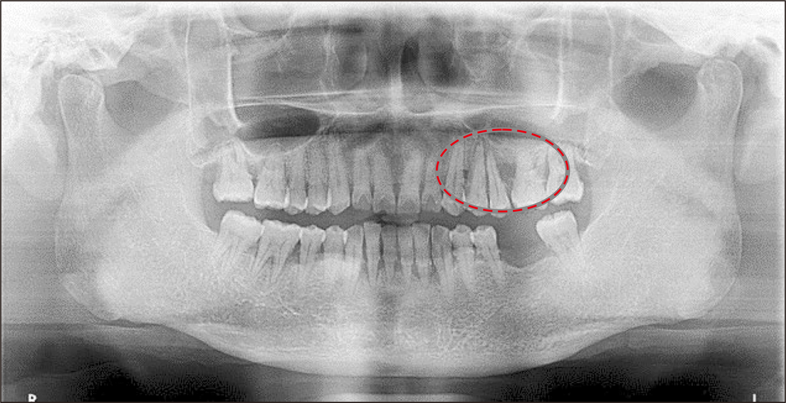
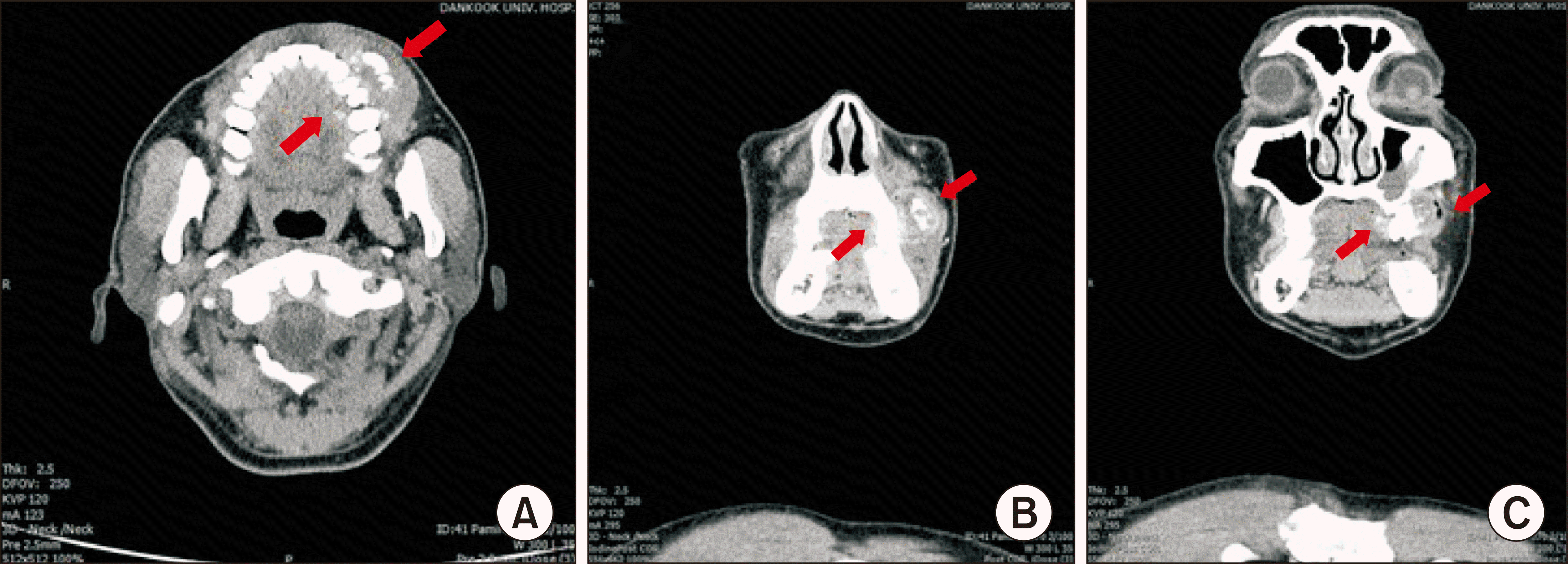
After approval from the Institutional Review Board (IRB) of Dankook University Dental Hospital (IRB No. DKUDH IRB 2024-04-004), a retrospective analysis was performed. A 63-year-old male presented himself to Dankook University Dental Hospital claiming that his upper left gum had been swollen for the previous 3 months and a mass had appeared. The patient did not have any specific medical history. Upon physical examination, facial swelling was observed with loss of the left nasolabial fold, while intra-orally, a sessile-based exophytic mass located in the vestibule of teeth #24-#26 was observed and was measured at 5.0×2.5 cm on the buccal side and 1.5×1.0 cm on the palatal side. The mass was bony-hard and erythematous but not mobile. The patient suffered no tenderness upon palpation, spontaneous pain, or bleeding. Pre-operative panoramic X-ray (Fig. 1) and CBCT images (Fig. 2) were collected for further evaluation. CBCT findings revealed a mass of approximately 4.8 cm arising from the left maxillary alveolar bone, with erosion of the adjacent maxillary alveolar bone, and intralesional calcifications, suggesting a probable tumorous lesion. Surgical removal of the mass was planned since it was large enough to deform the patient’s facial features and compromise the surrounding teeth and structures.
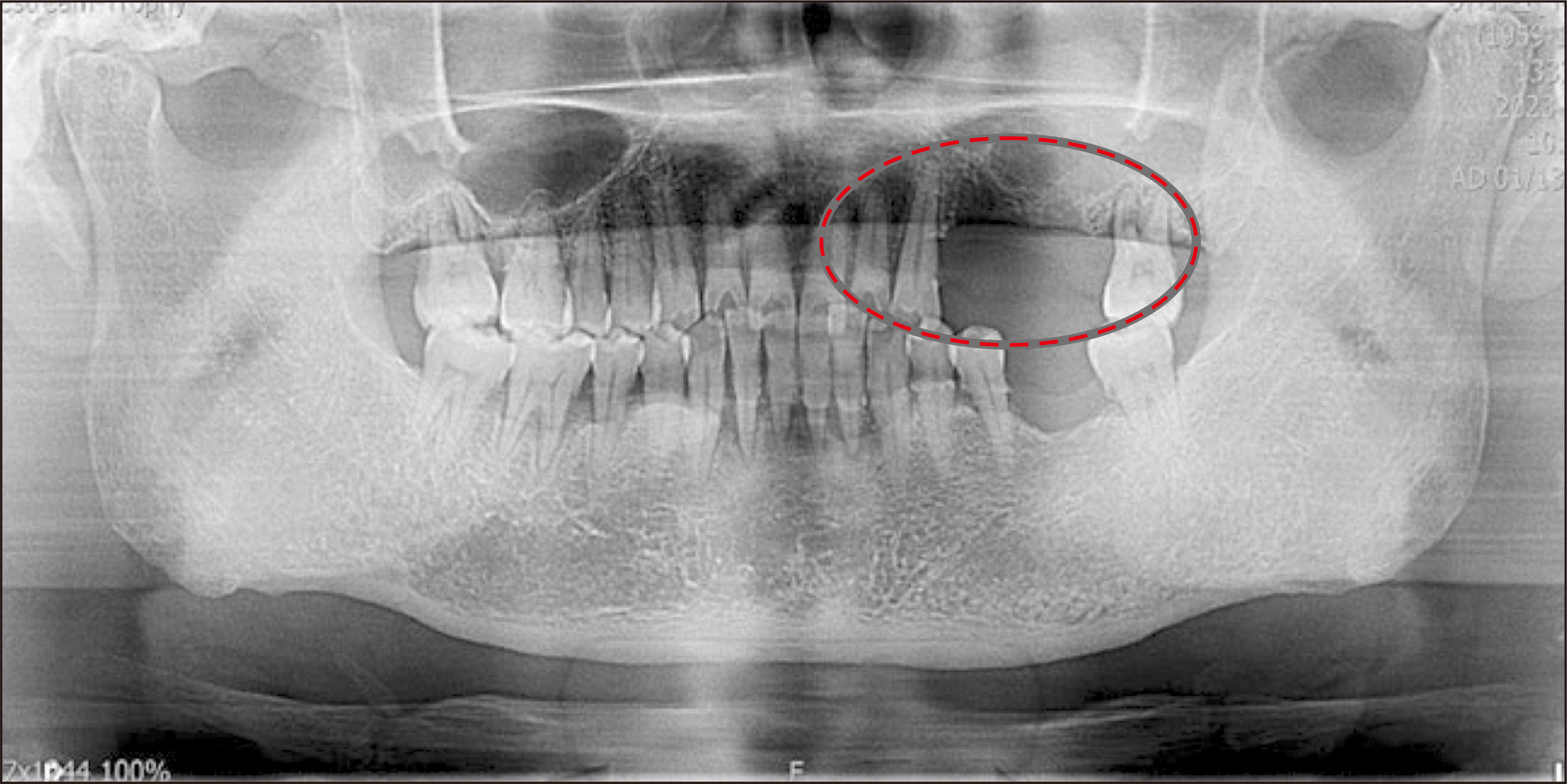
Complete resection of the mass along with extraction of teeth #24, #25, and #26 was performed under general anesthesia.(Fig. 3) The patient was admitted to the hospital post-surgery, intravenous antibiotics and analgesics were administered; the patients was discharged after 2 days without notable complications. After 1 week, stitches were removed and the excised area was in good healing state.(Fig. 4) Adequate re-epithelization was noted, and preservation of the alveolar ridge was successful.
Histopathological examination identified immature osteoids with irregular mineralization and a cellular stroma.(Fig. 5. A) Cementum-like material was observed in the fibrous connective tissue stroma.(Fig. 5. B) The lesions were moderately cellular with active, proliferating fibroblasts and dense fibrous stroma, with foci of calcified spherules corresponding to irregular bony trabeculae.(Fig. 5. C) These corresponding results were suggestive of POF.
POF is a type of benign tumor that falls into the group of fibro-osseous lesions. Fibro-osseous lesions include fibrous dysplasia, ossifying/cementifying fibroma, and periapical cemental dysplasia10-12. While it accounts for 3% of all oral tumors and 9.6% of all gingival lesions, people in their 20s to 40s are most susceptible and it shows consistent female predominance13,14. The higher prevalence of POF in females is thought to be related to hormonal influences, particularly estrogen and progesterone, as fluctuations in hormone levels can affect the production of gingival crevicular fluid15,16. While POF is reported to occur in both upper and lower gingiva, most studies identified the buccal surface of the anterior maxilla, especially around incisors and premolars, to be the most common site4. POF generally has a pedunculated or sessile base with well-delimited boundaries and shows gradual growth. Sizes vary widely, with an average size of 1-2 cm and a maximum diameter typically not exceeding 3 to 5 cm5,6,14. This report presented a relatively elderly male, aged 63, with a lesion located on the posterior maxilla, which is rather rare. Although the patient claimed that the mass had grown rapidly within 3 months, it seems more likely that it had been growing for a longer period. Additionally, it is possible that the patient’s habits of alcohol consumption and poor oral hygiene may have contributed to the condition.
Despite having quite distinct features, as described above, periodontal lesions like pyogenic granuloma and peripheral giant cell granuloma share similar pathological characteristics. This similarity necessitates the use of additional diagnostic tools, such as radiography and histopathological analysis6,13. Radiographic features of POF described by Eversole and Rovin3 include cortical bone expansion with well-defined lesion margins. Moreover, two distinct radiographic patterns could be identified: a unilocular radiolucent appearance, with or without radiopaque foci, and a multilocular radiolucent appearance. The presence of calcification within the lesion is commonly observed, as in the current case; however, it cannot be used as the sole criterion for differentiation due to variations between lesions. While preexisting bone structure typically remains unchanged, except for superficial concave defects caused by compression and occasional tooth displacement, larger lesions that have grown over time may sometimes involve erosion or even destruction of the bone surface4.
Histopathology of POF is characterized by fibrous connective tissue containing varying numbers of fibroblasts, along with the formation of different amounts of mineralized products such as bone components, both woven and lamellar bone, cementum-like materials, dystrophic calcification, or a combination of these elements4,6,17. Eversole and Rovin3 described four variations of keratotic products observed in ossifying lesions based on histological findings. These include ossified products composed of woven and lamellar trabeculae with dense bone deposits, spheroid-curvoid products scattered or exhibiting Sharpey’s fiber-like fringe at the margins, and dystrophic-appearing calcifications, typically spheroidal in shape, embedded in fasciculated or storiform stroma. Additionally, interconnected curvilinear trabeculae display dense deposits. In their study, 31% of the lesions consisted of extensive trabeculae and highly cellular fibrous tissue, while 47% were made up of mixed keratotic products. These findings align with the histopathological results in the current case, confirming the diagnosis of POF. Furthermore, Cavalcante et al.5 reported that lesions with a hypercellular mesenchymal component were more common, often featuring inflammatory infiltrates that were generally mild and chronic. In the reports, only 36.4% of cases exhibited ulceration in the epithelium. Since the mononuclear cells of the mesenchymal component originate from the periodontal ligament, Shrestha et al.4 reported that these cells can exhibit osteoblastic, cementoblastic, or fibroblastic characteristics due to cellular transformation triggered by irritants like dental calculus, orthodontic devices, and poorly fitted restorations. These studies support the etiological hypothesis that POF originates from the periodontal ligament. The findings of hypercellularity and inflammatory infiltrates, along with the cellular transformation of mesenchymal components in response to local irritants, align with the theory that POF arises from this specific tissue.
The standard treatment for POF is conservative local excision, mainly to prevent aesthetic discomfort due to its frequent occurrence in the maxillary anterior region18. This involves complete removal of the lesion to the adjacent periosteum or periodontal ligament, thorough scaling and root planing, and elimination of irritating factors to reduce the chances of recurrence4,6,17,19. Recurrence rates range from 8% to 20%, with a study by Cavalcante et al.5 and Lázare et al.6 reporting recurrence in 37 of 270 cases6. These high rates are often due to incomplete periosteum removal near the lesion’s pedicle and failure to eliminate local irritants, necessitating continuous follow-up.
This study has several limitations, including a lack of information on the identification and removal of potential irritative factors, as well as details on the treatments applied and their outcomes, such as mucogingival defects. In addition, most biopsy records did not document lesion recurrence, and the study experienced a loss of long-term follow-up, limiting the ability to assess treatment success and recurrence over time.
Although POF is an inflammatory reactive proliferative lesion, its significant enlargement can lead to alveolar bone destruction, complicating its differentiation from gingival malignancy. Accurate diagnosis depends on its characteristic histopathological features and potential underlying chronic mechanical stimuli. Effective treatment requires complete resection of the lesion, elimination of local irritants, and improvement of oral hygiene. This case presents a rare case of POF and aims to assist in its accurate diagnosis through radiological and histopathological methods, helping to distinguish it from other gingival reactive lesions and ensuring appropriate treatment and follow-up.
Notes
Authors’ Contributions
C.H.K., S.Y.A., and C.M.K. participated in performing the clinical treatment, and data collection. S.Y.A. and C.H.S. participated in writing the manuscript. C.H.K. helped to draft the manuscript. All authors read and approved the final manuscript.
References
1. Waldron CA. 1993; Fibro-osseous lesions of the jaws. J Oral Maxillofac Surg. 51:828–35. https://doi.org/10.1016/s0278-2391(10)80097-7. DOI: 10.1016/S0278-2391(10)80097-7. PMID: 8336219.

2. Shepherd SM. 1843; Alveolar exostosis. Am J Dent Sci. 4:53–4.
3. Eversole LR, Rovin S. 1972; Reactive lesions of the gingiva. J Oral Pathol. 1:30–8. DOI: 10.1111/j.1600-0714.1972.tb02137.x.

4. Shrestha A, Keshwar S, Jain N, Raut T, Jaisani MR, Sharma SL. 2021; Clinico-pathological profiling of peripheral ossifying fibroma of the oral cavity. Clin Case Rep. 9:e04966. https://doi.org/10.1002/ccr3.4966. DOI: 10.1002/ccr3.4966. PMID: 34691463. PMCID: PMC8513507.

5. Cavalcante IL, Barros CC, Cruz VM, Cunha JL, Leão LC, Ribeiro RR, et al. 2022; Peripheral ossifying fibroma: a 20-year retrospective study with focus on clinical and morphological features. Med Oral Patol Oral Cir Bucal. 27:e460–7. https://doi.org/10.4317/medoral.25454. DOI: 10.4317/medoral.25454. PMID: 35717619. PMCID: PMC9445604.

6. Lázare H, Peteiro A, Pérez Sayáns M, Gándara-Vila P, Caneiro J, García-García A, et al. 2019; Clinicopathological features of peripheral ossifying fibroma in a series of 41 patients. Br J Oral Maxillofac Surg. 57:1081–5. https://doi.org/10.1016/j.bjoms.2019.09.020. DOI: 10.1016/j.bjoms.2019.09.020. PMID: 31601435.

7. Tsiligkrou IA, Tosios KI, Madianos PN, Vrotsos IA, Panis VG. 2015; Oxytalan-positive peripheral ossifying fibromas express runt-related transcription factor 2, bone morphogenetic protein-2, and cementum attachment protein. An immunohistochemical study. J Oral Pathol Med. 44:628–33. https://doi.org/10.1111/jop.12275. DOI: 10.1111/jop.12275. PMID: 25359431.

8. Walters JD, Will JK, Hatfield RD, Cacchillo DA, Raabe DA. 2001; Excision and repair of the peripheral ossifying fibroma: a report of 3 cases. J Periodontol. 72:939–44. https://doi.org/10.1902/jop.2001.72.7.939. DOI: 10.1902/jop.2001.72.7.939. PMID: 11495143.

9. El Achkar VNR, Medeiros RDS, Longue FG, Anbinder AL, Kaminagakura E. 2017; The role of osterix protein in the pathogenesis of peripheral ossifying fibroma. Braz Oral Res. 31:e53. https://doi.org/10.1590/1807-3107bor-2017.vol31.0053. DOI: 10.1590/1807-3107bor-2017.vol31.0053. PMID: 28678972.

10. Koury ME, Regezi JA, Perrott DH, Kaban LB. 1995; "Atypical" fibro-osseous lesions: diagnostic challenges and treatment concepts. Int J Oral Maxillofac Surg. 24:162–9. https://doi.org/10.1016/s0901-5027(06)80094-9. DOI: 10.1016/S0901-5027(06)80094-9. PMID: 7608584.

11. Voytek TM, Ro JY, Edeiken J, Ayala AG. 1995; Fibrous dysplasia and cemento-ossifying fibroma. A histologic spectrum. Am J Surg Pathol. 19:775–81. DOI: 10.1097/00000478-199507000-00005. PMID: 7793475.
12. Slootweg PJ. 1996; Maxillofacial fibro-osseous lesions: classification and differential diagnosis. Semin Diagn Pathol. 13:104–12.
13. Verma E, Chakki AB, Nagaral SC, Ganji KK. 2013; Peripheral cemento-ossifying fibroma: case series literature review. Case Rep Dent. 2013:930870. https://doi.org/10.1155/2013/930870. DOI: 10.1155/2013/930870. PMID: 23365762. PMCID: PMC3556846.

14. Buchner A, Shnaiderman-Shapiro A, Vered M. 2010; Relative frequency of localized reactive hyperplastic lesions of the gingiva: a retrospective study of 1675 cases from Israel. J Oral Pathol Med. 39:631–8. https://doi.org/10.1111/j.1600-0714.2010.00895.x. DOI: 10.1111/j.1600-0714.2010.00895.x. PMID: 20456619.

15. Barot VJ, Chandran S, Vishnoi SL. 2013; Peripheral ossifying fibroma: a case report. J Indian Soc Periodontol. 17:819–22. https://doi.org/10.4103/0972-124x.124533. DOI: 10.4103/0972-124X.124533. PMID: 24554899. PMCID: PMC3917219.

16. Mishra AK, Maru R, Dhodapkar SV, Jaiswal G, Kumar R, Punjabi H. 2013; Peripheral cemento-ossifying fibroma: a case report with review of literature. World J Clin Cases. 1:128–33. https://doi.org/10.12998/wjcc.v1.i3.128. DOI: 10.12998/wjcc.v1.i3.128. PMID: 24303483. PMCID: PMC3845913.

17. Mergoni G, Meleti M, Magnolo S, Giovannacci I, Corcione L, Vescovi P. 2015; Peripheral ossifying fibroma: a clinicopathologic study of 27 cases and review of the literature with emphasis on histomorphologic features. J Indian Soc Periodontol. 19:83–7. https://doi.org/10.4103/0972-124x.145813. DOI: 10.4103/0972-124X.145813. PMID: 25810599. PMCID: PMC4365164.

18. Salaria SK, Gupta N, Bhatia V, Nayar A. 2015; Management of residual mucogingival defect resulting from the excision of recurrent peripheral ossifying fibroma by periodontal plastic surgical procedure. Contemp Clin Dent. 6(Suppl 1):S274–7. https://doi.org/10.4103/0976-237x.166832. DOI: 10.4103/0976-237X.166832. PMID: 26604587. PMCID: PMC4632236.

19. Cuisia ZE, Brannon RB. 2001; Peripheral ossifying fibroma--a clinical evaluation of 134 pediatric cases. Pediatr Dent. 23:245–8.
Fig. 1
Pre-operation panoramic view. Partial alveolar bone resorption was observed in the area of the upper left teeth (#24-#26), along with radiopaque particles that resemble dental calculus (red circle).

Fig. 2
Pre-operation cone beam computed tomography images. A. Axial view of peripheral ossifying fibroma (POF). Radiopaque calcified mass inside the lesion is observed (red arrows). B, C. Coronal view of POF. Note the sclerotic rim formation surrounding calcifications within the mass (red arrows).

Fig. 3
Clinical views during operation. A. A nodular and sessile base lesion in upper left gingiva, exhibiting erythematous areas on the buccal aspect and some ulcerative lesion on the palatal surface. B. After excision of the peripheral ossifying fibroma and teeth (#24-#26) closely related to the lesion. C. Application of fibrin sealant after sutures. D. Excised lesion measured 5.0×2.5 cm.

Fig. 4
Post-operation panoramic view. Postoperative view after removal of the peripheral ossifying fibroma and extraction of teeth #24-#26 (red circle).

Fig. 5
Histopathological findings. A. Immature osteoid with irregular mineralization and cellular stroma (H&E staining, ×40). B. Cementum-like material in fibrous connective tissue stroma (note the red arrows; H&E staining, ×100). C. Moderately cellular with active, proliferating fibroblast, dense fibrous stroma, with foci of calcified spherules corresponding to irregular bony trabeculae (H&E staining, ×200).


 PDF
PDF Citation
 Print
Print


 XML Download