

 PDF
PDF Citation
 Print
Print


I. Introduction
Although traumatic optic neuropathy (TON) is relatively rare, it can cause various degrees of visual function impairment and result in permanent damage1,2. One cause of TON is optic nerve canal fracture (OCF), which occurs in approximately 20% of patients with TON3. Direct compression of the optic nerve by bone fragments associated with OCF and rupture of the nerve fibers can cause TON and indirect injury to the optic nerve. Sources of indirect injury include circulatory failure, edema, and inflammation4,5.
TON is caused by application of strong external forces to the front of the head, especially to the lateral part of the eyebrow3, and is associated with approximately 0.5%-5% of head injuries and 2.5% of midfacial fractures2,6-8.
At present, the main treatment options for TON include observation, steroid administration, surgical decompression, or a combination of these modalities. However, no guidelines have been established for the treatment of TON1,9,10; for surgical decompression, protocol varies by institution based on surgical environment and experience. Trans-cranial and trans-facial surgical approaches are the classical methods. Interestingly, an endoscopic trans-nasal approach was reported as early as 1998, and suggested that the endoscopic approach has many benefits compared with the traditional approach in terms of recovery of function and cosmesis due to its minimally invasive characteristics11. Current advances in anatomical knowledge and instrumentation have offer endoscopic trans-nasal optic nerve decompression (ETOND) as the option that offers maximal visualization of the orbital apex and optic nerve. These advances are further enhanced by the aid of new medical tools like navigation guidance12.
In cases of TON accompanied by a midfacial fracture in our hospital, we collaborate with the Department of Ophthalmology, Otorhinolaryngology, and Head and Neck Surgery and the Department of Neurosurgery. In this study, we evaluated the feasibility of navigation-assisted ETOND for the treatment of TON based on retrospective clinical data from four consecutive patients with midfacial fractures. These patients underwent surgical treatment by oral and maxillofacial surgeons in collaboration with an endoscopic rhinologist. We also provided a literature review on pre-operative diagnosis and indications for navigation-assisted ETOND for treatment of TON.
Go to : 
II. Patients and Methods
1. Selection criteria for research patients
Patients who fulfilled the following eligibility criteria and did not fulfill any of the exclusion criteria were included in the study.
2. Eligibility criteria
Patients with midfacial fractures and TON who underwent diagnosis and surgical open reduction and internal fixation (ORIF) qualified for this study. These patients presented or were referred to the Department of Oral and Maxillofacial Surgery (OMFS), Maxillofacial Trauma Center of Shimane University Hospital between April 2021 and September 2023.
Patients who were treated conservatively for a TON were excluded, as were patients who underwent trans-cranial optic nerve decompression. Patients with a post-operative follow-up period less than 6 months were also excluded.
Six eligible patients were initially enrolled, but two who subsequently underwent trans-cranial optic nerve decompression were excluded. Therefore, the study includes evaluation of data from four consecutive patients.
3. Study method
The electronic medical records of the four patients were reviewed for treatment methods including timing of navigation-assisted ETOND, performance of ORIF surgery for midfacial fractures with or without orbital reconstruction, types of reconstruction materials used, and need for steroid therapy. In addition, post-operative complications and results of vision assessment were recorded. This study was approved by the Institutional Review Board of Shimane University Hospital (No. 20231107-1). The written informed consent was waived due to the retrospective review study.
4. Surgical procedure for endoscopic trans-nasal optic nerve decompression
Each patient was maintained in a supine position with the head in a neutral position and rotated 10°-15° to the right. During the surgical procedure, a navigation system (Stealth Station ENT; Medtronic) was used for image guidance. A gauze soaked in an adrenalin solution was placed in the nasal cavity to ensure vasoconstriction, and adrenaline was injected into the nasal mucosa. The surgical technique varied considerably according to the traumatic conditions. A standard endoscopic ethmoidectomy was performed. The natural ostium of the sphenoid sinus was identified, and sphenoidotomy was performed across the ethmoid to the sphenoid lateral wall and roof. The orbital apex was clearly identified using a 0° endoscope.
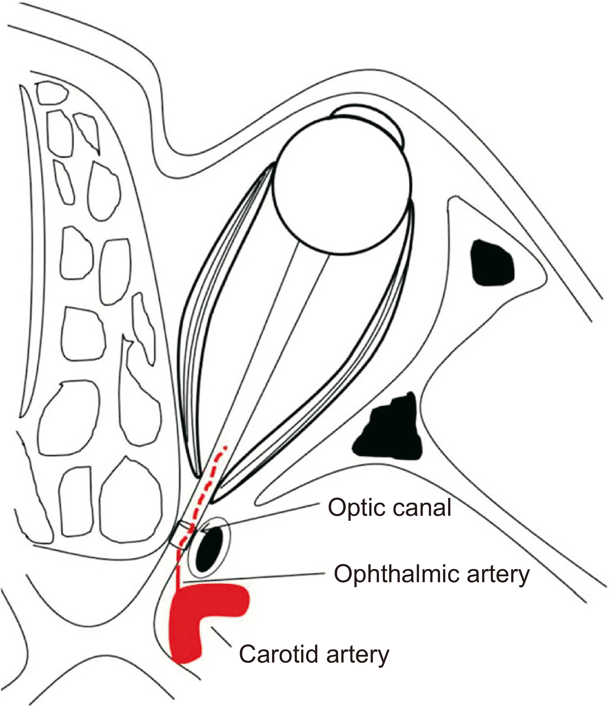
When bone fragments directly impeded the optic nerve, thinning of the nerve canal was performed with a diamond bur; and the fragments were removed using a curette and forceps. The posterior extent of decompression was determined using a navigation probe at the sphenoidal planum and internal carotid artery.(Fig. 1) Incising the optic nerve sheath was avoided and the nerve was preserved.
Go to :
III. Results
1. Demographic data and clinical management
The characteristics of the four patients, two men and two women, are described in Table 1. The average age of the patients was 75 years (range: 60-87 years). The TON in three patients was attributed to a fall, and one was due to a motor vehicle accident. At the initial visit, the Glasgow Coma Scale (GCS) score of three patients was 15 points for consciousness. In each case, 4 points were given for eye-opening (E), 5 points were assigned for verbal response (V), and 6 points were assessed for motor response (M). The fourth patient scored 14 points (E3V5M6). Soft tissue damage, swelling, and subcutaneous hemorrhage around the orbit on the affected side were observed in all four patients; and three patients had lacerations on the lateral eyebrow or upper eyelid on the affected side. Computed tomography (CT) identified OCF in all four patients. All patients presented with zygomaticomaxillary complex (ZMC) fractures on the affected side; two patients had isolated ZMC fractures, one had a ZMC fracture with a type I naso-orbito-ethmoid (NOE) fracture, and the other had a ZMC fracture with frontal bone and NOE type I fractures. All patients were evaluated pre-operatively for visual impairment by an ophthalmologist. Two patients perceived movement at 30 cm, and one had light perception. All three of these patients had relative afferent pupillary defects (RAPDs). The fourth patient had no loss of visual acuity despite the presence of an OCF on the CT image; he also had no RAPD. Visual impairment was present immediately after injury in the three affected and was delayed in one patient. All patients underwent navigation-assisted ETOND performed by an endoscopic rhinologist, either on or the day after ORIF for the midfacial fractures.
Table 1
Patient characteristics
![]()
2. Treatment methods, post-operative complications, and visual assessment
1) Timing of the navigation-assisted endoscopic trans-nasal optic nerve decompression
Navigation-assisted ETOND was performed by the same endoscopic rhinologist (T.S.) between the day of injury and post-trauma day 12. One patient received ETOND before ORIF, and three patients received ETOND and ORIF simultaneously. Patient #1 did not develop TON until the fifth day after injury; navigation-assisted ETOND was performed two days later. In Patient #2, in whom the optic nerve was directly affected by bone fragments caused by OCF according to CT imaging, navigation-assisted ETOND was performed on the same day of the injury.(Table 2)
Table 2
Treatment results
![]()
2) Open reduction and internal fixation surgery for midfacial fractures
In Patient #1, emergency navigation-assisted ETOND was performed two days before the ORIF; navigation-assisted ETOND was performed simultaneously with the ORIF in the other three patients on the day of injury, day 7 post-injury, and day 11 post-injury for Patients #2, #3, and #4, respectively.
3) Orbital reconstruction and the reconstruction materials
Orbital reconstruction with various materials was performed in three of the four patients according to the size of the defects13. The anterior bony wall of the maxillary sinus was used to reconstruct the orbital floor in Patient #1, autologous bone from the outer wall of the parietal bone was used in Patient #3, and a bioactive/bioresorbable sheet was used in Patient #4. In Patient #3, reduction of the medial orbital wall was performed during navigation-assisted ETOND.
4) Steroid therapy
Steroids were administered to two patients, one pre-operatively and one post-operatively as deemed necessary by an ophthalmologist. Patient #3 received steroid therapy and experienced recovery of visual acuity prior to the ORIF surgery.
5) Post-operative complications
Post-operative complications, such as olfactory dysfunction, bleeding, nasal septal perforation, or cerebrospinal fluid (CSF) rhinorrhea associated with navigation-assisted ETOND, were not observed in any of the patients.
6) Post-operative visual assessment
Patients #1 and #2 experienced visual improvement after navigation-assisted ETOND. Patients #3 and #4 who had no pre-operative visual impairment did not have visual loss associated with ORIF for midfacial fractures. The follow-up period ranged from 6 months to 33 months with a mean of 16.3 months.
3. Case presentation
1) Patient #1
The patient was a 60-year-old male who fell from a stepladder. He experienced traumatic injuries of multiple systems and was transported to the advanced trauma center of our hospital where he was referred to the Department of OMFS for examination and treatment of the maxillofacial trauma. No reduction in consciousness was observed. Marked left periorbital swelling, subcutaneous hemorrhage, and a laceration from the lateral eyebrow to the buccal area of the cheek were observed. CT revealed a left type I NOE fracture, left ZMC fractures, and OCF.(Fig. 2) No vision loss was initially observed, but a decrease to detection of hand motion at 30 cm was observed on the left side on the fifth day post-trauma. We referred the patient to an ophthalmologist who diagnosed TON. We promptly discussed the treatment with specialtists; on the seventh day post-trauma, an endoscopic rhinologist performed navigation-assisted ETOND. A trans-nasal endoscope was inserted through the left nostril, the posterior ethmoidal honeycomb was opened, and the filling hematoma was removed. Next, the left sphenoid sinus was opened, and a depressed bone fragment was detected in the distal optic nerve canal. This and other bone fragments were removed, and the optic nerve canal was decompressed.(Fig. 3. A-3. C) The operative time was 116 minutes, and there was minimal intra-operative bleeding. Post-operative CT showed successful removal of bone fragments around the optic nerve canal and release of the optic nerve.(Fig. 4. A) Vision started improving from the day after the surgery, and steroid therapy was initiated for three days post-operatively. On the 15th day post-injury, a surgeon performed ORIF for the midfacial fracture and orbital floor reconstruction using multiple incisions and a maxillary sinus approach. The incisions were hemi-coronal, left sub-tarsal, and oral vestibular.(Fig. 3. D-3. G) The ophthalmologist assessed eye movement and position using the traction test and confirmed the absence of snagging. The operative time was 218 minutes with minimal bleeding. Post-operative CT showed good repositioning, fixation, and reconstruction of the orbital floor.(Fig. 4. B-4. D) The patient’s vision improved gradually. At 6 months post-operatively, the standard logarithmic visual chart score was 0.03. Two years post-operatively, he is allowed to drive with the assistance of glasses, and the midfacial trauma is resolving.
 | Fig. 2Pre-operative computed tomography (CT) findings of Patient #1. A, B. Axial and coronal CT images showing a fracture of the lateral wall of the left optic nerve canal. The bone fragment compresses the left optic nerve within the nerve canal (yellow arrows). C, D. Coronal and three-dimensional CT images showing naso-orbito-ethmoid type I fracture, left zygomaticomaxillary complex fracture, and inferior wall fracture of the left orbit (yellow arrow).
|
 | Fig. 3Surgical view of Patient #1. A. During navigation-assisted endoscopic trans-nasal optic nerve decompression performed by an endoscopic rhinologist, a depressed bone fragment was identified in the distal part of the optic canal. B. Depressed bone fragments were filed. C. Decompression of the optic nerve canal was performed. The optic nerve sheath (white arrow) was visualized after bone removal. D. open reduction and internal fixation (ORIF) of midfacial fractures; the left zygomaticomaxillary complex fractures were treated by ORIF with a hemi-coronal incision approach. E. Bone was harvested from the anterior wall of the maxillary sinus. F. The harvested bone was shaped according to the morphology of the left orbital floor. G. The orbital floor was reconstructed using the harvested bone and fixed with micro-titanium screws.
|
 | Fig. 4Post-operative computed tomography (CT) findings of Patient #1. A. Axial CT showing removal and release of the bone fragments around the optic nerve canal (yellow arrow). B-D. Three-dimensional CT shows that the midfacial fracture, including orbital morphology, was adequately reconstructed (yellow arrows).
|
2) Patient #2
The patient was an 87-year-old female who fell in her bathroom. She was transported to the advanced trauma center on the following day and was referred to the department of OMFS for examination and treatment of the maxillofacial trauma. She had no decrease in consciousness, though marked swelling, subcutaneous hemorrhage around the orbit, and loss of vision relative to light perception were observed on the left side. CT revealed ZMC fracture and OCF (Fig. 5. A-5. D) for which we consulted members of the ear, nose, and throat (ENT) and ophthalmology departments.
 | Fig. 5Computed tomography (CT) findings and surgical view of Patient #2. A, B. Axial and coronal pre-operative CT showing a fracture of the lateral wall of the left optic nerve canal. A bone fragment was compressing the left optic nerve within the nerve canal (yellow arrows). C, D. Axial and three-dimensional pre-operative CT showing a left zygomaticomaxillary complex fracture with medial displacement of bone fragments. E. During navigation-assisted endoscopic trans-nasal optic nerve decompression performed by an endoscopic rhinologist, a depressed bone fragment was identified in the distal part of the optic nerve canal. F. Bone fragment removal. G, H. Axial and coronal post-operative CT showing the removal and release of the bone fragments around the optic nerve canal (yellow arrows). I. Three-dimensional post-operative CT shows that the zygomatic fragment was repositioned symmetrically.
|
An endoscopic rhinologist performed emergency navigation-assisted ETOND. A trans-nasal endoscope was inserted through the left nostril, and the left sphenoid sinus was exposed. Depressed bone fragments were detected in the lateral posterior part of the ethmoidal honeycomb and the distal part of the optic nerve canal. The bone fragments were removed, the optic nerve canal was decompressed, and the hematoma between the bone fragments and orbital periosteum was removed.(Fig. 5. E, 5. F) Simultaneously, ORIF for the left ZMC fracture was performed by an ophthalmic surgeon. The ZMC was manually reduced, and the zygomatic-maxillary buttresses and zygomatic-frontal sutures were fixed using a titanium plate. The ophthalmologist assessed eye movement and position using the traction test and confirmed the absence of snagging. The operative time was 235 minutes with minimal bleeding. Post-operative CT showed removal of the bone fragments around the optic nerve canal and release of the optic nerve. In addition, the zygomatic fragment was symmetrically repositioned.(Fig. 5. G-5. I) The patient’s vision had recovered enough for her to count fingers on the day following surgery, and vision had recovered to standard logarithmic visual chart 0.15 at eight days after surgery. One year after surgery, vision was restored to the same level as before the injury, and midfacial trauma was resolving.
3) Patient #3
The patient was a 78-year-old female with an injury attributed to a motor vehicle accident. She presented with a frontal bone fracture and a type I NOE fracture; left ZMC fracture; a left orbital blowout fracture, defect category type IV; and TON on the left side.(Fig. 6) Steroid therapy was initiated for three days immediately after the injury, and the patient’s visual acuity was restored. On the seventh day after the injury, ORIF for midfacial fracture, orbital reconstruction, and navigation-assisted ETOND were performed in collaboration with an endoscopic rhinologist. An ophthalmic surgeon performed ORIF for the midfacial fracture using bilateral coronal incisions, a left subtarsal incision, and an oral vestibular incision. An extraparietal plate was harvested for orbital floor reconstruction.(Fig. 7. A-7. D) Intraoperative cone-beam CT (CBCT) was performed, and the images confirmed proper reduction and fixation of the midfacial fracture. The data were transferred to the navigation system (Stealth Station ENT) (Fig. 7. E-7. H) and used during navigation-assisted ETOND and reduction of the left medial orbital wall fracture with nasal endoscopy.(Fig. 7. I, 7. J) The ophthalmic surgeon performed orbital floor reconstruction using a piece of the outer wall of the parietal bone shaped precisely using a pre-operatively designed model of the orbit. The operation time was 447 minutes with minimal bleeding. Eleven months after surgery, vision was restored to the same level as before the injury, and the midfacial injuries were resolving.(Fig. 8)
 | Fig. 6Pre-operative computed tomography (CT) findings of Patient #3. A. Axial CT showing a fracture of the frontal bone. B. Axial CT showing a fracture of the left zygomaticomaxillary complex. C, D. Coronal and axial CT showing a left type IV blowout fracture, medial orbital wall fracture, and optic nerve canal fracture (yellow arrow). E. Three-dimensional CT showing fracture of the frontal bone, a type I left naso-orbito-ethmoid fracture, and a left zygomaticomaxillary complex fracture.
|
 | Fig. 7Surgical view of Patient #3. A. ORIF of the midfacial fractures, frontal bone fracture, and left zygomaticomaxillary complex fracture were treated by ORIF with a bi-coronal incision. B. The zygomaticomaxillary buttress and the nasomaxillary buttress were treated by ORIF through an oral vestibular incision. C. The infraorbital rim was treated by ORIF through a sub-tarsal incision. D. The bone used for orbital floor reconstruction was harvested from the parietal exostosis. E. Workflow based on intra-operative CT to update the navigation system and navigation-assisted ETOND and intra-operative CT imaging using mobile-type cone-beam CT (3D Accuitomo M, Morita). F. Registration of the navigation system (Stealth Station ENT) using intraoperatively acquired CT data. G. Screen view during registration to the navigation system. H. Navigation-assisted ETOND by an endoscopic rhinologist. I. During the navigation-assisted ETOND performed by the endoscopic rhinologist, the posterior wall of the posterior ethmoid sinus was removed to gain access to the sphenoid sinuses (black arrows). J. Optic prominence (white arrow) with a view of the sphenoid sinus. (ORIF: open reduction and internal fixation, CT: computed tomography, ETOND: endoscopic trans-nasal optic nerve decompression)
|
 | Fig. 8Post-operative computed tomography (CT) findings of Patient #3. A, B. Three-dimensional construction and coronal CT showing that the midfacial fracture, including orbital morphology, were adequately reconstructed. C. Axial CT showing release of the bone on the medial side of the optic nerve canal (yellow arrow).
|
Go to :
IV. Discussion
TON is a severe, damaging, and debilitating complication of closed-head trauma. The hallmark of TON is loss of visual function that can manifest as subnormal visual acuity, visual field loss, or color vision dysfunction. Visual acuity loss associated with TON can be partial or complete and temporary or permanent1,2.
The main treatment options for TON are simple observation, steroid administration, surgical decompression, or some combination of these. However, there is no consensus on first-line treatment13.
The rationale for optic nerve decompression through partial removal of the optic nerve canal is to limit the detrimental effects of compression and restore nerve function. Decompression of the optic nerve lowers intracanalicular pressure, enables the removal of impinging bone fragments, and allows restoration of nerve function14. Various approaches have been reported for optic nerve decompression and can be broadly classified into trans-cranial and trans-nasal approaches10,15,16. In the past, surgery was performed in patients with evident fractures of the optic nerve canal because craniotomy approaches have a high rate of complications17. However, ETOND is a novel and innovative surgical treatment that has gained popularity with endoscopic rhinology specialists15. ETOND is the proposed gold standard, providing many benefits including reduced mortality, faster patient recovery, and minimal invasion, allowing a better cosmetic outcome when performed by an experienced endoscopic rhinologist5,18. However, only the inner one-third of the optic nerve canal can be accessed with ETOND; and the trans-cranial approach to the superior and lateral walls of the optic nerve canal depends on the trans-cranial method19. Sufficient decompression cannot be achieved in patients in whom the bone fragment is in the superior or lateral wall of the optic nerve canal. In these cases, the use of the trans-cranial approach is warranted19.
A system for navigation allows the surgeon to visualize the actual position of surgical instruments in real time on a monitor that displays the patient’s three-dimensional CT data20. The use of navigation for areas in which surgical approaches are challenging or anatomical attention is required provides confidence in the use of this approach. Image-guided navigation can help surgeons pinpoint the target area when facing anatomical deformation caused by trauma or tumor lesion12. These systems have recently evolved to improve precision and simplify the surgical procedure by minimizing intraoperative invasiveness. The development of navigation-assisted surgery has improved execution and predictability, allowing greater precision during endoscopic rhinology, neurosurgery, and oral and maxillofacial surgery20,21. Navigation-assisted ETOND is a feasible, safe, practical, and minimally invasive approach12,22. In this study, using image-guided navigation, the optic nerve was located accurately in all patients.
No intra-operative or post-operative complications, such as CSF leakage or ophthalmoplegia, were observed. However, several factors influence the accuracy of a navigation system, including the imaging data resolution, registration precision, and computer algorithm accuracy; and image drift highly affects system accuracy23. Considering the repair of complex maxillofacial fractures with bone segment movement, intraoperative topographic changes cause discrepancies between the pre-operative image data and the surgical site24. Recently, intraoperative CBCT, C-arm, and O-arm systems have been introduced and employed in clinical practice25; and the effectiveness of a navigation system using intraoperative CT images was demonstrated in orthopedic surgery involving complicated movements26. In our Patient #3, we explained the intra-operative changes in the anatomic morphology close to the optic nerve canal and orbit by reduction of the midfacial fracture. Intraoperative CBCT was performed following ORIF for the midfacial fracture. The navigation system was updated based on the data and used for navigation-assisted ETOND and orbital reconstruction. To the best of our knowledge, this was the first use of this method in oral and maxillofacial surgery. Further studies in more patients are warranted to establish its feasibility.
Many authors have suggested that ETOND should be performed if vision fails to improve after massive systemic administration of steroids or if progressive visual loss occurs during steroid therapy2,5,15,27. Conflicting opinions exist regarding the optimal timing for surgical treatment. Several studies have suggested that surgery within three days obtains the greatest benefit by preventing long-term damage to the nerve2,10,28. Emanuelli et al.17 reported a significant difference in outcome based on initiation of surgical treatment within 12 hours of the injury. In their systematic data review, Dhaliwal et al.10 reviewed 24 studies and concluded that >50% of patients benefitted from surgery regardless of the timing.
However, if OCF is radiologically evident and the displaced bone fragments are impinging the optic nerve, early ETOND surgery is strongly recommended to prevent permanent nerve damage1-3,9. Patients #1 and #2 of our study showed vision impairment and OCF on CT images; and we performed ETOND in collaboration with an endoscopic rhinologist immediately after the development of TON. Patient #3 showed OCF, but no obvious displaced bone fragments were impinging on the optic nerve; initial steroid therapy was successful, and the patient’s vision was restored. Patient #4 did not show any loss of visual acuity despite the presence of an OCF on the CT image. However, a case of TON after surgery for a ZMC fracture has been reported6. Therefore, to prevent exacerbation of TON due to inadvertent passive movement or migration of bone fragments caused by ORIF, ETOND was performed simultaneously with ORIF for the midfacial fracture.
Two patients with pre-operative visual impairment showed visual acuity improvement, and two patients who underwent preventive optic neurotomy showed no deterioration post-operatively. Initial visual acuity is a strong predictor of prognosis; and an initial vision assessment of no light perception indicates poor prognosis for recovery of normal vision. Therefore, treatment is not indicated in these cases29,30. However, the reported efficacy rate was 63.6% when optic nerve canal release surgery was performed fewer than three days from onset27. Thus, because of the disadvantages of surgery, treatment of patients with no light perception requires careful consideration.
Early diagnosis and treatment of TON are beneficial and should be known by oral and maxillofacial surgeons. In patients with facial trauma and TON, prompt examination, diagnosis, and treatment are imperative, highlighting the importance of collaboration among relevant hospital departments.
Go to :
V. Conclusion
We described four consecutive patients with midfacial fractures and TON who were treated surgically in close collaboration with ophthalmic and ENT surgeons. The feasibility of navigation-assisted ETOND performed by an endoscopic rhinologist was confirmed. To effectively manage maxillofacial trauma with TON, oral and maxillofacial surgeons should be familiar with its diagnosis and treatment.
Go to :
 XML Download