Journal List > Ewha Med J > v.47(4) > 1516088863

Jeong, Goh, and Kwak: Challenges and opportunities to integrate artificial intelligence in radiation oncology: a narrative review

Abstract

Artificial intelligence (AI) is rapidly transforming various medical fields, including radiation oncology. This review explores the integration of AI into radiation oncology, highlighting both challenges and opportunities. AI can improve the precision, efficiency, and outcomes of radiation therapy by optimizing treatment planning, enhancing image analysis, facilitating adaptive radiation therapy, and enabling predictive analytics. Through the analysis of large datasets to identify optimal treatment parameters, AI can automate complex tasks, reduce planning time, and improve accuracy. In image analysis, AI-driven techniques enhance tumor detection and segmentation by processing data from CT, MRI, and PET scans to enable precise tumor delineation. In adaptive radiation therapy, AI is beneficial because it allows real-time adjustments to treatment plans based on changes in patient anatomy and tumor size, thereby improving treatment accuracy and effectiveness. Predictive analytics using historical patient data can predict treatment outcomes and potential complications, guiding clinical decision-making and enabling more personalized treatment strategies. Challenges to AI adoption in radiation oncology include ensuring data quality and quantity, achieving interoperability and standardization, addressing regulatory and ethical considerations, and overcoming resistance to clinical implementation. Collaboration among researchers, clinicians, data scientists, and industry stakeholders is crucial to overcoming these obstacles. By addressing these challenges, AI can drive advancements in radiation therapy, improving patient care and operational efficiencies. This review presents an overview of the current state of AI integration in radiation oncology and insights into future directions for research and clinical practice.

Introduction

Background

Radiation oncology has seen significant advancements in recent decades, driven by the introduction of several innovative technologies. These include intensity-modulated radiation therapy, stereotactic radiosurgery, stereotactic body radiation therapy, image-guided radiation therapy (IGRT), and respiratory beam control. These technologies have increased the precision of treatments [1], reduced side effects [2,3], and improved patient outcomes [46]. Furthermore, the advent of various imaging modalities such as CT, MRI, and PET scans, coupled with significant developments in computational resources, has greatly expanded the capabilities of radiation oncology. These imaging modalities provide complementary information: CT scans deliver detailed anatomical information, MRI offers exceptional soft tissue contrast, and PET scans provide metabolic insights into tumors. By integrating these modalities, radiation oncologists can achieve more accurate tumor localization, more precise treatment planning, and better treatment adaptation, thereby further improving patient care and outcomes.
Intensity-modulated radiation therapy enables the modulation of radiation beams to conform more precisely to the shape of the tumor, thereby protecting surrounding healthy tissues and reducing complications [79]. However, this technique requires meticulous treatment planning and the implementation of rigorous quality assurance protocols, which can be challenging [10,11]. Stereotactic radiosurgery and stereotactic body radiation therapy administer high doses of radiation with sub-millimeter accuracy to treat small, well-defined tumors in the brain and body, respectively. The primary challenge in these therapies is managing patient movement and ensuring accurate targeting. These issues can be addressed using advanced imaging technologies and motion management techniques [12,13]. IGRT employs advanced imaging technologies to enhance the precision of radiation delivery by compensating for patient movement and anatomical changes during treatment [14]. The advantage of IGRT is its ability to adapt treatment in real-time. However, challenges include integrating imaging and treatment systems and maintaining consistent image quality [15]. Respiratory gating aligns radiation delivery with the patient's breathing cycle, minimizing exposure to moving organs such as the lungs and liver. The main challenges associated with respiratory gating include the need for advanced equipment and software, as well as requiring patient cooperation [16].
Integrating artificial intelligence (AI) into radiation oncology is becoming increasingly important due to various social and healthcare trends [17]. The global population is aging, which leads to a higher incidence of cancer in older age groups. As life expectancy increases, so does the demand for effective and efficient cancer treatments, placing significant burdens on healthcare systems worldwide [18]. To manage the increasing patient load effectively, it is essential to adopt advanced technologies [19].
Moreover, healthcare systems are under pressure to improve patient outcomes while controlling costs [20]. AI has the potential to meet these challenges by improving the accuracy of radiation therapy, shortening treatment durations, and reducing side effects. This can result in better resource utilization and heightened patient satisfaction [21]. Additionally, AI supports personalized medicine by offering treatments customized to the unique characteristics of each patient. By analyzing extensive datasets from diverse sources, AI can assist in creating more effective treatment plans that are specifically tailored to the genetic, phenotypic, and lifestyle factors of individual patients [22,23].
The necessity and justification for AI in radiation oncology extend beyond these practical improvements. The complexity and variability inherent in cancer treatments require sophisticated decision-making tools capable of analyzing vast amounts of data in real time—capabilities that surpass human limits. AI excels in integrating and interpreting multi-dimensional data from various imaging modalities and patient records, significantly enhancing clinical decision-making. This reduces errors and improves treatment outcomes. Additionally, AI supports continuous learning and adaptation in treatment protocols, allowing them to evolve with new medical insights and tailored to individual patient responses. Therefore, the integration of AI not only enhances efficiency but also elevates the standard of personalized patient care, rendering it an essential component in contemporary radiation oncology.

Objectives

This review provides a comprehensive overview of the current state of AI integration in radiation oncology, exploring both the benefits and challenges associated with its implementation.

Ethics statement

As this study is a literature review, it did not require institutional review board approval or individual consent.

Opportunities for artificial intelligence integration

Treatment planning

AI offers significant opportunities in various facets of radiation oncology, such as treatment planning, image analysis, adaptive radiation therapy (ART), and predictive analytics. In these domains, AI can enhance clinical workflows, shorten treatment durations, and deliver more personalized and effective treatments.
AI can optimize radiation treatment planning by automating complex tasks, reducing planning time, and enhancing accuracy [24]. Machine learning algorithms can analyze extensive datasets to identify optimal treatment parameters, potentially leading to personalized treatment plans that maximize efficacy while minimizing side effects [25]. Over the past few decades, AI has been progressively integrated into radiation treatment planning. Initially, simple rule-based systems supported clinical decision-making. As computational power and data availability have increased, more sophisticated machine learning algorithms, including artificial neural networks, have been developed. These algorithms, which initially supported basic tasks, have now evolved to handle complex treatment planning scenarios. Current research in AI for radiation treatment planning focuses on developing algorithms that can analyze large datasets to identify optimal treatment parameters. Machine learning models are trained using historical treatment data, patient outcomes, and imaging data to enhance the precision and accuracy of treatment plans [26]. Studies have demonstrated that AI can significantly reduce the time required for planning while maintaining or improving the quality of the plans. Research is also exploring how to integrate AI with ART to continuously update treatment plans based on real-time patient data [27]. The automated generation of radiation fluence is a critical component of treatment planning, determining the intensity and distribution of radiation beams to achieve the desired dose distribution within the target area while sparing healthy tissues. AI has played a crucial role in improving the accuracy and efficiency of fluence map optimization. Deep learning techniques can predict optimal fluence patterns based on patient-specific anatomical and dosimetric data, reducing the complexity and time required for manual adjustments [28,29].
Several commercial products have incorporated AI into radiation treatment planning. For example, Varian's Ethos Therapy [30,31] and Elekta's MOSAIQ Plaza [32] utilize AI to improve treatment planning and adaptive therapy. These systems automate aspects of the planning process, optimize dose distribution, and adjust treatment plans in response to anatomical changes or variations in tumor size during treatment.

Image analysis

AI-driven image analysis can significantly enhance the accuracy of tumor detection and segmentation [33]. Deep learning algorithms are capable of processing imaging data from various modalities, including CT, MRI, and PET scans, to provide precise tumor delineation, which is crucial for effective radiation targeting [34,35]. The journey of AI-based image analysis in radiation oncology is marked by significant milestones. Initially, basic image processing techniques were employed to improve image quality and enhance tumor visualization. With the advent of machine learning, more advanced algorithms were developed to automate tumor detection and segmentation tasks. Early methodologies heavily relied on handcrafted features, but they were soon surpassed by deep learning algorithms capable of autonomously learning features from data [36]. Current research in AI-based image analysis focuses on increasing the precision and efficiency of tumor detection and segmentation [37,38]. Deep learning models, particularly convolutional neural networks, are widely used to analyze imaging data from CT, MRI, and PET scans [39]. These models have demonstrated an exceptional ability to distinguish tumors from adjacent healthy tissues, often surpassing the accuracy and consistency of human experts [40]. Additionally, research in multimodal image analysis indicates that AI can combine data from various imaging modalities to enhance diagnostic accuracy and inform treatment planning [41].
The segmentation of tumors and surrounding normal organs is a critical task in radiation therapy, requiring accurate delineation of tumor boundaries and normal tissues to ensure effective treatment planning [42]. Manual segmentation is traditionally labor-intensive and prone to inter-observer variability. However, AI-based segmentation techniques streamline this process, providing consistent and rapid results. Advanced algorithms, such as U-Net and its variants, have become the standard for medical image segmentation, known for their ability to accurately identify tumor boundaries [43]. These models utilize an encoder-decoder architecture to capture the complex spatial hierarchies within images, ensuring precise segmentation. Commercial applications of AI-based image analysis in radiation oncology are already making an impact. For example, IBM Watson for Oncology leverages AI to analyze medical images and provide insights for treatment planning [44]. Similarly, Varian's ARIA oncology information system integrates AI to enhance treatment planning and execution [45]. Siemens Healthineers also offers AI-based tools for advanced image analysis and interpretation within its syngo.via platform [46].

Adaptive radiation therapy

In ART, treatment plans are adjusted based on changes in patient anatomy and tumor size throughout the course of treatment. AI improves ART by swiftly analyzing imaging data and making real-time adjustments to the treatment plan, thus increasing the accuracy and effectiveness of the treatment. The concept of ART has significantly evolved over the past few decades. Initially, radiation treatment plans were static and did not accommodate anatomical changes during the treatment course. As technology progressed, the necessity for more adaptive approaches became apparent, leading to the development of ART [47]. Early implementations of ART involved periodic imaging and manual adjustments, which were time-consuming and could not be performed in real-time. Current research and development in ART focus on utilizing AI to automate and improve the adaptability of treatment plans [48]. AI-based ART systems employ advanced imaging technologies, such as daily cone-beam CT scans, to monitor tumor size and anatomical changes in patients. Machine learning algorithms then analyze these imaging data to predict anatomical changes and adjust radiation dose distributions accordingly. This capability for real-time adaptation ensures that radiation doses are precisely targeted to the tumor, minimizing exposure to surrounding healthy tissues and enhancing overall treatment outcomes [4951].
Several commercial products have integrated AI to improve ART. Notably, Varian's Ethos Therapy system and Elekta's Unity system stand out. Varian's Ethos utilizes AI to analyze daily imaging and dynamically adjust treatment plans, offering personalized therapy for each session [49]. Similarly, Elekta's Unity combines a high-field MRI scanner with a linear accelerator to provide real-time imaging and adaptation during treatment sessions [52]. These systems mark significant progress in the field of commercial ART, facilitating more accessible and practical real-time adaptive therapy. As we look to the future, ART is poised to continue its evolution through the further integration of AI and advanced imaging technologies.

Predictive analytics

AI can analyze historical patient data to predict treatment outcomes and potential complications. Predictive analytics can guide clinical decision-making, allowing for more informed and personalized treatment strategies [53,54]. The application of predictive analytics in radiation oncology has advanced significantly over the years. Initially, treatment decisions were primarily based on empirical data and clinical experience. With the advent of data collection and storage technologies, large databases became available, enabling the identification of patterns and correlations through statistical methods. However, traditional methods were limited in handling complex, high-dimensional data. Current predictive analytics research focuses on leveraging AI to analyze large-scale historical patient data [55]. Machine learning algorithms, including deep learning and ensemble methods, are used to predict various clinical outcomes, including tumor response, survival rates, and potential side effects. These models process diverse data types, including demographic information, genetic profiles, imaging data, and treatment histories, to provide comprehensive predictions. Studies have shown that AI-based predictive models can outperform traditional statistical methods in terms of accuracy and robustness [56].
Several commercial products have integrated AI-based predictive analytics to support clinical decision-making in radiation oncology. For instance, IBM Watson for Oncology utilizes AI to analyze patient data and provide evidence-based treatment recommendations [57]. Similarly, the RayStation treatment planning system employs machine learning models to predict patient-specific treatment outcomes and optimize treatment plans [58]. Looking ahead, predictive analytics is poised to revolutionize clinical practice in radiation oncology. Researchers are focusing on improving the interpretability of AI models, which will enable clinicians to better understand and trust their predictions. Furthermore, the integration of predictive analytics with other AI-based technologies, such as image analysis and ART, is expected to streamline treatment workflows and improve efficiency. The adoption of federated learning, which allows AI models to be trained on data from multiple institutions without the need to share patient data, is anticipated to improve the generalizability and reliability of predictive models. The elements discussed in the "Opportunities for AI Integration" section are depicted in Fig. 1.
Fig. 1.

The radiation therapy procedures, tasks performed during the procedures, and subsequent related tasks. The black background signifies the aspects related to opportunities for AI integration, indicating that these AI integrations are influencing the entire radiation therapy process. The tasks within the radiation therapy process that are expected to change or be affected by AI in the near future are presented in black text. AI, artificial intelligence.

emj-47-4-53-g1

Challenges in artificial intelligence integration

Integrating AI into radiation oncology involves multiple challenges that need to be addressed to fully harness its potential. These challenges encompass technical, clinical, and ethical aspects, necessitating collaboration among researchers, clinicians, and policymakers.

Data quality and quantity

The effectiveness of AI models is heavily influenced by the quality and quantity of the data used for training [5961]. Challenges such as inconsistent data quality, missing data, and limited access to large annotated datasets are significant obstacles [62]. AI algorithms depend on robust datasets that accurately reflect diverse patient populations and clinical scenarios. However, acquiring such datasets is often challenging due to privacy concerns, variations in data collection protocols, and the absence of standardized data formats. Research has indicated that biases in training data can result in AI models that fail to generalize effectively across different patient groups. It is crucial to ensure data quality and to develop techniques for managing missing or incomplete data to build dependable AI systems. Moreover, large datasets from multiple institutions are necessary to train models that are effective in various clinical environments. Collaborative initiatives to share data while safeguarding patient privacy are vital for the progress of AI in radiation oncology [63].

Interoperability and standardization

Radiation oncology systems frequently utilize various software and data formats, which can lead to interoperability issues [64]. To facilitate the seamless integration of AI tools across different platforms and institutions, it is crucial to standardize data formats and protocols [65]. The lack of interoperability may impede the efficient deployment of AI technologies, as it poses challenges in integrating and analyzing data from disparate systems. These interoperability challenges are further exacerbated by the use of diverse imaging modalities, treatment planning systems, and electronic health records in radiation oncology. Developing common data standards, such as Digital Imaging and Communications in Medicine (DICOM), is essential for enabling data exchange and integration [66,67]. Encouraging the widespread adoption of these standards throughout the industry will help surmount obstacles to AI implementation and enhance the effectiveness of AI tools in clinical settings.

Regulatory and ethical considerations

Integrating AI into radiation oncology introduces several regulatory and ethical challenges, including concerns about patient privacy [68], data security, and the necessity for rigorous validation of AI models. Regulatory frameworks need to adapt to address these issues and ensure AI is used safely and effectively in clinical settings. Current regulations may not adequately capture the complexities of AI technologies, necessitating updates to existing guidelines and the creation of new standards. Ethical considerations are also paramount in the integration of AI [6971]. It is crucial to obtain patient consent and safeguard data privacy, particularly when handling sensitive health information. Additionally, the potential for AI to reinforce existing biases in healthcare must be addressed, as biased algorithms could worsen health disparities. Developing AI models that are transparent and explainable will foster trust between clinicians and patients, facilitating their understanding and acceptance of AI-driven decisions.

Clinical implementation and adoption

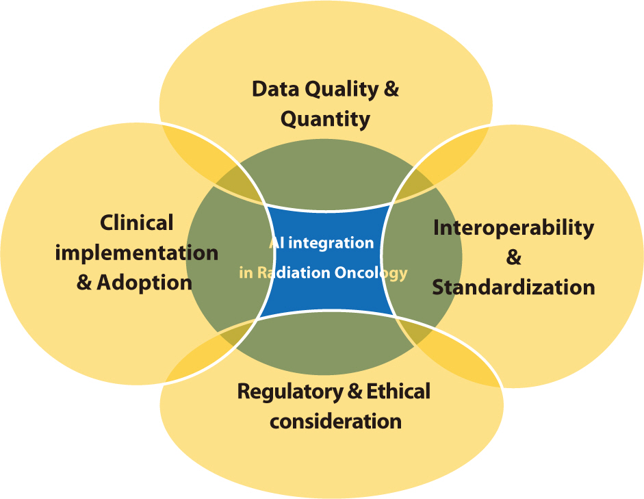
Adopting AI tools in clinical settings necessitates significant changes in workflows and staff training. Resistance to change and skepticism regarding the reliability of AI can impede adoption [72]. It is crucial to implement comprehensive training programs and clearly communicate the benefits of AI to overcome these barriers. Research indicates that involving clinicians in the development and implementation of AI tools is essential for meeting clinical needs and ensuring seamless integration into existing workflows [7375]. Providing ongoing education and support for healthcare professionals can foster trust in AI technologies and promote their adoption. Demonstrating the clinical and economic advantages of AI through pilot studies and real-world applications can also secure support from stakeholders. The issues discussed in the section "Challenges in AI Integration" are illustrated in Fig. 2.
Fig. 2.

The challenges involved in integrating AI into radiation oncology, which are likely to be highly complex and closely interconnected. This indicates the need for careful planning and consideration at an early stage to address these issues before they become entangled. AI, artificial intelligence.

emj-47-4-53-g2

Future directions

The future of AI in radiation oncology promises significant transformations in patient care. As AI technologies advance, several critical areas are poised to propel progress within the field. These areas encompass the creation of more advanced AI models, the integration of AI with other emerging technologies, the development of stringent standards and regulations, and a focus on collaborative and multidisciplinary approaches.
Future AI development will concentrate on creating more sophisticated models that can manage complex, high-dimensional data [76]. Advances in deep learning and reinforcement learning will enable the creation of models capable of predicting treatment outcomes with greater accuracy and adapting to new data in real time. These models will benefit from the ongoing expansion of available data, including multimodal datasets that integrate imaging, genomics, and clinical information. These comprehensive datasets will enable the development of personalized treatment plans specifically tailored to individual patients [77,78].
The integration of AI with other emerging technologies, such as radiomics, genomics, and wearable health devices, is expected to revolutionize radiation oncology [77]. Radiomics extracts a large number of features from medical images, which AI then uses to predict disease progression and treatment responses. Genomics offers insights into the genetic makeup of tumors, facilitating more targeted and effective treatments [79]. Wearable health devices continuously monitor patients' health indicators, supplying data that enables AI to dynamically adjust treatment plans in real-time [80].
To fully realize the potential of AI in radiation oncology, it is crucial to standardize and ensure interoperability across systems and institutions. The development and adoption of common data standards, such as DICOM, will facilitate the exchange and integration of data [81,82]. Additionally, it is essential to establish standardized protocols for the development, validation, and deployment of AI models to ensure that these tools are safe, reliable, and effective [83]. The International Electrotechnical Commission (IEC) is also working to establish standards that ensure the quality, safety, and reliability of AI in medical devices [84].
Collaboration among researchers, clinicians, data scientists, and industry stakeholders is essential for advancing AI in radiation oncology [76,85]. Multidisciplinary teams can leverage diverse expertise to create AI tools that address clinical needs and integrate smoothly into current workflows [86]. Collaborative research initiatives and shared databases facilitate the pooling of data and resources, which accelerates the development of robust AI models [82]. Promoting open science and data sharing while ensuring patient privacy will foster innovation and advance the field [87].
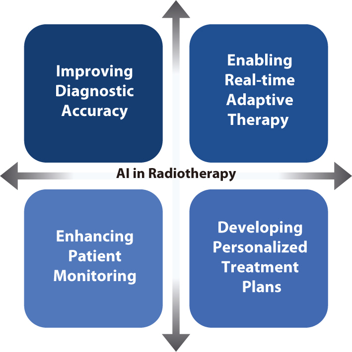
Developing explainable AI models is crucial for fostering trust between clinicians and patients [88]. Explainable AI offers insights into the decision-making processes of AI algorithms, thereby facilitating clinicians' understanding and validation of AI-based recommendations [89]. Increasing the transparency of AI systems will address concerns about bias and errors, ultimately encouraging the adoption of AI technologies in clinical settings [90,91]. AI has the potential to significantly improve the quality of clinical care while increasing efficiency. For instance, studies have demonstrated that AI can reduce the time needed for treatment planning and image analysis, allowing clinicians to devote more attention to patient care [92,93]. AI-based predictive analytics can identify patients at high risk of complications, enabling proactive interventions that improve outcomes [94,95]. Moreover, AI's capacity to continuously learn and adapt from new data ensures that treatment strategies are consistently updated with the latest medical knowledge and technological advancements [96]. In clinical settings, AI technologies have the potential to drive several key advancements in radiation oncology. These advancements include improving diagnostic accuracy, enabling real-time adaptive therapy, enhancing patient monitoring, and developing personalized treatment plans, all of which contribute to better patient outcomes and operational efficiencies [97101].
Improving diagnostic accuracy: AI-based image analysis increasing the accuracy of tumor detection and segmentation, thereby improving the precision of radiation targeting. This increased precision minimizes damage to surrounding healthy tissues and increases treatment efficacy.
Enabling real-time adaptive therapy: AI rapidly analyzes daily imaging data and adjusts treatment plans in real time, enhancing the effectiveness of ART. This capability ensures that radiation doses are precisely targeted to the tumor, accommodating anatomical changes or variations in tumor size throughout the treatment course.
Enhancing patient monitoring: Wearable health devices coupled with AI analytics enable continuous monitoring of patients' health indicators. This allows timely interventions when adverse changes are detected. Such a predictive approach aids in managing side effects and enhances overall treatment outcomes.
Developing personalized treatment plans: AI analyzes extensive datasets to identify patterns and predict individual responses to different treatment modalities. This capability allows clinicians to create treatment plans that are tailored to each patient's unique genetic and clinical profile, thereby maximizing efficacy and minimizing side effects.
Looking ahead, integrating AI into clinical practice in radiation oncology necessitates careful consideration of ethical, regulatory, and practical issues. However, the potential benefits, such as improved patient outcomes and operational efficiencies, make it a worthwhile endeavor. Collaborative efforts among multidisciplinary teams, the establishment of robust standards, and the continued advancement of AI technologies will shape the future of radiation oncology, ultimately transforming patient care. The themes explored in the "Future Directions" section are visualized in Fig. 3.
Fig. 3.

Advancements anticipated to result from the integration of AI into radiation therapy. These significant advancements are expected to lead to enhanced diagnostic accuracy, seamless patient monitoring, real-time therapeutic adaptations, and the delivery of highly personalized treatment, thereby substantially improving outcomes for many cancer patients. AI, artificial intelligence.

emj-47-4-53-g3

Conclusion

The integration of AI into radiation oncology offers significant opportunities to enhance the precision, efficiency, and outcomes of treatments. As AI technologies continue to advance, their potential to transform various aspects of radiation therapy becomes increasingly apparent. This review has highlighted key areas where AI can make substantial contributions, including treatment planning, image analysis, ART, and predictive analytics. Each area showcases AI's ability to improve clinical workflows, shorten treatment times, and deliver more personalized and effective treatments.
Despite these promising advancements, several challenges must be addressed to fully realize the potential of AI in radiation oncology. Data quality and quantity are critical issues because robust and comprehensive datasets are necessary for effective AI models. Ensuring interoperability and standardization across different systems and institutions is also essential to facilitate seamless integration and data exchange. Additionally, regulatory and ethical considerations must be carefully addressed to protect patient privacy and ensure the safe deployment of AI technologies in clinical environments.
Collaboration among researchers, clinicians, data scientists, and industry stakeholders is essential for overcoming these challenges. By forming multidisciplinary teams, diverse expertise can be harnessed to create AI tools that not only meet clinical needs but also integrate seamlessly into existing workflows. Initiatives that promote collaborative research and shared databases will facilitate the pooling of data and resources, thereby accelerating the development of robust AI models. Encouraging open science and data sharing, while simultaneously protecting patient privacy, will drive innovation and advance the field. Moreover, the development of explainable AI models is crucial for promoting trust between clinicians and patients. Explainable AI offers insights into the decision-making processes of AI algorithms, enabling clinicians to more easily understand and validate AI-based recommendations. Increasing the transparency of AI systems will address concerns about bias and errors, ultimately encouraging the adoption of AI technologies in clinical practice.
The future of AI in radiation oncology is bright, and ongoing research and development are poised to overcome current challenges and unlock new opportunities. AI-driven improvements in diagnostic accuracy, real-time adaptive therapy, patient monitoring, and personalized treatment plans are poised to revolutionize the field, enhancing patient care and operational efficiencies. By emphasizing collaborative research, continuous learning, and patient-centric approaches, the field can surmount these challenges and realize transformative advancements in radiation therapy.

Authors' contributions

Project administration: Kwak J

Conceptualization: Kwak J

Methodology & data curation: Jeong C, Goh YM, Kwak J

Funding acquisition: not applicable

Writing – original draft: Jeong C, Goh YM, Kwak J

Writing – review & editing: Jeong C, Goh YM, Kwak J

Conflict of interest

No potential conflict of interest relevant to this article was reported.

Funding

Not applicable.

Data availability

Not applicable.

Acknowledgments

Not applicable.

Supplementary materials

Not applicable.

References

1. Chun SG, Hu C, Komaki RU, Timmerman RD, Schild SE, Bogart JA, et al. Long-term prospective outcomes of intensity modulated radiotherapy for locally advanced lung cancer: a secondary analysis of a randomized clinical trial. JAMA Oncol. 2024; 10(8):1111–1115. DOI: 10.1001/jamaoncol.2024.1841. PMID: 38935373.
crossref
2. Fathy MM, Hassan BZ, El-Gebaly RH, Mokhtar MH. Dosimetric evaluation study of IMRT and VMAT techniques for prostate cancer based on different multileaf collimator designs. Radiat Environ Biophys. 2023; 62(1):97–106. DOI: 10.1007/s00411-022-01011-2. PMID: 36576578. PMCID: PMC9950215.
crossref
3. Mohan G, Ayisha Hamna TP, Jijo AJ, Saradha Devi KM, Narayanasamy A, Vellingiri B. Recent advances in radiotherapy and its associated side effects in cancer: a review. J Basic Appl Zool. 2019; 80(1):1–10. DOI: 10.1186/s41936-019-0083-5.
4. Zaorsky NG, Harrison AS, Trabulsi EJ, Gomella LG, Showalter TN, Hurwitz MD, et al. Evolution of advanced technologies in prostate cancer radiotherapy. Nat Rev Urol. 2013; 10(10):565–579. DOI: 10.1038/nrurol.2013.185. PMID: 24018567.
crossref
5. Lim DH. Localized intracranial germinoma: is it time to re-define target volume for whole ventricular irradiation? Radiat Oncol J. 2023; 41(2):59–60. DOI: 10.3857/roj.2023.00423. PMID: 37403347. PMCID: PMC10326506.
crossref
6. Yu JI. Myxoid liposarcoma: a well-defined clinical target variant in radiotherapy for soft tissue sarcoma. Radiat Oncol J. 2022; 40(4):213–215. DOI: 10.3857/roj.2022.00598. PMID: 36606298. PMCID: PMC9830037.
crossref
7. Teh BS, Woo SY, Butler EB. Intensity modulated radiation therapy (IMRT): a new promising technology in radiation oncology. Oncol. 1999; 4(6):433–442. DOI: 10.1634/theoncologist.4-6-433.
crossref
8. Scaringi C, Agolli L, Minniti G. Technical advances in radiation therapy for brain tumors. Anticancer Res. 2018; 38(11):6041–6045. DOI: 10.21873/anticanres.12954. PMID: 30396918.
crossref
9. Park SY, Kim J, Chun M, Ahn H, Park JM. Assessment of the modulation degrees of intensity-modulated radiation therapy plans. Radiat Oncol. 2018; 13:1–8. DOI: 10.1186/s13014-018-1193-9. PMID: 30545396. PMCID: PMC6293636.
10. Tanaka Y, Hashimoto M, Ishigami M, Nakano M, Hasegawa T. Development of a novel delivery quality assurance system based on simultaneous verification of dose distribution and binary multi-leaf collimator opening in helical tomotherapy. Radiat Oncol. 2023; 18(1):180. DOI: 10.1186/s13014-023-02366-6. PMID: 37919745. PMCID: PMC10621123.
11. Mandava A, Koppula V, Kandati M, Raju KVVN. Synchronous radiation-induced enterovesical and enterocervical fistulas in carcinoma of the uterine cervix. Radiat Oncol J. 2023; 41(4):297–300. DOI: 10.3857/roj.2023.00500. PMID: 38185935. PMCID: PMC10772593.
crossref
12. Kavanagh BD, Timmerman RD. Stereotactic radiosurgery and stereotactic body radiation therapy: an overview of technical considerations and clinical applications. Hematol Oncol Clin. 2006; 20(1):87–95. DOI: 10.1016/j.hoc.2006.01.009. PMID: 16580558.
crossref
13. Jia-Mahasap B, Madla C, Sripan P, Chitapanarux I, Tharavichitkul E, Chakrabandhu S, et al. Stereotactic radiosurgery for limited brain metastasis using three different techniques: helical tomotherapy, volumetric modulated arc therapy, and cone-based LINAC radiosurgery. Radiat Oncol J. 2022; 40(4):232–241. DOI: 10.3857/roj.2022.00136. PMID: 36606300. PMCID: PMC9830036.
crossref
14. Jaffray DA, Siewerdsen JH. Cone-beam computed tomography with a flat-panel imager: initial performance characterization. Med Phys. 2000; 27(6):1311–1323. DOI: 10.1118/1.599009. PMID: 10902561.
crossref
15. Balter JM, Kessler ML. Imaging and alignment for image-guided radiation therapy. J Clin Oncol. 2007; 25(8):931–937. DOI: 10.1200/JCO.2006.09.7998. PMID: 17350941.
16. Kim J, Lee H, Wu HG, Chie EK, Kang HC, Park JM. Development of patient-controlled respiratory gating system based on visual guidance for magnetic-resonance image-guided radiation therapy. Med Phys. 2017; 44(9):4838–4846. DOI: 10.1002/mp.12447. PMID: 28675492.
crossref
17. Korreman S, Eriksen JG, Grau C. The changing role of radiation oncology professionals in a world of AI–just jobs lost–or a solution to the under-provision of radiotherapy? Clin Transl Radiat Oncol. 2021; 26:104–107. DOI: 10.1016/j.ctro.2020.04.012. PMID: 33364449. PMCID: PMC7752957.
crossref
18. Yang W, Williams JH, Hogan PF, Bruinooge SS, Rodriguez GI, Kosty MP, et al. Projected supply of and demand for oncologists and radiation oncologists through 2025: an aging, better-insured population will result in shortage. J Oncol Pract. 2014; 10(1):39–45. DOI: 10.1200/JOP.2013.001319. PMID: 24443733.
crossref
19. Kim TH. Has the growing evidence of radiotherapy for hepatocellular carcinoma increased the use of radiotherapy in elderly patients? Radiat Oncol J. 2023; 41(3):141–143. DOI: 10.3857/roj.2023.00710. PMID: 37793622. PMCID: PMC10556838.
crossref
20. Orszag PR. US health care reform: cost containment and improvement in quality. JAMA. 2016; 316(5):493–495. DOI: 10.1001/jama.2016.9876. PMID: 27400156.
21. Sheng K. Artificial intelligence in radiotherapy: a technological review. Front Med. 2020; 14(4):431–449. DOI: 10.1007/s11684-020-0761-1. PMID: 32728877.
22. Chakravarty K, Antontsev V, Bundey Y, Varshney J. Driving success in personalized medicine through AI-enabled computational modeling. Drug Discov Today. 2021; 26(6):1459–1465. DOI: 10.1016/j.drudis.2021.02.007. PMID: 33609781.
crossref
23. Weerarathna IN, Kamble AR, Luharia A. Artificial intelligence applications for biomedical cancer research: a review. Cureus. 2023; 15(11):e48307. DOI: 10.7759/cureus.48307.
24. Yoo S, Sheng Y, Blitzblau R, McDuff S, Champ C, Morrison J, et al. Clinical experience with machine learning-based automated treatment planning for whole breast radiation therapy. Adv Radiat Oncol. 2021; 6(2):100656. DOI: 10.1016/j.adro.2021.100656. PMID: 33748540. PMCID: PMC7966969.
25. Wang C, Zhu X, Hong JC, Zheng D. Artificial intelligence in radiotherapy treatment planning: present and future. Technol Cancer Res Treat. 2019; 18:1533033819873922. DOI: 10.1177/1533033819873922. PMID: 31495281. PMCID: PMC6732844.
26. Kiser KJ, Fuller CD, Reed VK. Artificial intelligence in radiation oncology treatment planning: a brief overview. J Med Artif Intell. 2019; 2:9. DOI: 10.21037/jmai.2019.04.02.
crossref
27. Blumenfeld P, Arbit E, Den R, Salhab A, Falick Michaeli T, Wygoda M, et al. Real world clinical experience using daily intelligence-assisted online adaptive radiotherapy for head and neck cancer. Radiat Oncol. 2024; 19(1):43. DOI: 10.1186/s13014-024-02436-3. PMID: 38555453. PMCID: PMC10981810.
28. Wang W, Sheng Y, Wang C, Zhang J, Li X, Palta M, et al. Fluence map prediction using deep learning models – direct plan generation for pancreas stereotactic body radiation therapy. Front Artif Intell. 2020; 3:68. DOI: 10.3389/frai.2020.00068. PMID: 33733185. PMCID: PMC7861344.
29. Cai W, Ding S, Li H, Zhou X, Dou W, Zhou L, et al. Automatic IMRT treatment planning through fluence prediction and plan fine-tuning for nasopharyngeal carcinoma. Radiat Oncol. 2024; 19(1):39. DOI: 10.1186/s13014-024-02401-0. PMID: 38509540. PMCID: PMC10956235.
30. Byrne M, Archibald-Heeren B, Hu Y, Teh A, Beserminji R, Cai E, et al. Varian ethos online adaptive radiotherapy for prostate cancer: early results of contouring accuracy, treatment plan quality, and treatment time. J Appl Clin Med Phys. 2022; 23(1):e13479. DOI: 10.1002/acm2.13479. PMID: 34846098. PMCID: PMC8803282.
crossref
31. Archambault Y, Boylan C, Bullock D, Morgas T, Peltola J, Ruokokoski E, et al. Making on-line adaptive radiotherapy possible using artificial intelligence and machine learning for efficient daily re-planning. Med Phys Int J. 2020; 8(2):77–86.
32. Massat MB. Oncology EMRs: more than a patient record. Appl Radiat Oncol. 2016; (3):38–40. DOI: 10.37549/ARO1105.
crossref
33. Rayed ME, Islam SMS, Niha SI, Jim JR, Kabir MM, Mridha MF. Deep learning for medical image segmentation: state-of-the-art advancements and challenges. Inform Med Unlocked. 2024; 47:101504. DOI: 10.1016/j.imu.2024.101504.
34. Hooshangnejad H, Feng X, Huang G, Zhang R, Kelly K, Chen Q, et al. EXACT-Net: EHR-guided lung tumor auto-segmentation for non-small cell lung cancer radiotherapy. arXiv: 2402.14099 [Preprint]. 2024.
35. Atiya SU, Ramesh NVK. Enhancing non-small cell lung cancer radiotherapy planning: a deep learning-based multi-modal fusion approach for accurate GTV segmentation. Biomed Signal Process Control. 2024; 92:105987. DOI: 10.1016/j.bspc.2024.105987.
36. Rai HM. Cancer detection and segmentation using machine learning and deep learning techniques: a review. Multimed Tools Appl. 2024; 83(9):27001–27035. DOI: 10.1007/s11042-023-16520-5.
crossref
37. Abo-El-Rejal A, Ayman SE, Aymen F. Advances in breast cancer segmentation: a comprehensive review. Acadlore Trans AI Mach Learn. 2024; 3(2):70–83. DOI: 10.56578/ataiml030201.
38. Liu X, Qu L, Xie Z, Zhao J, Shi Y, Song Z. Towards more precise automatic analysis: a systematic review of deep learning-based multi-organ segmentation. BioMed Eng OnLine. 2024; 23(1):52. DOI: 10.1186/s12938-024-01238-8. PMID: 38851691. PMCID: PMC11162022.
39. Zi Y, Wang Q, Gao Z, Cheng X, Mei T. Research on the application of deep learning in medical image segmentation and 3D reconstruction. Acad J Sci Technol. 2024; 10(2):8–12. DOI: 10.54097/0h77ge77.
crossref
40. Wang TW, Hong JS, Chiu HY, Chao HS, Chen YM, Wu YT. Standalone deep learning versus experts for diagnosis lung cancer on chest computed tomography: a systematic review. Eur Radiol. 2024; May. 22. [Epub]. DOI: 10.1007/s00330-024-10804-6. PMID: 38777902. PMCID: PMC11519296.
41. Santhakumar G, Takale DG, Tyagi S, Anitha R, Tiwari M, Dhanraj JA. Analysis of multimodality fusion of medical image segmentation employing deep learning. In:. Joshi K, Kumar Gupta S, editors. editors. Human cancer diagnosis and detection using exascale computing. Beverly: Scrivener;2024. p. p. 171–183. DOI: 10.1002/9781394197705.ch11.
crossref
42. Wu Y, Luo X, Xu Z, Guo X, Ju L, Ge Z, et al. Diversified and personalized multi-rater medical image segmentation. arXiv: 2403.13417 [Preprint]. 2024; DOI: 10.1109/CVPR52733.2024.01090.
43. Yu L, Min W, Wang S. Boundary-aware gradient operator network for medical image segmentation. IEEE J Biomed Health Inform. 2024; 28(8):4711–4723. DOI: 10.1109/JBHI.2024.3404273. PMID: 38776204.
44. Eisenstein M. AI assistance for planning cancer treatment. Nature. 2024; 629(8014):S14–S16. DOI: 10.1038/d41586-024-01431-8. PMID: 38811703.
crossref
45. Kehayias CE, Yan Y, Bontempi D, Quirk S, Bitterman DS, Bredfeldt JS, et al. Prospective deployment of an automated implementation solution for artificial intelligence translation to clinical radiation oncology. Front Oncol. 2024; 13:1305511. DOI: 10.3389/fonc.2023.1305511. PMID: 38239639. PMCID: PMC10794768.
46. Rayn K, Gokhroo G, Jeffers B, Gupta V, Chaudhari S, Clark R, et al. Multicenter study of pelvic nodal autosegmentation algorithm of Siemens Healthineers: comparison of male versus female pelvis. Adv Radiat Oncol. 2024; 9(2):101326. DOI: 10.1016/j.adro.2023.101326. PMID: 38405314. PMCID: PMC10885554.
47. Schwartz DL, Dong L. Adaptive radiation therapy for head and neck cancer: can an old goal evolve into a new standard? J Oncol. 2011; 2011(1):690595. DOI: 10.1155/2011/690595. PMID: 20847944. PMCID: PMC2933914.
48. Glide-Hurst CK, Lee P, Yock AD, Olsen JR, Cao M, Siddiqui F, et al. Adaptive radiation therapy (ART) strategies and technical considerations: a state of the ART review from NRG oncology. Int J Radiat Oncol Biol Phys. 2021; 109(4):1054–1075. DOI: 10.1016/j.ijrobp.2020.10.021. PMID: 33470210. PMCID: PMC8290862.
crossref
49. Liu H, Schaal D, Curry H, Clark R, Magliari A, Kupelian P, et al. Review of cone beam computed tomography based online adaptive radiotherapy: current trend and future direction. Radiat Oncol. 2023; 18(1):144. DOI: 10.1186/s13014-023-02340-2. PMID: 37660057. PMCID: PMC10475190.
50. Wang YF, Price MJ, Elliston CD, Munbodh R, Spina CS, Horowitz DP, et al. Enhancing safety in AI-driven cone beam CT-based online adaptive radiation therapy: development and implementation of an interdisciplinary workflow. Adv Radiat Oncol. 2024; 9(3):101399. DOI: 10.1016/j.adro.2023.101399. PMID: 38292890. PMCID: PMC10823112.
51. Sibolt P, Andersson LM, Calmels L, Sjöström D, Bjelkengren U, Geertsen P, et al. Clinical implementation of artificial intelligence-driven cone-beam computed tomography-guided online adaptive radiotherapy in the pelvic region. Phys Imaging Radiat Oncol. 2021; 17:1–7. DOI: 10.1016/j.phro.2020.12.004. PMID: 33898770. PMCID: PMC8057957.
52. Winkel D, Bol GH, Kroon PS, van Asselen B, Hackett SS, Werensteijn-Honingh AM, et al. Adaptive radiotherapy: the Elekta Unity MR-linac concept. Clin Transl Radiat Oncol. 2019; 18:54–59. DOI: 10.1016/j.ctro.2019.04.001. PMID: 31341976. PMCID: PMC6630157.
53. Badawy M, Ramadan N, Hefny HA. Healthcare predictive analytics using machine learning and deep learning techniques: a survey. J Electr Syst Inf Technol. 2023; 10(1):40. DOI: 10.1186/s43067-023-00108-y.
54. Lee Y, Choi HJ, Kim H, Kim S, Kim MS, Cha H, et al. Feasibility of artificial intelligence-driven interfractional monitoring of organ changes by mega-voltage computed tomography in intensity-modulated radiotherapy of prostate cancer. Radiat Oncol J. 2023; 41(3):186–198. DOI: 10.3857/roj.2023.00444. PMID: 37793628. PMCID: PMC10556843.
crossref
55. Mijderwijk HJ, Steiger HJ. Predictive analytics in clinical practice: advantages and disadvantages. In:. Staartjes VE, Regli L, Serra C, editors. editors. Machine learning in clinical neuroscience: foundations and applications. Cham: Springer;2022. p. p. 263–268. DOI: 10.1007/978-3-030-85292-4_30. PMID: 34862550.
56. Huang Y, Li J, Li M, Aparasu RR. Application of machine learning in predicting survival outcomes involving real-world data: a scoping review. BMC Med Res Methodol. 2023; 23(1):268. DOI: 10.1186/s12874-023-02078-1. PMID: 37957593. PMCID: PMC10641971.
57. Somashekhar SP, Sepúlveda MJ, Puglielli S, Norden AD, Shortliffe EH, Kumar CR, et al. Watson for oncology and breast cancer treatment recommendations: agreement with an expert multidisciplinary tumor board. Ann Oncol. 2018; 29(2):418–423. DOI: 10.1093/annonc/mdx781. PMID: 29324970.
crossref
58. Tsang DS, Tsui G, Santiago AT, Keller H, Purdie T, Mcintosh C, et al. A prospective study of machine learning-assisted radiation therapy planning for patients receiving 54 Gy to the brain. Int J Radiat Oncol Biol Phys. 2024; 119(5):1429–1436. DOI: 10.1016/j.ijrobp.2024.02.022. PMID: 38432285.
crossref
59. Budach L, Feuerpfeil M, Ihde N, Nathansen A, Noack N, Patzlaff H, et al. The effects of data quality on machine learning performance. arXiv: 2207.14529 [Preprint]. 2022.
60. Whang SE, Roh Y, Song H, Lee JG. Data collection and quality challenges in deep learning: a data-centric AI perspective. VLDB J. 2023; 32(4):791–813. DOI: 10.1007/s00778-022-00775-9.
crossref
61. Aldoseri A, Al-Khalifa KN, Hamouda AM. Re-thinking data strategy and integration for artificial intelligence: concepts, opportunities, and challenges. Appl Sci. 2023; 13(12):7082. DOI: 10.3390/app13127082.
crossref
62. Choi HS, Song JY, Shin KH, Chang JH, Jang BS. Developing prompts from large language model for extracting clinical information from pathology and ultrasound reports in breast cancer. Radiat Oncol J. 2023; 41(3):209–216. DOI: 10.3857/roj.2023.00633. PMID: 37793630. PMCID: PMC10556835.
crossref
63. Arasteh ST, Lotfinia M, Nolte T, Saehn M, Isfort P, Kuhl C, et al. Preserving privacy in domain transfer of medical AI models comes at no performance costs: the integral role of differential privacy. arXiv: 2306.06503 [Preprint]. 2023.
64. Kessel KA, Combs SE. Data management, documentation and analysis systems in radiation oncology: a multi-institutional survey. Radiat Oncol. 2015; 10:1–6. DOI: 10.1186/s13014-015-0543-0. PMID: 26572494. PMCID: PMC4647666.
65. Hughes N, Kalra D. Data standards and platform interoperability. In:. He W, Fang Y, Wang H, editors. editors. Real-world evidence in medical product development. Cham: Springer;2023. p. p. 79–107. DOI: 10.1007/978-3-031-26328-6_6. PMCID: PMC10098370.
crossref
66. Bukowski M, Farkas R, Beyan O, Moll L, Hahn H, Kiessling F, et al. Implementation of eHealth and AI integrated diagnostics with multidisciplinary digitized data: are we ready from an international perspective? Eur Radiol. 2020; 30:5510–5524. DOI: 10.1007/s00330-020-06874-x. PMID: 32377810. PMCID: PMC7476980.
crossref
67. Caffery LJ, Rotemberg V, Weber J, Soyer HP, Malvehy J, Clunie D. The role of DICOM in artificial intelligence for skin disease. Front Med. 2021; 7:619787. DOI: 10.3389/fmed.2020.619787. PMID: 33644087. PMCID: PMC7902872.
68. Murdoch B. Privacy and artificial intelligence: challenges for protecting health information in a new era. BMC Med Ethics. 2021; 22:1–5. DOI: 10.1186/s12910-021-00687-3. PMID: 34525993. PMCID: PMC8442400.
69. Pesapane F, Volonté C, Codari M, Sardanelli F. Artificial intelligence as a medical device in radiology: ethical and regulatory issues in Europe and the United States. Insights Imaging. 2018; 9:745–753. DOI: 10.1007/s13244-018-0645-y. PMID: 30112675. PMCID: PMC6206380.
crossref
70. Hasan HE, Jaber D, Khabour OF, Alzoubi KH. Ethical considerations and concerns in the implementation of AI in pharmacy practice: a cross-sectional study. BMC Med Ethics. 2024; 25(1):55. DOI: 10.1186/s12910-024-01062-8. PMID: 38750441. PMCID: PMC11096093.
71. Mohammad Amini M, Jesus M, Fanaei Sheikholeslami D, Alves P, Hassanzadeh Benam A, Hariri F. Artificial intelligence ethics and challenges in healthcare applications: a comprehensive review in the context of the European GDPR mandate. Mach Learn Knowl Extr. 2023; 5(3):1023–1035. DOI: 10.3390/make5030053.
crossref
72. Duong MT, Rauschecker AM, Rudie JD, Chen PH, Cook TS, Bryan RN, et al. Artificial intelligence for precision education in radiology. Br J Radiol. 2019; 92(1103):20190389. DOI: 10.1259/bjr.20190389. PMID: 31322909. PMCID: PMC6849670.
73. Talwar S, Dhir A, Islam N, Kaur P, Almusharraf A. Resistance of multiple stakeholders to e-health innovations: integration of fundamental insights and guiding research paths. J Bus Res. 2023; 166:114135. DOI: 10.1016/j.jbusres.2023.114135.
74. van de Sande D, Van Genderen ME, Smit JM, Huiskens J, Visser JJ, Veen RER, et al. Developing, implementing and governing artificial intelligence in medicine: a step-by-step approach to prevent an artificial intelligence winter. BMJ Health Care Inform. 2022; 29(1):e100495. DOI: 10.1136/bmjhci-2021-100495. PMID: 35185012. PMCID: PMC8860016.
75. Brady AP, Allen B, Chong J, Kotter E, Kottler N, Mongan J, et al. Developing, purchasing, implementing and monitoring AI tools in radiology: practical considerations. A multi-society statement from the ACR, CAR, ESR, RANZCR & RSNA. Can Assoc Radiol J. 2024; 75(2):226–244. DOI: 10.1177/08465371231222229. PMID: 38251882.
crossref
76. Huynh E, Hosny A, Guthier C, Bitterman DS, Petit SF, Haas-Kogan DA, et al. Artificial intelligence in radiation oncology. Nat Rev Clin Oncol. 2020; 17(12):771–781. DOI: 10.1038/s41571-020-0417-8. PMID: 32843739.
crossref
77. Kawamura M, Kamomae T, Yanagawa M, Kamagata K, Fujita S, Ueda D, et al. Revolutionizing radiation therapy: the role of AI in clinical practice. J Radiat Res. 2024; 65(1):1–9. DOI: 10.1093/jrr/rrad090. PMID: 37996085. PMCID: PMC10803173.
78. Cui S, Ten Haken RK, El Naqa I. Integrating multiomics information in deep learning architectures for joint actuarial outcome prediction in non-small cell lung cancer patients after radiation therapy. Int J Radiat Oncol Biol Phys. 2021; 110(3):893–904. DOI: 10.1016/j.ijrobp.2021.01.042. PMID: 33539966. PMCID: PMC8180510.
crossref
79. Grass GD, Mills MN, Scott JG, Eschrich SA, Torres-Roca J. Genomics and radiomics: tools to see the unseen to personalize radiation therapy. Appl Radiat Oncol. 2019; 8:9–22. DOI: 10.37549/ARO1213.
80. McGregor BA, Vidal GA, Shah SA, Mitchell JD, Hendifar AE. Remote oncology care: review of current technology and future directions. Cureus. 2020; 12(8):e10156. DOI: 10.7759/cureus.10156.
81. Harvey H, Glocker B. A standardised approach for preparing imaging data for machine learning tasks in radiology. In:. Ranschaert ER, Morozov S, Algra PR, editors. editors. Artificial intelligence in medical imaging: opportunities, applications and risks. Cham: Springer;2019. p. p. 61–72. DOI: 10.1007/978-3-319-94878-2_6.
crossref
82. Wahid KA, Glerean E, Sahlsten J, Jaskari J, Kaski K, Naser MA, et al. Artificial intelligence for radiation oncology applications using public datasets. Semin Radiat Oncol. 2022; 32(4):400–414. DOI: 10.1016/j.semradonc.2022.06.009. PMID: 36202442. PMCID: PMC9587532.
crossref
83. Fraser AG, Biasin E, Bijnens B, Bruining N, Caiani EG, Cobbaert K, et al. Artificial intelligence in medical device software and high-risk medical devices: a review of definitions, expert recommendations and regulatory initiatives. Expert Rev Med Devices. 2023; 20(6):467–491. DOI: 10.1080/17434440.2023.2184685. PMID: 37157833.
crossref
84. Beckers R, Kwade Z, Zanca F. The EU medical device regulation: implications for artificial intelligence-based medical device software in medical physics. Phys Med. 2021; 83:1–8. DOI: 10.1016/j.ejmp.2021.02.011. PMID: 33657513.
crossref
85. Amann J, Blasimme A, Vayena E, Frey D, Madai VI. Explainability for artificial intelligence in healthcare: a multidisciplinary perspective. BMC Med Inform Decis Mak. 2020; 20:310. DOI: 10.1186/s12911-020-01332-6. PMID: 33256715. PMCID: PMC7706019.
86. Dwivedi YK, Hughes L, Ismagilova E, Aarts G, Coombs C, Crick T, et al. Artificial intelligence (AI): multidisciplinary perspectives on emerging challenges, opportunities, and agenda for research, practice and policy. Int J Inf Manag. 2021; 57:101994. DOI: 10.1016/j.ijinfomgt.2019.08.002.
87. Mauthner NS, Parry O. Open access digital data sharing: principles, policies and practices. Soc Epistemol. 2013; 27(1):47–67. DOI: 10.1080/02691728.2012.760663.
88. Knapič S, Malhi A, Saluja R, Främling K. Explainable artificial intelligence for human decision support system in the medical domain. Mach Learn Knowl Extr. 2021; 3(3):740–770. DOI: 10.3390/make3030037.
crossref
89. Band SS, Yarahmadi A, Hsu CC, Biyari M, Sookhak M, Ameri R, et al. Application of explainable artificial intelligence in medical health: a systematic review of interpretability methods. Inform Med Unlocked. 2023; 40:101286. DOI: 10.1016/j.imu.2023.101286.
90. Mensah GB. Artificial intelligence and ethics: a comprehensive review of bias mitigation, transparency, and accountability in AI Systems. ResearchGate. [Preprint]. 2023.
91. Khanna S, Srivastava S. Patient-centric ethical frameworks for privacy, transparency, and bias awareness in deep learning-based medical systems. Appl Res Artif Intell Cloud Comput. 2020; 3(1):16–35.
92. Vaassen F, Hazelaar C, Vaniqui A, Gooding M, van der Heyden B, Canters R, et al. Evaluation of measures for assessing time-saving of automatic organ-at-risk segmentation in radiotherapy. Phys Imaging Radiat Oncol. 2020; 13:1–6. DOI: 10.1016/j.phro.2019.12.001. PMID: 33458300. PMCID: PMC7807544.
crossref
93. Habuza T, Navaz AN, Hashim F, Alnajjar F, Zaki N, Serhani MA, et al. AI applications in robotics, diagnostic image analysis and precision medicine: current limitations, future trends, guidelines on CAD systems for medicine. Inform Med Unlocked. 2021; 24:100596. DOI: 10.1016/j.imu.2021.100596.
94. Bates DW, Saria S, Ohno-Machado L, Shah A, Escobar G. Big data in health care: using analytics to identify and manage high-risk and high-cost patients. Health Aff. 2014; 33(7):1123–1131. DOI: 10.1377/hlthaff.2014.0041. PMID: 25006137.
crossref
95. Shah V. AI in mental health: predictive analytics and intervention strategies. J Environ Sci Technol. 2022; 1(2):55–74.
96. Kim N, Chun J, Chang JS, Lee CG, Keum KC, Kim JS. Feasibility of continual deep learning-based segmentation for personalized adaptive radiation therapy in head and neck area. Cancers. 2021; 13(4):702. DOI: 10.3390/cancers13040702. PMID: 33572310. PMCID: PMC7915955.
crossref
97. van Leeuwen KG, de Rooij M, Schalekamp S, van Ginneken B, Rutten MJCM. How does artificial intelligence in radiology improve efficiency and health outcomes? Pediatr Radiol. 2022; 52:2087–2093. DOI: 10.1007/s00247-021-05114-8. PMID: 34117522. PMCID: PMC9537124.
crossref
98. Brock KK. Adaptive radiotherapy: moving into the future. Semin Radiat Oncol. 2019; 29(3):181–184. DOI: 10.1016/j.semradonc.2019.02.011. PMID: 31027635. PMCID: PMC7219982.
crossref
99. Kalweit G, Valiña LG, Mastroleo I, Klett A, Boedecker J, Mertelsmann R, et al. AI as an always-available oncologist: a vision for AI-optimized cancer therapy based on real-time adaptive dosing at the patient level. J Sci Humanit Arts. 2024; 11(1):1–12. DOI: 10.17160/josha.11.1.975.
100. Ahmed A, Aziz S, Abd-alrazaq A, Farooq F, Sheikh J. Overview of artificial intelligence–driven wearable devices for diabetes: scoping review. J Med Internet Res. 2022; 24(8):e36010. DOI: 10.2196/36010. PMID: 35943772. PMCID: PMC9399882.
101. Johnson KB, Wei-Qi W, Weeraratne D, Frisse ME, Misulis K, Rhee K, et al. Precision medicine, AI, and the future of personalized health care. Clin Transl Sci. 2021; 14(1):86–93. DOI: 10.1111/cts.12884. PMID: 32961010. PMCID: PMC7877825.
TOOLS
Similar articles