INTRODUCTION
Airway management in infants and children is unique and challenging because of their small size and different proportions of anatomical structures compared to adults [
1]. Anatomical differences in the pediatric airway make intubation procedures using direct or indirect laryngoscopy more difficult. The position of the larynx in children is more anterior and cephalad than that in adults. This laryngeal position forms a sharp angle between the base of the tongue and the upper opening of the larynx. A relatively large tongue limits the manipulation of an endotracheal tube (ETT) in the oral cavity. Attachment of the inferior part of the hyoepiglottic ligament may also cause difficulty in placing the tip of the laryngoscope in the vallecula. In addition, trauma-related airway edema increases the risk of airway obstruction by exponentially increasing airway resistance. Physiological differences in children also lead to an increased risk of hypoxemia and rapid desaturation [
2,
3].
Without intervention, clinicians have approximately three minutes to secure a child's airway before oxygen saturation drops to 95%, although this depends on the pathophysiology, the patient’s condition, and many other factors [
3]. Desaturation occurs more rapidly in younger children [
4]. First-attempt successful intubation is a crucial parameter in the airway management of pediatric patients. Total intubation time also contributes to the occurrence of an adverse event. Literature shows the importance of timing in pediatric airway management.
Various efforts have been made to improve intubation success rates on the first attempt, including the use of video laryngoscopy (VL) and stylets. Utilizing a stylet significantly reduces the intubation duration compared to using a bougie or no stylet. However, inadequate stylet use can cause airway trauma [
5]. The flexible stylet can be used to manipulate the ETT into various shapes, including a hockey stick [
6]. Various manipulations of the ETT angle using stylets were found to significantly shorten intubation time. However, blinded clinical trials in pediatric populations with a unique airway anatomy are lacking [
7,
8]. A study of 302 newborns found that stylet-assisted intubation resulted in a higher first-attempt successful intubation rate than intubation without a stylet, but the difference was not significant [
9]. Intubation using an ETT manipulated into a spiral shape has been reported to facilitate the placement of the ETT into the glottis opening in infants with Pierre-Robin sequences [
1]. Additionally, in a randomized clinical trial of infants for endotracheal intubation using a glidescope, it was found that the use of a spiral ETT improved the accuracy of ETT placement in the center of the glottis opening, resulting in a shorter total tube handling time compared to a standard hockey stick ETT [
10]. There is potential for using spiral ETT in the airway management of pediatric patients with VL. However, studies on the use of spiral ETT in intubating pediatric patients with Macintosh-type VL are lacking. Therefore, we conducted a clinical trial to evaluate whether using a spiral ETT improves intubation success rates compared to ETT without a stylet in pediatric patients aged one month to six years using the McGrath
TM VL.
MATERIALS AND METHODS
The study protocol was approved by the ethical committee from the Faculty of Medicine, Universitas Indonesia - dr.
Cipto Mangunkusumo Hospital (approval no. KET-979/UN2.F1/ETIK/PPM.00.02/2021). Written informed consent was obtained from parents or guardians before patients’ participation in the study. All methods were conducted according to the hospital’s standard guidelines and regulations as well as the Helsinki Declaration of 1975.
Subjects and randomization
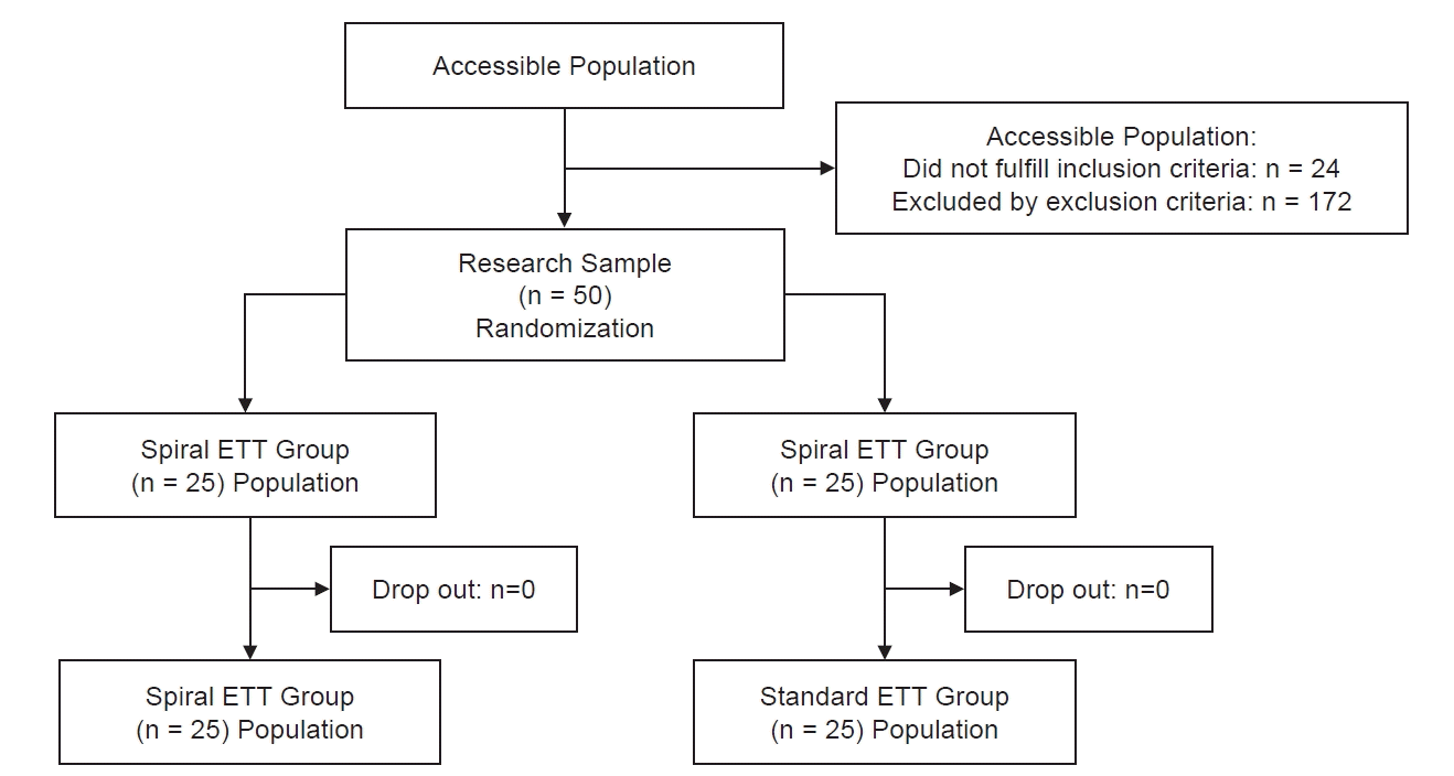
This randomized clinical trial compared the efficacy of spiral ETT versus no-stylet ETT/standard ETT using the McGrathTM VL. We included 50 children aged one month to six years old with an American Society of Anesthesiologists physical status of I-III, scheduled for elective surgery under general anesthesia with endotracheal intubation at the institution. We excluded patients who were already intubated or had a tracheostomy or were suspected of having a foreign body or mass in the airway, abdominal distension, obesity, craniomaxillofacial disorder, respiratory problems, or a history of difficult intubation. Patients were also excluded if they had congenital anomalies that could compromise the airway, including cleft lip and palate, or a history of hypersensitivity to the standard induction drugs specified in the study protocol.
Samples were selected using a consecutive sampling method. Randomization was performed by a research assistant when the patient arrived at the operating theatre using a computer-based block-permuted randomization technique. The subjects were assigned to two blocks/groups: spiral ETT or no-stylet ETT, with an allocation ratio of 1:1. The randomization results were written on paper, inserted into an envelope, and opened before the anesthesia induction process was started.
ETT preparation
The anesthesia team in the OR chose the size of the ETT appropriate for the patient using Cole’s formula [(Age/4) + 4] [
11]. The research team prepared the ETT according to the allocation group described in the envelope. Standard anesthesia was then induced. Spiral ETT was performed manually by inserting a soft stylet inside the cuff ETT (Rusch). Spiral formation was performed using both hands, with the right hand at the end of the ETT connector and the left hand at the base of the ETT, starting with a clockwise rotation from the end of the connector to the base of the ETT, adjusted with the laryngoscope. The ETT was rotated 90° using a tool developed by the researcher to achieve the same angulation as each spiral ETT (
Fig. 1). There was no noticeable difference in the number of spirals for the different tube sizes. The spiral was maintained in the shape of the letter S, and not in a coil shape. The procedure was performed without touching the distal three-quarters of the ETT by keeping the part inside the plastic to keep the ETT clean. The ETT was constructed before anesthesia induction and given to the intubator immediately before intubation.
Intubation and data collection
Anesthesia induction procedures were performed according to standard hospital procedures, followed by laryngoscopy and endotracheal intubation. Four second-year anesthesia residents performed the intubation procedure during the pediatric anesthesia rotation. They were familiar with using the McGrath
TM VL and inserting an ETT with or without a stylet. All residents had similar experiences using the McGrath
TM VL. Second-year residents learned about pediatric airway management during pediatric anesthesia rotations. In this case, we expected that they would not be experts in endotracheal intubation of children. Laryngoscopy using McGrath
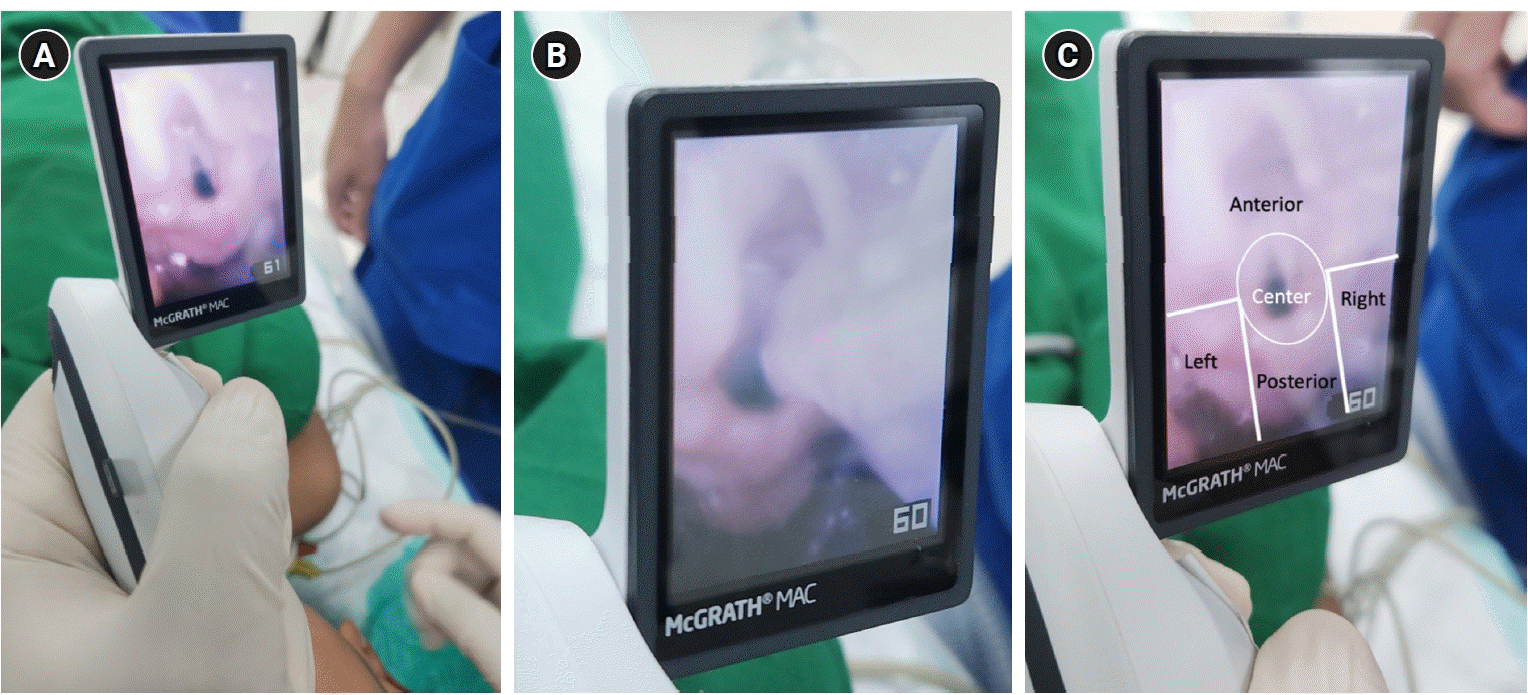
TM VL was performed until the best view of the larynx was obtained (Cormack-Lehane 1 or 2). Two researchers observed the view from the VL during laryngoscopy and endotracheal intubation, and the ETT position was declared when approaching the larynx (
Fig. 2). The intubation process was recorded using video to obtain accurate time measurements. ETT placement was confirmed using VL visualization, followed by symmetrical breath sounds of the lung, and finally by the presence of normal end-tidal carbon dioxide waves. Each confirmation method was performed by the same residents to reduce the time variation between methods. Any difficulties encountered during the intubation process that could cause the subject to fall into the dropout criteria was handled, and appropriate action was taken by alerting the difficult airway team when the intubator encountered two failed intubation attempts with the VL.
Total tube handling time was defined as the time (in seconds) after the larynx was well-visualized with the VL until all appropriate parts of the ETT entered the larynx, meaning that the tip of the ETT was inserted into the trachea until it reached the black line on the ETT. Total intubation time was defined as the time (in seconds) after the laryngoscope touched the lips until confirmation of ETT placement by capnography. Time was assessed using a stopwatch and recorded to a precision of one decimal place. ETT placement accuracy involved placing the ETT tip in the glottic rim area. An independent anesthetist assessed the tube placement accuracy. The subjects were assessed for the presence of tube-related airway trauma, post-extubation hoarseness, and complications.
Statistics
The data were verified and processed using SPSS Version 21.0 (IBM Co.). The sample size was calculated using the following formula (statistical superiority design with the primary outcome measure as a continuous variable) [
12]:
Where,
N = size per group
z = the standard normal deviation of α and β (derived from 1 - power of 80%). Hence, α = 0.05,
β = 0.2
δ = a clinically acceptable margin = 2.8 seconds (from Min et al. [
10] study)
S= polled standard deviation of both groups = 5
Thus, this clinical trial required a minimum of 50 participants.
Data were processed using descriptive and analytical methods. Analysis was performed according to the manufacturer’s protocol. Data normality was tested using the Kolmogorov-Smirnov test (normally distributed with a P value > 0.05). Categorical data were analyzed using the Chi-square or Fisher’s exact test, while numerical data were analyzed using an independent t-test or Mann-Whitney test (if the data distribution was not normal). Results were considered significant if the P value was < 0.05.
DISCUSSION
Our study found that intubation using a spiral ETT resulted in a significantly shorter mean total tube handling time than using the standard ETT. However, no statistically significant differences were found in the mean total intubation time or successful first-attempt intubation rate between the spiral and standard ETT. Spiral ETT demonstrated a significantly higher central placement accuracy than standard ETT. No adverse effects were observed after intubation with either ETT type. The use of a spiral ETT was first reported in a case report by Lillie et al. [
1], who utilized a spiral ETT to intubate a 40-week-old child with a suspected Pierre-Robin sequence. The patient had a small mandible and large tongue, causing altered respiratory distress. A spiral ETT was used after three failed intubation attempts using a Glidescope and standard ETT. The spiral ETT provided a sufficient angle to pass through the inlet of the anterior vocal cord and avoid contact with the glottis [
1]. The angle of the ETT tip was adjusted to the angle of the blade used, as in the standard ETT. The spiral ETT provided a sufficient angle to position the ETT in the center of the upper laryngeal inlet [
10]. We used the no-stylet ETT as the control group because this is the standard practice in our institution, particularly for non-anesthesiologists.
In this study, the spiral ETT group had a younger median age and higher mean body weight than the standard ETT group. Nikhar et al. [
13] reported that intubation predictors were most influenced by height (P = 0. 001), followed by age (P = 0.04). Lower height and age were associated with lower geniohyoid, thyromental, and sternomental distances, leading to increased intubation difficulty. A study by Heinrich et al. [
14] on 11,219 children showed that younger age was associated with a higher Mallampati score (III/IV), in which the Mallampati III/IV group was significantly associated with difficult intubation (P < 0.001). In addition, American Society of Anesthesiologists class III/IV and low body mass index were significantly associated with difficult intubation (P < 0.001) [
14].
This study found a significant relationship when comparing the average total tube handling time between the spiral and standard ETT, where the spiral ETT showed a shorter average time than the standard ETT. Similar results were shown in a study on 86 infants and neonates in South Korea by Min et al. [
10]; the average total tube handling time in the spiral ETT group was significantly different from the standard ETT group (15.4 ± 4.7 vs. 18.2 ± 5.3 seconds, P = 0.012). However, a three to five-second duration was not clinically significant in reducing complications during endotracheal intubation in children. In contrast, the total intubation time showed no statistically significant results between the two types of ETT groups, although the average spiral ETT was shorter than the standard ETT (46.5 ± 5.2 vs. 48.4 ± 4.9 seconds; t = 1.3; P = 0.205). To date, no study has compared the total intubation time of spiral ETT with that of other types of ETT. A meta-analysis by O'Shea et al. [
9], which compared the orotracheal intubation process using an ETT with and without a stylet in infants, showed that there was no significant difference between stylet use and total intubation time (P = 0.23), where the median total duration time in the ETT group with a stylet was 43 seconds (IQR 30–60 seconds), and the ETT group without a stylet was 38 seconds (IQR 27–57 seconds). Another study by Omur et al. [
6] comparing intubation procedures with various types of stylets and without stylets on mannequins showed that the total intubation time with various types of stylets showed significantly different results (P = 0.009), where the shortest total intubation time was obtained in the ETT group with a D-blade type stylet (37.4 ± 13.3 seconds) and the longest total intubation time was identified in the ETT group without a stylet (55.0 ± 19.3 seconds).
A difference in significance between total intubation time and total tube handling time is possible, although by definition, total tube handling time is one of the components of total intubation time. This significant difference is thought to result from the lower strength of significance (P = 0.049); therefore, a slight difference in time outside the total tube handling time can cause a decrease in the significance of total intubation time to the point of insignificance. One factor that can cause differences in time outside of the total tube handling time is operator difference. Operator difference was defined as the difference in the ability of each operator to perform intubation, including the processes of laryngoscopy and ETT insertion. For example, some operators tended to slowly visualize the glottis with a laryngoscope, leading to a longer total intubation time, but could insert an ETT quickly, which contributed to a shorter total tube handling time, and vice versa.
Successful intubation in one attempt is associated with a reduced risk of complications during intubation and reduced time needed for intubation, which, in turn, reduces the exposure time of health workers to potential pathogens [
15]. The relationship between the number of attempts and ETT type was not significant (P = 0.208). However, the data revealed that the percentage of successful first-attempt intubations in the spiral ETT group was greater than that in the standard ETT group (80% vs. 64%). The results showed that the use of a spiral ETT increased the percentage of successful first-attempt ET intubations. Min et al. [
10] also demonstrated similar results, where the ETT type was not significantly associated with one intubation attempt, with the percentage of one attempt in the spiral ETT group being greater than that in the standard ETT group (100% vs. 98%).
In this study, spiral ETT was significantly associated with greater central placement accuracy than standard ETT (84% vs. 52%; P = 0.015). A study by Min et al. [
10] also demonstrated similar results; spiral ETT had a significantly higher central placement accuracy than standard ETT (88% vs. 47%; P < 0.001). In intubation with standard ETT, off-center placement of the ETT tip indicated contact with the perilaryngeal region in 48% of the subjects, and most of the contact occurred on the right side of the perineal region, followed by the posterior and anterior regions [
10]. The presence of such contact may cause swelling of the tissues around the larynx, which may prolong intubation time [
10]. Similarly, Lilie, et al. [
1] reported that twisting the stylet into a spiral shape may improve the maneuverability of the ETT and its central placement. In comparison, a meta-analysis by O'Shea et al. [
9] showed that local trauma to the tissues around the endotracheal region, which suggests contact between the ETT and the area around the larynx, was not significantly different between the two groups (10% with a stylet vs. 13% without a stylet; P = 0.49). Thus, although not significant, intubation with a stylet can reduce the risk of complications [
9]. We realize that gentle placement of an ETT without a stylet was unlikely to induce trauma. However, this becomes a significant concern when the intubation procedure is performed in a complex environment, such as an emergency department, and the operator is not an expert in ET intubation in children. Various adverse effects can occur during and after intubation, including dental trauma, lip laceration, aspiration, mucosal bleeding around the larynx, pneumothorax, and hoarseness [
16]. In this study, the side effects assessed were airway trauma (oropharynx, dental, croup) and desaturation to less than 90%. Our study found no adverse events in patients who underwent spiral or standard ETT intubation. Similarly, Jaber et al. [
15] showed that the use of stylets in ETT was not significantly associated with intubation-related complications; the percentage of complications in ETT with stylets was lower than that in ETT without stylets (38.7% vs. 40.2%; P = 0.64). In the same study, the incidence of traumatic injury in both types of ETT groups was not significantly related, with the incidence in the ETT group with a stylet being greater than that in the ETT without a stylet (4% vs. 3.6%; P = 0.76) [
15].
Several limitations are identified in this research. First, this study was not blinded; even though the research team tried to use recorded videos to assess time and accuracy, real blinding could not be carried out completely. In addition, intubation was not performed by a single surgeon. This could lead to the possibility of bias in the intubation procedures performed owing to differences in abilities between operators, even though their educational levels were the same. This difference in operators is likely to cause an insignificant comparison between the total intubation time and the type of ETT used. Third, this study did not use a conventional angled stylet, which is commonly used in practice, for comparison. Thus, the reason for the lack of a difference in the total intubation time between the spiral ETT and no-stylet groups remains unclear, as it is difficult to differentiate whether this is due to operator variability or the time required to remove the stylet. Fourth, this study was performed in pediatric patients with normal airways, which could mask the benefits of using a spiral ETT in difficult intubation cases. The use of the spiral ETT may not be generalizable to other intubation devices, including conventional laryngoscopes or other types of video laryngoscopes, which may require further study. Overall, this study showed that the total intubation time between spiral and no-stylet ETTs differed by approximately two seconds; however, the clinical relevance of this result could be challenged, as there was no clinically meaningful time difference between the two techniques. However, this study emphasizes that the spiral ETT can be used as an alternative to decrease intubation time while preventing airway trauma in children under 6 years of age.
In conclusion, this study demonstrates the effectiveness of using a spiral ETT for reducing total tube handling time compared to a standard ETT (without a stylet) in children aged one month to six years old using VL. Using a spiral ETT also provides a higher accuracy of ETT placement at the larynx inlet than the standard ETT without a stylet, thereby decreasing the possibility of airway trauma. However, the spiral ETT did not significantly increase the first attempt at a successful intubation rate or reduce the total intubation time in children aged one month to six years old.


 PDF
PDF Citation
 Print
Print


 XML Download