Journal List > Anesth Pain Med > v.19(Suppl 1) > 1516088827

Ozgen: Neuromuscular blockade monitoring in pediatric patients

Abstract

Neuromuscular blocking agents (NMBAs), a cornerstone of pediatric anesthesia, facilitate intubation and muscle relaxation during surgery. However, NMBAs can also lead to serious complications including respiratory depression, residual paralysis, and prolonged recovery. Neuromuscular monitoring (NMM) in pediatric patients is therefore essential, as it is in adults, to ensure adequate paralysis during surgery and prompt recovery from NMBA-induced paralysis. This article aimed to provide a comprehensive overview of NMM in pediatric anesthesia including the various available methods, their advantages and disadvantages, and the importance of a standardized NMM approach.

INTRODUCTION

Neuromuscular blocking agents (NMBAs) are essential tools in anesthesia, improving surgical exposure, facilitating intubation, and reducing airway complications to optimize conditions under general anesthesia [1]. However, inadequate reversal or excessive neuromuscular blockade (NMB) can lead to serious complications including respiratory insufficiency, aspiration pneumonia, prolonged hospital stay, and increased hospitalization costs [2,3]
Neuromuscular monitoring (NMM) is crucial in assessing neuromuscular function and the depth of NMB in pediatric surgical patients. Accurate monitoring is vital for ensuring patient safety, optimizing anesthesia management, and minimizing complications. However, in the pediatric anesthesia literature, data regarding the principles, guidelines, techniques, and considerations for the optimal means of quantitative monitoring of train-of-four (TOF) in children, the incidence of residual NMB, and its impact on pediatric postoperative outcomes are scarce [4-7]. This review aimed to discuss the various methods available for monitoring NMB, the advantages and disadvantages of each method, and the importance of using a standardized NMM approach to ensure patient safety.
Despite the vast literature recommending the routine use of NMM [8,9], its use is inconsistent among clinicians in clinical practice. This may be due to an overconfidence in current anesthetic practices, a preference for intuition, or the inability to correctly interpret NMM results. In an international survey of more than 2500 anesthesiologists, 20% Europeans, 10% Americans, and 0% New Zealand and Australian anesthetists routinely used NMM. Moreover, 83% of the respondents stated that they had never used NMM devices and 72% stated that they had no NMM equipment. Furthermore, only 33% routinely administer NMBA reversal agents [9]. Some clinicians find the routine monitoring of NMB unnecessary. While the lack of knowledge or equipment, and the inability to use the available equipment are other common causes of ‘keeping the NMM in the drawer of the anesthesia machine,’ some anesthetists did not trust the equipment, and in some centers, quantitative monitors are rarely available. Additionally, older devices require calibration and are impractical and difficult to use [9].

OBJECTIVES AND BENEFITS OF NEUROMUSCULAR MONITORING IN PEDIATRIC PATIENTS

In pediatric anesthesia, NMM is essential to achieve the following five main goals throughout the course of anesthesia whenever NMBAs are used:
1. Ensuring sufficient NMB for successful intubation and adequate exposure during surgical procedures. NMM applied before intubation and throughout surgery ensures sufficient NMB is maintained to facilitate ideal intubation conditions, alleviate coughing or movement, and optimize surgical exposure by muscle relaxation [10].
2. Monitoring adequate NMB reversal can reduce the risk of respiratory complications. Proper NMB reversal on completion of surgery is critical for preventing residual NMB and its associated complications. Residual NMB can cause respiratory insufficiency and aspiration pneumonia. These complications may be more detrimental in pediatric patients because of their fragile respiratory physiology [3].
3. Avoiding unnecessary NMB by continuous monitoring allows for the titration of NMBAs, thus, preventing excessive blockade and its associated risks.
4. Guiding the administration of reversal agents to ensure prompt recovery from NMB. Residual NMB can cause muscle weakness, discomfort, and anxiety, which may be more pronounced in pediatric patients who cannot verbalize or define the problem. Adequate and effective reversals also lead to shorter recovery times. Residual NMB can prolong recovery and increase the need for postoperative ventilation. Residual NMB reversal is paramount in young pediatric patients, especially in infants in whom signs may not be noticed [3,11].
5. Providing safe and reliable criteria for extubation readiness. NMB should be adequately reversed to ensure spontaneous respiration and prevent residual neuromuscular weakness.
To ensure safe practice, anesthetists rely on state-of-the-art technologies and confirm vital parameters through monitoring, as well as their clinical expertise to maintain patient homeostasis, and NMM should be considered an essential component of anesthetic management [11]. The administration of an NMB antagonist, such as sugammadex or neostigmine, waiting for several minutes, examining the clinical signs, and then, extubating without confirming adequate recovery is similar to administering vasoactive drugs and assessing their efficacy only by palpating an artery for a powerful pulse, without rechecking the blood pressure and confirming success. Using a tactile method, such as palpating for a thumb response to a TOF stimulation with a peripheral nerve stimulator (PNS), and subjectively determining whether adequate recovery has been achieved is comparable to palpating the carotid artery during volume resuscitation.

NEUROMUSCULAR MONITORING TECHNIQUES IN PEDIATRIC PATIENTS

The three primary methods of NMM include clinical signs, qualitative evaluation (nerve stimulators), and quantitative techniques.

Clinical signs

Clinical signs of NMB include muscle weakness, loss of muscle tone, and fasciculation. Clinical signs at the end of NMB include a head lift of 5 s, grip strength, leg lift of 5 s, biting, maximum inspiratory pressure > 50 cmH2O, and hip elevation in infants [11]. However, these signs are subjective and may be unreliable, particularly in pediatric patients. The protective airway reflexes may still be impaired despite positive clinical signs of NMB reversal. Hence, NMM must consider neuromuscular recovery that allows the patient to breathe spontaneously [11].
The safest way to determine the reversal of neuromuscular transmission is to monitor NMB. Monitoring the depth of NMB can be performed using either qualitative or quantitative nerve stimulation [2].

Types of nerve stimulation

Using qualitative monitoring, the indirectly evoked muscle response to nerve stimulation by a PNS is evaluated visually or by sensations. Nerve stimulators provide an objective and accurate assessment of NMB [2,12]. They work by stimulating a peripheral nerve and measuring the resulting muscle response.
The most common types of nerve stimulators include:

1. Single twitch

This nerve stimulator has a supramaximal stimulus frequency of 0.1–1.0 Hz. Its clinical value is limited. However, it may be useful for baseline assessment of neuromuscular function before NMBA administration. A single twitch was used to determine the time to the onset of neuromuscular blockade or to assess the status of the depolarizing NMB.

2. TOF stimulation

This is the gold standard for NMB monitoring. TOF provides information about the depth of NMB and the degree of recovery after reversal. TOF stimulation involves delivering a train of four electrical stimuli to a peripheral nerve, observing the evoked muscle responses, and measuring the amplitude of the resulting muscle twitch. The stimulation pattern consists of 4 twitches at a frequency of 2 Hz. A stimulation-free interval of at least 10 s is allowed between successive TOF stimulation patterns. The loss of the fourth response is indicative of 75–80% NMB. Sufficient NMB for surgical procedures is maintained until the reappearance of 2–4 responses. TOF count is the number of twitches elicited before fading, whereas TOF ratio (TOFR) is the ratio of the fourth to the first twitch. A TOF count of 0 indicates a deep blockade. A TOFR of 1 indicates full recovery, a TOFR of 0.7 is suggestive of adequate diaphragmatic recovery, a TOFR of > 0.9 indicates adequate pharyngeal muscle function, and a TOFR of 0.8 or less indicates the need for reversal [12]. A TOFR of 0.7 at the adductor pollicis muscle demonstrates satisfactory recovery of neuromuscular function. However, this NMB level is associated with the subjective symptoms of profound weakness [13] and objective signs of upper respiratory muscle and swallowing dysfunction [14,15]. The current standard for acceptable strength recovery is a TOFR of at least 0.90 in the adductor pollicis muscle. Responses to TOF stimulation may be evaluated subjectively (qualitatively) with direct visual observation and counting of muscle twitches following stimulation with a PNS. It can also be assessed objectively (quantitatively) by measuring the number of twitches in the TOF sequence (TOF count or TOFC), or by measuring the twitch height of the first TOF twitch (T1), the height of the 4th twitch (T4), and calculating the T4/T1 ratio or TOFR (Table 1) [2].
Monitoring the TOF response following the administration of NMBAs remains essential in guiding the timing of redosing and evaluating the efficacy of NMB reversal [16,17].

3. Tetanic stimulation (TS)

High-frequency (50–200 Hz) stimulation is applied for 5 s. A fading effect is observed with incomplete NMB recovery. The sensitivity and specificity of TS in detecting residual curarization are 70% and 50%, respectively [12].

4. Post-tetanic count (PTC)

This technique is used to assess deep NMB when TOF responses are absent. In other words, it is a tactile and visual evaluation of deep non-depolarizing NMB that does not respond to TOF. PTC involves the application of a tetanic stimulus, followed by a single nerve stimulus [12]. TS of 50 Hz for 5 s followed by 1 Hz supramaximal stimulation after a gap of 3s, is applied. The number of twitches elicited after a tetanic stimulus indicates the degree of NMB. Ideally, the PTC should be zero if a very deep NMB is desired. When the return of TOF is imminent, 5–7 responses are detectable.

5. Double burst stimulation (DBS)

This method is more sensitive than TOF stimulation for detecting residual NMB. DBS involves delivering two bursts of electrical stimuli at 50 Hz with an interval of 750 ms to a peripheral nerve and observing the evoked muscle responses. Bursts consist of 2–3 impulses, combined with impulse series 3/3 and 3/2. Fading of the second impulse series compared to the first one indicates incomplete NM recovery and is comparable to a TOF of < 0.6. DBS is more sensitive for the tactile evaluation of residual blockade [12].
TOF and PTC are the most useful methods for detecting deep and moderate NMBs (Table 1 and Fig. 1).

Quantitative NMM

Postoperative residual blockade is observed at TOF < 0.9 in 10–28% of patients [8], and it is suggested that when NMBAs are used, quantitative NMM is mandatory to optimize intubation time, monitor intraoperative muscle relaxation, determine adequate pharmacologic reversal agents, and reduce postoperative residual paralysis. With quantitative monitoring, the evaluation of the indirectly evoked muscle response is done by a device that displays the TOFR in ‘real-time.’ Quantitative monitoring may employ different technologies including mechanomyography (MMG), electromyography (EMG) [18], acceleromyography (AMG) [19] and kinemyography (KMG). Given the potential for inaccuracies in visual inspection when using qualitative monitoring, the use of quantitative devices during the intraoperative care of adults has increased [4,20-24].

1. EMG

EMG measures the electrical activity of muscles and provides a real-time assessment of NMB. Unlike TOF stimulation, EMG provides continuous monitoring of NMB, allowing for the real-time assessment of muscle relaxation. However, EMG requires specialized equipment and expertise, making it less widely used than TOF stimulation [21-23].

2. MMG

Although not commercially available, every new device is compared to MMG. MMG is the gold standard of historical importance and requires careful calibration [21].

3. AMG

The TOF-Watch (Organon) was the first device to be widely used in clinical practice. It has been evaluated against the ‘gold standard’ MMG. However, it is no longer commercially available. AMG measures the acceleration of muscle movement, providing a more objective NMB assessment than qualitative TOF stimulation using nerve stimulators. AMG is the most widely used NMM, with the de facto standard of clinical care, is easy to handle, and is suitable for any free-moving muscle. AMG objectively measures the response to neurostimulation using a transducer fixed to the muscle of interest. Traditionally, standard electrocardiogram (ECG) electrodes are placed over the ulnar nerve and acceleration of the adductor pollicis muscle is measured. This configuration is very similar to employing a PNS in the hand, except for the additional transducer fixed to the thumb, foot (flexor hallucis brevis), and face (orbicularis oculi/corrugator supercilii), although not preferred or recommended [4,12,21,24-26].
For accurate measurements, the thumb must be allowed to move freely, and when the arms are placed under surgical drapes and are inaccessible during surgery, the NMM may not be accurate. Two or three serial TOF measurements at 15-s intervals should always be taken before deciding on an action (e.g., need for top-up doses, determining block level, and ensuring recovery after reversal).
Unlike the MMG and EMG, the baseline TOF ratio often exceeds 1.0 (100%), with some values reaching as high as 1.4 (140%). This is known as ‘reverse fade,’ and its cause is not fully understood, but it might be due to the thumb’s elastic recoil not going back to the baseline after each TOF stimulus. Therefore, AMG devices must be set to a baseline (supramaximal) value before an NMB and adjusted to reach a target recovery ratio of 0.9. For instance, if the baseline value is 1.2, a recovery ratio of 0.9 would need a measurement of 1.08. To avoid complications after surgery, recovery to at least 0.95 is necessary with AMG, and the European Society of Anesthesiology and Intensive Care (ESAIC) guidelines advise aiming for 1.0 when using uncalibrated ratios. Newer AMG devices have improved accuracy as they utilize sensors that measure movement in three directions, accounting for thumb movements in response to ulnar nerve stimulation. Some manufacturers assert that their products do not require calibration. In practical terms, the TOFR should be close to 1.0 when using AMG acknowledging that even with this level of recovery, the majority (> 75%) of postsynaptic receptors may remain blocked. Nevertheless, AMG monitors are readily accessible, easy to apply and operate. Importantly, a substantial body of evidence has demonstrated that they outperform clinical and qualitative recovery methods based on PNS [4].
With AMG, the TOFR overestimation is at least 0.15, with a baseline TOFR of > 1.0. Classic AMG options include the TOF-WatchR (Organon), Infinity Trident NMT PodR (Drager), 3D AMGs like TOFscanR (Drager Technologies), Stimpod NMS 450R (Xavant Technology), and Mindray NM transmission transducerR (Mindray Co.) [21,25,26].

4. KMG

KMG is closely related to AMG as a monitoring modality. KMG monitoring includes a piezoelectric sensor placed in the groove between the thumb and index finger, which bends when the APM contracts following ulnar nerve stimulation. Although it is easy to use, it is only used for the ulnar nerve adductor pollicis muscle group. Additionally, free thumb movement and good strip placement between the fingers are required [20].
Previous reports demonstrated wide limits of agreement between MMG and KMG. Similar to the AMG, the KMG is dependent on the ability of the thumb to move freely during surgical positioning. Patient movement during emergence can also affect KMG monitoring, as does the repositioning of the sensor over the course of the perioperative period. Currently, the only available KMG-based device is incorporated into anesthesia workstations (e.g., Datex Ohmeda NMTR) [20,21,25].
The advantages and disadvantages of the quantitative monitoring methods are summarized in Table 2.
Quantitative TOF monitoring has not been used widely in infants and children primarily because of the lack of effective equipment. In a recent study, a novel commercially available EMG-based monitor known as the TetraGraph™ (Senzime AB) and a novel pediatric array known as the TetraSens™ (Senzime AB) Pediatric, with a pediatric-sized self-adhesive sensor, had been assessed (Fig. 2) [25,27,28]. The TetraGraph™ is a commercially available EMG-based TOF monitor, which provides electrical stimulation over a peripheral nerve, and then, directly measures the amplitude (muscle action potential) of the evoked responses of the innervated muscle, providing a quantitative measurement of the muscle response to the stimulus. The measurement and quantification of the EMG response eliminates the subjective evaluation required for visual observation of twitches. Clinical studies have also demonstrated that quantitative measurements using the TetraGraph provide a more sensitive and reproducible measure of the degree of NMB and recovery than visual measurements using a PNS [24,29]. The TetraSensTM sensor is indicated for pediatric patients younger than 8 years of age. It is able to monitor EMG-based neuromuscular transmissions in infants and children in whom AMG and/or visual observations of the TOF response may not be possible (Fig. 2) Automatic detection of neuromuscular stimulating parameters, such as supramaximal current intensity level and baseline amplitude of the muscle action potential, is possible in pediatric patients including those weighing less than 15 kg or in instances where there is limited access to the extremity being monitored [28]. The application of this novel sensor in infants and children weighing less than 10-15 kg has led to consistently successful NMM, which was previously deemed not possible. However, a difference in the recovered amplitude should be considered (the recovered amplitude is statistically less in patients ≤ 15 kg). This may be related to differences in the maturation of neuromuscular transmission [29], which has been previously noted when evaluating TOF using an AMG with a force displacement transducer [20].
Quantitative NMB monitors, such as the Tetragraph™ (Senzime AB), Twitchview (Seattle), and TOFScanR (Drager Technologies), are able to display PTC and TOFR in real-time. When TOF is zero, AutoPTC is activated. TOF-Watch SX (Organon) is as accurate as TOFScan when calibrated but may overestimate the TOFR. In contrast, the TOFScan is 3D, easily understandable and no calibration is required [30].
As previously mentioned, AMG necessitates that the target muscle (typically the thumb) move freely for response measurement or be visible for subjective assessment. This requirement may limit its use in procedures in which the patient’s arms are restricted by surgical drapes or are not freely visible. The risk of postoperative residual NMB has been shown to be 5-fold higher when a PNS and the supramaximal current are not determined [31]. Notably, with AMG NMM, the baseline TOFR may be > 100% [32-34]. Considering this falsely elevated baseline, the recovering TOFR must be normalized to the starting baseline ratio to avoid inaccuracies when assessing NMBA reversal and the degree of residual NMB. This inaccuracy has not been observed with EMG-based technologies like the TetraGraph™. When compared to visual monitoring, the TetraGraph™ evaluates fade by calculating the TOFR, thus allowing for intraoperative titration of additional NMBA doses, as well as documentation of effective antagonism (TOFR > 0.90). This aims to prevent residual paralysis and its clinical consequences [10,11]. Although potential or residual NMB should be assessed in the post-anesthesia care unit (PACU), its clinical impact has not been evaluated in studies thus far, as it is very difficult, and possibly ethically questionable, to apply NMM in awake patients.
Most pediatric pharmacokinetic and pharmacodynamic studies conducted before the mid-1990s utilized either MMG (Grass FT-03 or Grass FT-10 force–displacement transducer, Grass Instrument Co.) or EMG (Relaxograph, Datex). MMG is still considered the research standard for NMM. However, recent evidence has demonstrated that EMG correlates well with MMG, and is reliable, reproducible, and easier to use in clinical research [4,23]. Importantly, MMGs are no longer manufactured for clinical use. However, reference MMG-based monitors are available for validating new EMG-based monitors [23]. AMG, such as the TOF-Watch SX (Organon), has been used in pediatric studies. The TOF-Watch was the first device widely used in clinical practice. It has been evaluated against the ‘gold standard’ (MMG). AMG use in pediatric patients poses similar limitations as in the adult population including TOFR overestimation (inverse fade) when compared with MMG and EMG and the “staircase” phenomenon associated with ST stimulation [35]. AMG monitors also require a significant amount of time (5–10 min) for stabilization of the baseline signal, although a 5 s tetanic stimulation may prevent the staircase phenomenon and shorten the signal stabilization requirement [29]. Neonates and infants, aged less than 1 year, may require a manual increase in the gain of the transducer to obtain a first twitch in the TOF sequence (T1) of 100% [25].
Recent reports have described the successful use of EMG in pediatric patients [36-38]. There currently are no studies that have determined the supramaximal current requirements as a function of age, but in pediatric patients weighing 20–60 kg, the current intensity at a pulse width of 0.2 ms was 30 mA in 3%; 40 mA in 42%; 50 mA in 28%; and 60 mA in 22% of patients, while the average baseline amplitude of the response was 7.5 mV [38]. Minimum current amplitudes necessary for the visual detection of contractions are approximately 20 mA (with a range of 10–45 mA), which is similar to those in adults [39]. The small sizes of pediatric patients impose technical challenges regarding appropriate equipment. Lead placement and electrode size may affect the current density and, therefore, the required charge for a supramaximal response. Temperature changes can also affect the EMG electrical signal amplitude, but to a lesser extent than when using MMG or AMG [4].

STANDARDIZED APPROACH TO NMM IN PEDIATRIC PATIENTS

A standardized approach is essential to ensure consistent and reliable NMM in all patients [4]. However, monitoring NMB and neuromuscular management in pediatric patients can be challenging because of several factors.

Physiological characteristics

Children have unique physiological characteristics that differentiate them from adults. The immature neuromuscular system, and varying pharmacokinetics and pharmacodynamics, require specific consideration when monitoring pediatric neuromuscular function. Pediatric patients may respond differently to NMBAs, making accurate monitoring critical. Infants (children aged 1 year and younger) are more sensitive to NMBAs because of their developing neuromuscular system [29]. Regarding the immaturity of the neuromuscular junction, premature babies, neonates, and infants less than two months of age lack muscle strength and have lower acetylcholine stores. In this specific population, there is a significant fade with increasing stimulation rates from 20 to 50 to 100 Hz. An increase in frequency does not cause an increase in post-tetanic facilitation, and a 5 s, 100 Hz tetanic stimulation generally leads to post-tetanic exhaustion [4,29]. The smaller muscle mass in pediatric patients renders it more difficult to assess muscle twitch [4,29].

Variability

Variability in drug metabolism and limited pharmacokinetic data in pediatric patients should also be considered. NMBA metabolism can vary significantly among pediatric patients, making it difficult to predict the duration of action. The pharmacokinetics of NMBAs in neonates and infants may also differ from those of NMBAs in older children and adults. Moreover, there is a relative lack of comprehensive pharmacokinetic data for this population, making precise dosing challenging. Factors, such as organ immaturity, altered protein binding, and different rates of drug metabolism and elimination contribute to the variability in drug response. Furthermore, gestational age, postnatal age, weight, and coexisting medical conditions affect the sensitivity and metabolism of these drugs. The lack of a standardized response makes it challenging to establish universal guidelines and reference values for NMM in neonates and infants [29].

Anatomical differences

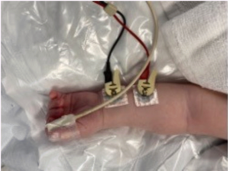
The anatomy of pediatric neuromuscular junctions may differ from that of adults, which can affect nerve stimulation responses. The placement of monitoring electrodes on a small arm, often obscured by intravenous and arterial lines, can be challenging (Fig. 3).

Clinical assessment

Clinical assessment is also challenging in infants and children because of communication difficulties and noncompliance with instructions. Approximately 28% of children may experience a postoperative residual block, which can go unnoticed due to communication difficulties [7-9]. Importantly, complications due to residual block can be detrimental given that children have smaller oxygen reserves and are more vulnerable to airway collapse [29].

The lack of standardized guidelines

Compared with adult patients, there are relatively few standardized guidelines and reference values for NMM in neonates. The limited data on the normal neonatal response to NMB agents makes it difficult to interpret the monitoring data accurately. Consequently, clinical decisions regarding the administration and reversal of these drugs often rely on expert opinions and clinical judgment.

Technical difficulties

The small size and anatomical differences of neonates present technical challenges for the accurate diagnosis of NMM. The placement of electrodes on small muscle groups, such as the adductor pollicis, can be challenging. Additionally, obtaining consistent and artifact-free signals can be difficult because of various factors, such as movement, poor skin-electrode contact, and electrical interference from other medical equipment. These technical limitations can affect the reliability and accuracy of NMM [4,29].

Influence of coexisting conditions

Neonates often have coexisting medical conditions, such as premature birth, congenital abnormalities, or neuromuscular diseases, which can influence their baseline neuromuscular function. These conditions can affect the sensitivity and response to NMBAs, thus affecting the accurate interpretation of the monitoring data. Individualized assessment and management are necessary in these cases [29].

Relevance of monitoring data

The clinical relevance of NMM in neonates remains a topic of ongoing research and debate. Although NMM is commonly used in adult patients to guide drug administration and reversal, a direct correlation between monitoring data and clinical outcomes in neonates is not well-established. The relationship between NMB and adverse respiratory events is non-linear. Furthermore, the need for prolonged postoperative ventilation in neonates is complex and influenced by multiple factors beyond the monitoring data.

Other individual differences

Individual variations in distribution volume, muscle mass, clearance of NMB agents, and age-dependent maturation of the NM junction all contribute to differences in the initial TOFR, and normalization may also adjust the inherent TOFR.
Considering these unique pediatric features, the use of neostigmin to reverse deep NMB may not lead to effective or adequate recovery, and thus, recurarization may be required [3]. Recurarization may also be seen clinically after using inadequate doses of sugammadex, and inadequate antagonism, subsequent fatigue rather than recurrence of NMB may have detrimental consequences, especially in pediatric patients. Consequently, NMM should be applied whenever NMBAs are used.
The methods to overcome the specific challenges of NMM in children and obtain the best measurements from NMM are as follows:

1. Skin preparation

Properly cleansed skin, rubbed with an abrasive, is essential prior to placing surface electrodes for supramaximal stimulation. The skin surface should also be allowed to dry completely before electrode placement [4]

2. Correct placement of stimulation electrodes

The correct placement of the electrodes is important to ensure that the nerve is stimulated with the selected current (Fig. 3). The size of the conducting area of the stimulating electrode is also important. With a large conducting area, it may be difficult or impossible to obtain supramaximal stimulation because a sufficient current density cannot be obtained in the nerve underlying the stimulating electrode. Typically, the contact area of the stimulating electrode is 7–11 mm in diameter. The distance between the centers of the two electrodes is 3–6 cm. Finally, although the polarity of the stimulating electrodes is also important, it is recommended that the negative electrode be placed distally to optimize the nerve stimulation response [3,36].
1. Pediatric-sized nerve-stimulating electrodes made of silver/silver chloride (Ag/AgCl) are utilized. The negative electrode is always distal to the positive electrode [33]. An alternative position is to place the positive electrode on the ulnar groove at the elbow to improve ulnar nerve depolarization. Electrodes are placed on the supinated palm while extended passively [4].
2. Two electrodes should be placed along the ulnar nerve [2].
3. The negative (black) electrode is positioned distal to the styloid process of the radius [2,38].
4. The arm should be fixed to an arm board to minimize muscle movements (e.g., adductor pollicis brevis).
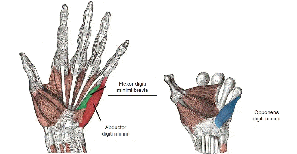
5. The adductor pollicis brevis muscle originates from two heads (oblique head from the second and third metacarpal bones, and transverse head from the third metacarpal bone) and inserts at the base of the proximal phalanx of the thumb. Contraction of the adductor pollicis brevis brings the tip of the thumb to the center of the palm. However, adjacent muscles, such as the opponens pollicis and flexor pollicis brevis can also contribute to similar movements. Due to the shallow skin-to-nerve and nerve-to-nerve distances in pediatric patients, using a standard ‘3 cm’ distance between the positive and negative electrodes may inadvertently stimulate the median nerve, which innervates the opponens pollicis and flexor pollicis brevis. This distance determines the penetration depth. With the electrodes being relatively far apart in the relatively short pediatric forearm, nearby muscles may also react. Strapping prevents the movement of only four fingers, not the thumb. The opponens pollicis and flexor pollicis brevis cannot be restricted to mere strapping. Moreover, the deep parts of the opponens pollicis and flexor pollicis brevis in 20% of the population are innervated by the ulnar nerves C8 and T1. Erroneous extra-apposition of the thumb due to the mixed effect of unwanted muscle movement is likely to occur in pediatric patients with smaller hands and a greater proximal nerve-to-nerve distance. Since this may cause unwanted acceleration, normalization is required when assessing 3D acceleration in children (Fig. 4) [38].
6. To establish a baseline NMB level before administering NMBAs, calibration should be performed before NMBA administration but not after induction.
7. Individual differences in distribution volume, muscle mass, clearance of NMBAs, and age-dependent maturation of neuromuscular junctions contribute to differences in the initial TOFR and normalization adjusts the inherent TOFR.
8. While changes in core temperature influence the pharmacodynamics and pharmacokinetics of NMBAs, variations in peripheral temperature at the NMM site can also impact the response to nerve stimulation. Neonates are highly sensitive to changes in body temperature, which can affect neuromuscular function and subsequently affect monitoring accuracy. Hypothermia can potentiate the effects of neuromuscular blocking agents, leading to increased sensitivity and prolonged blockade duration. However, hyperthermia can also result in drug resistance. Therefore, maintaining stable body temperature is crucial for reliable NMM in neonates. The temperature should be maintained constant; a central temperature above 35°C and peripheral temperature at the monitored muscle above 32°C is recommended [4,39-42].
9. For a meaningful comparison between technologies (AMG, EMG, and MMG), the AMG monitor must be calibrated. For AMG, the final recovery TOFR must be normalized to the baseline ratio unless tetanic pre-stimulation is performed [4,30].
10. To expand the validity of the TOFscan to infants and neonates, a smaller pediatric hand sensor is required [38].
11. The application of a 5-s, 50-Hz tetanic stimulation prior to calibration may prevent the staircase phenomenon and shorten the signal stabilization period [4,28].
12. A preload can be considered for neonates and infants; however, a clear description of the technique must be presented when a study is planned [4].

RECOMMENDATIONS

Several organizations, including the World Federation of Societies of Anesthesiologists (WFSA) and Société Française d'Anesthésie Réanimation (SFAR), recommend incorporating objective NMM into daily pediatric anesthesia practices [8]. NMM should be used to evaluate the effect of NMBAs. NMM should always be monitored to ensure sufficient NMB and reversal. In the first European Society of Anesthesiology and Intensive Care (ESAIC) guidelines on the perioperative management of neuromuscular block, the use of ulnar nerve stimulation and quantitative NMM at the adductor pollicis muscle to exclude residual paralysis is recommended, and sugammadex is recommended to antagonize deep, moderate, and shallow NMB induced by aminosteroidal agents (rocuronium and vecuronium). To continue the quantitative monitoring of NMB until a TOFR of more than 0.9 has been attained (starting from spontaenous recovery (i.e. TOF ratio > 0.2) with neostigmine-based reversal) is also recommended [43-46].
NMB causes residual postoperative weakness. Although quantitative monitoring may not completely prevent residual NMB, it is well established that it helps mitigate such risks. An unacceptable number of patients had suffered from respiratory distress during recovery, although 20–40% of these patients had been reversed. Pharyngeal dysfunction, increased risk for aspiration and pneumonia, acute respiratory events (hypoxemia, airway obstruction), the need for tracheal intubation, discomfort for patients and surgeons, and increased stay in PACU can be prevented by using quantitative NMM whenever a non-depolarizing muscle relaxant is used and documenting TOF in the anesthetic record is necessary.
It is better to keep things as simple as possible. The NMM should be handy, have an easy-to-understand interface, have greater acceptance among clinicians, and not have a steep learning curve for routine use in clinical practice. Acquiring the latest and most expensive quantitative monitor is unlikely to solve the problem of undetected postoperative residual NMB. Similarly, a device left in the drawers of the anesthesia machine will not help pediatric patients recover safely [45]. Anesthetists clinicians must embrace the routine use of NMM devices, which are available and practical, and has been shown to effectively monitor NMB, preventing unwanted adverse effects and detrimental complications of NMBAs in the clinical setting.

CONCLUSION

Accurate and safe monitoring of NMB is essential during pediatric anesthesia to prevent complications and improve patient outcomes. NMB should be monitored throughout the course of anesthesia using appropriate techniques, such as TOF stimulation and PTC. Healthcare providers should be familiar with the benefits, challenges, and recommendations of NMM in pediatric patients.
Failure to follow well-established guidelines for the clinical use of NMBAs is common.
What we need is not more complicated monitoring, but the application of well-established criteria. Every anesthetist should monitor NMB in pediatric patients whenever NMBA is administered, and appropriate reversal is of concern.
NMM is an indispensable tool in pediatric anesthesia, ensuring that patients receive optimal NMB for surgical procedures while minimizing the risk of complications.

Notes

FUNDING

None.

ACKNOWLEDGMENTS

Prof Jin-Tae Kim and his team for their organizing the excellent ASPA2023 Meeting.

CONFLICTS OF INTEREST

No potential conflict of interest relevant to this article was reported.

DATA AVAILABILITY STATEMENT

The datasets generated during and/or analyzed during the current study are available from the corresponding author on reasonable request.

REFERENCES

1. Duţu M, Ivaşcu R, Tudorache O, Morlova D, Stanca A, Negoiţă S, et al. Neuromuscular monitoring: an update. Rom J Anaesth Intensive Care. 2018; 25:55–60.
2. Klucka J, Kosinova M, Krikava I, Stoudek R, Toukalkova M, Stourac P. Residual neuromuscular block in paediatric anaesthesia. Br J Anaesth. 2019; 122:e1–2.
crossref
3. Cammu G, De Witte J, De Veylder J, Byttebier G, Vandeput D, Foubert L, et al. Postoperative residual paralysis in outpatients versus inpatients. Anesth Analg. 2006; 102:426–9.
crossref
4. Rodney G, Raju P, Brull SJ. Neuromuscular block management: evidence-based principles and practice. BJA Edu. 2024; 24:13–22.
crossref
5. Fuchs-Buder T, Brull SJ, Fagerlund MJ, Renew JR, Cammu G, Murphy GS, et al. Good clinical research practice (GCRP) in pharmacodynamic studies of neuromuscular blocking agents III: The 2023 Geneva revision. Acta Anaesthesiol Scand. 2023; 67:994–1017.
crossref
6. Scheffenbichler FT, Rudolph MI, Friedrich S, Althoff FC, Xu X, Spicer AC, et al. Effects of high neuromuscular blocking agent dose on post-operative respiratory complications in infants and children. Acta Anaesthesiol Scand. 2020; 64:156–67.
crossref
7. Raval AD, Uyei J, Karabis A, Bash LD, Brull SJ. Incidence of residual neuromuscular blockade and use of neuromuscular blocking agents with or without antagonists: A systematic review and meta-analysis of randomized controlled trials. J Clin Anesth. 2020; 64:109818.
crossref
8. Carvalho H, Verdonck M, Cools W, Geerts L, Forget P, Poelaert J. Forty years of neuromuscular monitoring and postoperative residual curarisation: A meta-analysis and evaluation of confidence in network meta-analysis. Br J Anaesth. 2020; 125:466–82.
crossref
9. Thilen SR, Weigel WA, Todd MM, Dutton RP, Lien CA, Grant SA, et al. 2023 American Society of Anesthesiologists practice guidelines for monitoring and antagonism of neuromuscular blockade: a report by the American Society of Anesthesiologists task force on neuromuscular blockade. Anesthesiology. 2023; 138:13–41.
crossref
10. Yang L, Yang D, Liu C, Zuo Y. Application of neuromuscular monitoring in pediatric anesthesia: a survey in China. J Perianesth Nurs. 2020; 35:658–60.e1.
11. Vanlinthout LE, Geniets B, Driessen JJ, Saldien V, Lapré R, Berghmans J, et al. Neuromuscular-blocking agents for tracheal intubation in pediatric patients (0-12 years): a systematic review and meta-analysis. Pediatr Anesth. 2020; 30:401–14.
crossref
12. Naguib M, Brull SJ, Johnson KB. Conceptual and technical insights into the basis of neuromuscular monitoring. Anaesthesia. 2017; 72 Suppl 1:16–37.
crossref
13. Murphy GS. Neuromuscular monitoring in the perioperative period. Anesth Analg. 2018; 126:464–8.
crossref
14. Kopman AF, Yee PS, Neuman GG. Relationship of the train-of-four fade ratio to clinical signs and symptoms of residual paralysis in awake volunteers. Anesthesiology. 1997; 86:765–71.
crossref
15. Eikermann M, Groeben H, Hüsing J, Peters J. Accelerometry of adductor pollicis muscle predicts recovery of respiratory function from neuromuscular blockade. Anesthesiology. 2003; 98:1333–7.
16. Eriksson LI, Sundman E, Olsson R, Nilsson L, Witt H, Ekberg O, et al. Functional assessment of the pharynx at rest and during swallowing in partially paralyzed humans: simultaneous videomanometry and mechanomyography of awake human volunteers. Anesthesiology. 1997; 87:1035–43.
17. Brull SJ, Kopman AF. Current status of neuromuscular reversal and monitoring: Challenges and opportunities. Anesthesiology. 2017; 126:173–90.
18. Viby-Mogensen J, Claudius C. Evidence-based management of neuromuscular block. Anesth Analg. 2010; 111:1–2.
crossref
19. Carter JA, Arnold R, Yate PM, Flynn PJ. Assessment of the Datex Relaxograph during anesthesia and atracurium induced neuromuscular blockade. Br J Anaesth. 1986; 58:1447–52.
20. Jensen E, Viby-Mogensen J, Bang U. The accelograph: a new neuromuscular transmission monitor. Acta Anaesthesiol Scand. 1988; 32:49–52.
crossref
21. Motamed C, Kirov K, Combes X, Duvaldestin P. Comparison between the Datex-Ohmeda M-NMT module and a force-displacement transducer for monitoring neuromuscular blockade. Eur J Anaesthesiol. 2003; 20:467–9.
crossref
22. Bowdle A, Bussey L, Michaelsen K, Jelacic S, Nair B, Togashi K, et al. Counting train-of-four twitch response: Comparison of palpation to mechanomyography, acceleromyography, and electromyography. Br J Anaesth. 2020; 124:712–7.
crossref
23. Todd MM, Hindman BJ, King BJ. The implementation of quantitative electromyographic neuromuscular monitoring in an academic anesthesia department. Anesth Analg. 2014; 119:323–31.
crossref
24. Bowdle A, Bussey L, Michaelsen K, Jelacic S, Nair B, Togashi K, et al. A comparison of a prototype electromyograph vs. a mechanomyograph and an acceleromyograph for assessment of neuromuscular blockade. Anaesthesia. 2020; 75:187–95.
crossref
25. Nemes R, Lengyel S, Nagy G, Hampton DR, Gray M, Renew JR, et al. Ipsilateral and simultaneous comparison of responses from acceleromyography and electromyography-based neuromuscular monitors. Anesthesiology. 2021; 135:597–611.
crossref
26. Driessen JJ, Robertson EN, Booij LHD. Acceleromyography in neonates and small infants: Baseline calibration and recovery of the responses after neuromuscular blockade with rocuronium. Eur J Anaesth. 2005; 22:11–5.
crossref
27. Espinal LM, Kalsotra S, Rice-Weimer J, Kitio SA, Tobias JD. Tolerance to preoperative placement of electrodes for neuromuscular monitoring using the Tetragraph™. Saudi J Anaesth. 2024; 18:205–10.
crossref
28. Renew JR, Hernandez‑Torres V, Logvinov I, Nemes R, Nagy G, Li Z, et al. Comparison of the TetraGraph and TOFscan for monitoring recovery from neuromuscular blockade in the post‑anesthesia care unit. J Clin Anesth. 2021; 71:110234.
29. Kalsotra S, Rice-Weimer J, Tobias JD. Intraoperative electromyographic monitoring in children using a novel pediatric sensor. Saudi J Anaesth. 2023; 17:378–82.
crossref
30. Goudsouzian NG. Maturation of neuromuscular transmission in the infant. Br J Anaesth. 1980; 52:205–14.
crossref
31. Unterbuchner C, Werkmann M, Ziegleder R, Kraus S, Seyfried T, Graf B, et al. Shortening of the twitch stabilization period by tetanic stimulation in acceleromyography in infants, children and young adults (STSTS-Study): a prospective randomised, controlled trial. J Clin Monit Comput. 2020; 34:1343–9.
crossref
32. Thilen SR, Hansen BE, Ramaiah R, Kent CD, Treggiari MM, Bhananker SM. Intraoperative neuromuscular monitoring site and residual paralysis. Anesthesiology. 2012; 117:964–72.
crossref
33. Bowdle A, Michaelsen K. Quantitative twitch monitoring:What works best and how do we know? Anesthesiology. 2021; 135:558–61.
34. Brull SJ, Silverman DG. Real time versus slow-motion train-of-four monitoring: A theory to explain the inaccuracy of visual assessment. Anesth Analg. 1995; 80:548–51.
35. Bowdle A, Jelacic S. Progress towards a standard of quantitative twitch monitoring. Anaesthesia. 2020; 75:1133–5.
crossref
36. Zhou ZJ, Wang X, Zheng S, Zhang XF. The characteristics of the staircase phenomenon during the period of twitch stabilization in infants in TOF mode. Paediatr Anaesth. 2013; 23:322–7.
37. Owusu-Bediako K, Munch R, Mathias J, Tobias JD. Feasibility of intraoperative quantitative neuromuscular blockade monitoring in children using electromyography. Saudi J Anaesth. 2022; 16:412–8.
crossref
38. Soffer OD, Kim A, Underwood E, Hansen A, Cornelissen L, Berde C. Neurophysiological assessment of prolonged recovery from neuromuscular blockade in the neonatal intensive care unit. Front Pediatr. 2020; 8:580.
39. Yhim HB, Jang YE, Lee JH, Kim EH, Kim JT, Kim HS. Comparison of the TOFscan and TOF-Watch SX during pediatric neuromuscular function recovery: a prospective observational study. Perioper Med (Lond). 2021; 10:45.
40. Heier T, Caldwell JE, Sessler DI, Miller RD. The effect of local surface and central cooling on adductor pollicis twitch tension during nitrous oxide/isoflurane and nitrous oxide/fentanyl anesthesia in humans. Anesthesiology. 1990; 72:807–11.
41. Eriksson LI, Lennmarken C, Jensen E, Viby-Mogensen J. Twitch tension and train-of-four ratio during prolonged neuromuscular monitoring at different peripheral temperatures. Acta Anaesthesiol Scand. 1991; 35:247–52.
42. Smith DC, Booth JV. Influence of muscle temperature and forearm position on evoked electromyography in the hand. Br J Anaesth. 1994; 72:407–10.
43. Heier T, Caldwell JE. Impact of hypothermia on the response to neuromuscular blocking drugs. Anesthesiology. 2006; 104:1070–80.
crossref
44. Radkowski P, Barańska A, Mieszkowski M, Dawidowska-Fidrych J, Podhorodecka K. Methods for clinical monitoring of neuromuscular transmission in anesthesiology: a review. Int J Gen Med. 2024; 17:9–20.
45. Habercorn S, Faulk JD. Monitoring Depth of Neuromuscular Blockade. [Internet]. 2024. [updated 2023 Apil 14; cited 2023 Sep 4]. Available from https://www.openanesthesia.org/keywords/monitoring-depth-of-neuromuscular-blockade/.
46. Fuchs-Buder T, Romero CS, Lewald H, Lamperti M, Afshari A, Hristovska AM, et al. Peri-operative management of neuromuscular blockade: A guideline from the European Society of Anaesthesiology and Intensive Care. Eur J Anaesthesiol. 2023; 40:82–94.
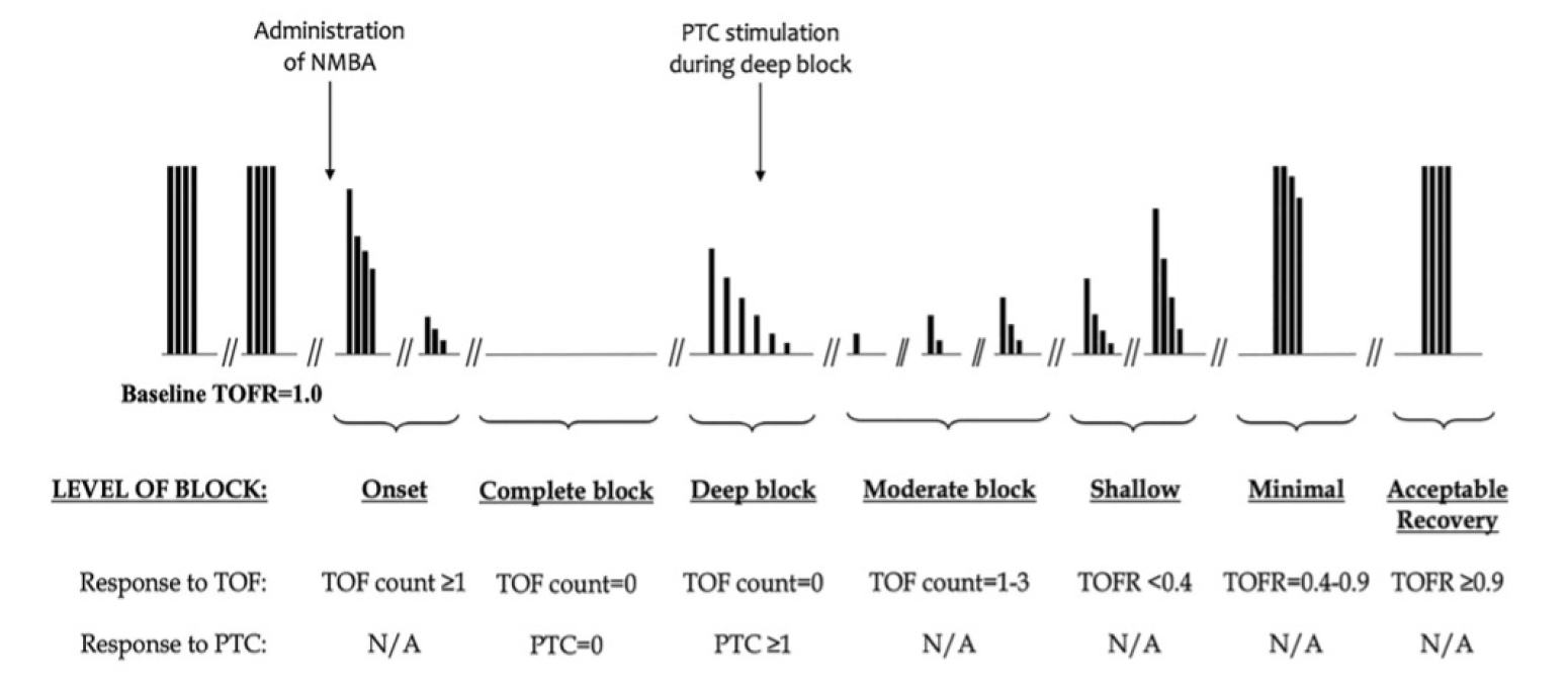
Fig. 1.
NMB after a normal intubating dose of a non-depolarizing NMBA, as classified using PTC and TOF stimulation. NBM: neuromuscular blockade, NMBA: neuromuscular blocking agents, PTC: post-tetanic count, TOF: train-of-four, N/A: non applicable. Adapted from the article of Heier and Caldwell (Anesthesiology 2006; 104: 1070-8) [43].
apm-23158f1.tif
Fig. 2.
(A) The adult and pediatric versions of the TetraSens™ self-adhesive sensor. The adult sensor measures approximately 9 inches in length compared to 7 inches for the pediatric sensor. The skin was prepped with an alcohol swab and the sensor was placed. (B) The pediatric sensor or recording electrodes (TetraSens™ Pediatric) was placed on the palmar surface of the adductor pollicis muscle and its insertion on the medial aspect of the proximal phalanx of the thumb. The stimulating electrodes were placed along the ulnar nerve on the volar surface of the forearm. After the sensor was placed on the patient, the electrodes were connected using the cord to the TetraGraph™ (Senzime AB). The device automatically determines the stimulating current necessary for maximal muscle contraction, to ensure consistent recruitment of all muscle fibers. Adapted from the article of Heier and Caldwell (Anesthesiology 2006; 104: 1070-8) [43].
apm-23158f2.tif
Fig. 3.
Traditional NMM placement in pediatric patient. NMM: neuromuscular monitoring.
apm-23158f3.tif
Fig. 4.
Anatomy of the muscles innervated by the ulnar nerve.
apm-23158f4.tif
Table 1.
NMB and Its Objective and Subjective Evaluations
Level of block Depth of block Objective measurement Subjective Evaluation
Level 5 Complete PTC = 0
Level 4 Deep PTC >1, TOFC = 0
Level 3 Moderate TOFC = 1-3
Level 2b Shallow TOFR < 0.4 TOFC = 4 & fade detected
Level 2a Minimal TOFR = 0.4-0.89 TOFC = 4 & fade not detectable
Level 1 Adequate Recovery TOFR > 0.9 Cannot be determined

NMB: neuromuscular blockade, PTC: Post-tetanic count; TOFC: Train-of-four count; TOFR: Train-of-four ratio.

Table 2.
The Advantages and Disadvantages of MMG, KMG, AMG, and EMG
Monitor Advantages Disadvantages
MMG Considered the ‘gold standard’, as results are precise and reliable. Inconvenient set-up process. Not manufactured for clinical use (research uses only).
KMG Simple set-up that does not require an external display or calibration. Can only be deployed at the adductor pollicis muscle.
AMG Can be deployed on any free-moving muscle, including locations on the hand, foot, or face. Cannot be deployed on immobilized muscles. Greatest accuracy requires device calibration prior to NMBA.
EMG Considered as accurate and reliable as MMG. Can be deployed in a variety of locations, including immobilized muscles. Subject to electrical interference. Accuracy reduced by low muscle temperature. Greatest accuracy requires device calibration prior to NMBA.

MMG: mechanomyography, KMG: kinemyography, AMG: acceleromyography, EMG: electromyography.

TOOLS
Similar articles