Journal List > Anesth Pain Med > v.19(Suppl 1) > 1516088826

Kim, Lee, Lee, Kim, Koh, Ro, and Kim: Feasibility and accuracy of pediatric core temperature measurement using an esophageal probe inserted through the gastric lumen of a second-generation supraglottic airway device: a prospective observational study

Abstract

Background

Accurate core temperature measurement in children is crucial; however, measuring esophageal temperature (TE) using a supraglottic airway device (SAD) can be challenging. Second-generation SADs, which have a gastric channel, can measure TE, and reduce gastric air volume. This study aimed to compare TE, measured using a probe inserted through the SAD gastric channel, with tympanic membrane (TTM) and forehead (TZHF) temperatures, measured using a zero-heat-flux cutaneous thermometer, with rectal temperature (TR).

Methods

Temperature was recorded at 10-min intervals from 10 min after probe insertion until completion of surgery. We performed an equivalence test to evaluate whether the TE, TTM, and TZHF were equivalent to TR, with a margin of 0.3°C. Additionally, intraclass correlation coefficients (ICC) were calculated to assess the reliability of TE and TR at each time point.

Results

We included 41 patients in the final analysis. In all patients, the esophageal probe was successfully inserted through the gastric channel of the SAD. When assessing agreement with TR as a reference, TE demonstrated equivalent results at all time points (P < 0.001 at 0, 10, 20, 30, and 40-min intervals and P = 0.018 at the 50-min interval), except at the completion of surgery (P = 0.697). TE also demonstrated good reliability with TR as a reference throughout the surgery (ICC > 0.75).

Conclusions

In children with SAD insertion, TE can be accurately and feasibly measured through the SAD’s gastric channel, making it suitable for routine application.

INTRODUCTION

Inadvertent perioperative hypothermia is relatively common in patients undergoing general anesthesia and may significantly impact patient outcomes adversely [1]. The risk of perioperative hypothermia is higher in pediatric patients than in adult patients, owing to their proportionately larger body surface area and impaired physiological compensatory mechanisms [2,3]. Adequate core temperature measurement is a prerequisite for detecting and controlling core temperature in pediatric patients during surgery.
Core temperature can be measured at various sites using different techniques. The gold standard for thermoregulation in pediatric patients remains rectal temperature (TR) measurement [4]. However, this technique can cause complications such as rectal perforation, increased blood pressure, and decreased partial pressure of oxygen [5]. As an alternative, esophageal temperature (TE), which allows continuous monitoring with minimal adverse effects, is commonly employed in pediatric patients.
The supraglottic airway device (SAD) is widely used in patients undergoing general anesthesia. However, its use makes measuring TE challenging because it fills the oral cavity. Most second-generation SADs, including AuraGain, i-Gel, Supreme, and ProSeal laryngeal mask airways, which are commonly used nowadays, have a gastric channel that facilitates access to the esophagus and stomach [6]. The gastric channel provides a passage for TE probe insertion, enabling gastric decompression through removal of air in the stomach resulting from mask ventilation.
In this study, we aimed to assess the feasibility and accuracy of core temperature measurement using a TE probe inserted through the gastric lumen of a second-generation SAD in pediatric patients. Furthermore, we aimed to examine correlations between TE and tympanic membrane (TTM) and forehead (TZHF) cutaneous temperatures in pediatric patients, using TR as the reference. Additionally, we assessed the gastric decompression effect obtained by performing suction through the TE probe inserted through the SAD.

MATERIALS AND METHODS

Ethical considerations

This prospective observational study was approved by the Institutional Review Board of Asan Medical Center (Seoul, Korea) (IRB# 2020-1695) in November 2020. Written informed consent was obtained from the legal guardians of each participating patient on the day before surgery. This study was registered at ClinicalTrials.gov (NCT05705206, Principal Investigator: Ha-Jung Kim). This manuscript adheres to the applicable Strengthening the Reporting of Observational Studies in Epidemiology (STROBE) statement. Our study was conducted in accordance with the Ethical Principles for Medical Research Involving Human Subjects, as outlined in the Helsinki Declaration.

Patients

Patients aged < 6 years, weighing 10–30 kg, and with an American Society of Anesthesiologists physical status of Ⅰ–Ⅲ who were scheduled to undergo general anesthesia using a SAD were enrolled in this study conducted between March 2021 and March 2022. Patients who refused to participate had tumors of the esophagus or esophageal varices, had a monitoring device inserted through the esophagus intraoperatively, had ear inflammation, had a congenital anomaly of the rectum, or were judged ineligible to participate by the medical staff for other reasons, were excluded from the study.

Anesthesia

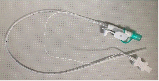
The patient’s preoperative, morphometric, and demographic characteristics were recorded in the ward before consent was obtained. The patients were instructed to fast from midnight the day before surgery. Upon arrival in the operating room, patients were monitored using pulse oximetry, electrocardiography, and noninvasive blood pressure measurements. Preoxygenation was performed, and thiopental (4–5 mg/kg) was administered. When the eyelash reflex subsided, mask ventilation was performed with 5–6% sevoflurane and 100% oxygen for 3 min. Neuromuscular blocking agents were not administered. If the depth of anesthesia was considered sufficient, a second-generation SAD (Ambu® AuraGain™) was placed, and a TE probe of 9 Fr (standard transverse [ST] probe, S&S MED) was inserted through the gastric channel (Fig. 1). The locations of the SAD and TE probe were confirmed using sonography [7]. Temperature was measured using a non-invasive zero-heat-flux (ZHF) cutaneous thermometer (3MTM Bair HuggerTM Temperature Patient Sensor, Arizant Healthcare Inc.) attached to the patient’s lateral forehead and a TR probe (E-temp, Ewha Biomedics) [8]. The temperature of the operating room was controlled using an air conditioner, and a forced-air warming system (Bair Hugger, Augustine Medical Inc.) was used to maintain normothermic body temperature in each patient.

Outcome assessment

The primary objective was to assess the feasibility and accuracy of body temperature measurement using a TE probe inserted through the gastric channel of a SAD. As secondary objectives, TTM and TZHF were compared, with TR as the reference. Additionally, suction functionality of the TE probe inserted through the SAD was evaluated.
TE, TR, and TZHF were measured 10 min after probe insertion or attachment to ensure consistency by allowing the external thermistor of the device to reach equilibrium with the ambient temperature. TTM was measured using a Braun ThermoScan 6 (Braun Thermoscan 4020, Braun GmbH) by the same operator for each patient, and two measured values were averaged to enhance the objectivity of the measurement. We measured the temperature at 10-min intervals throughout the surgery. All measurements were performed and recorded by a single anesthesiologist.
Sonography of the gastric antrum was conducted by the attending anesthesiologist using a GE Logiq P9 ultrasound machine with a 12-MHz linear probe (GE Healthcare). The appearance of acoustic shadows or comet-tails after air was injected through the gastric channel was considered to indicate gastric insufflation [9]. Subsequently, negative pressure was applied to the TE probe to decompress the stomach, and the effects were evaluated via sonography. Once air was removed, the attending anesthesiologist performed sonography to confirm the successful removal and qualitatively assess the absence of air [10,11]. The antrum was considered empty if it appeared flat, with the anterior and posterior walls in close proximity. 

RESULTS

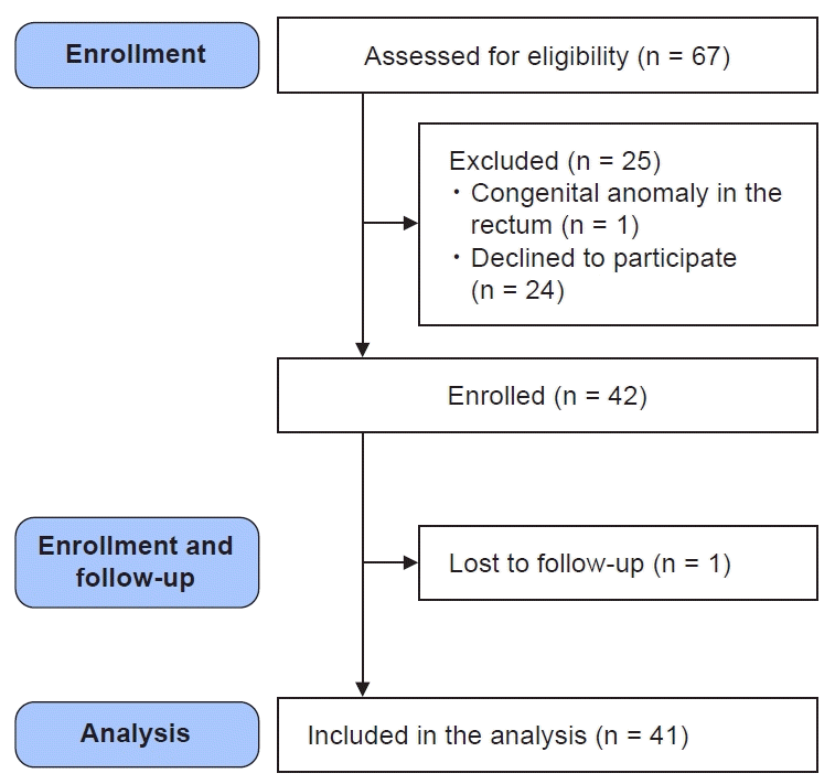
A total of 67 pediatric patients, aged < 6 years and weighing 10–30 kg, who were scheduled to undergo general anesthesia, were assessed for eligibility. Among them, one who had a congenital anomaly of the rectum and 24 whose guardians declined participation were excluded. Subsequently, the remaining 42 patients were enrolled. However, one patient was excluded from the final analysis due to an error in the thermometer device. The STROBE flow diagram of patient selection and dropout is presented in Fig. 2. The patients’ demographic characteristics and perioperative variables are presented in Table 1. Table 2 shows the range of body temperature data according to the measurement sites during anesthesia.
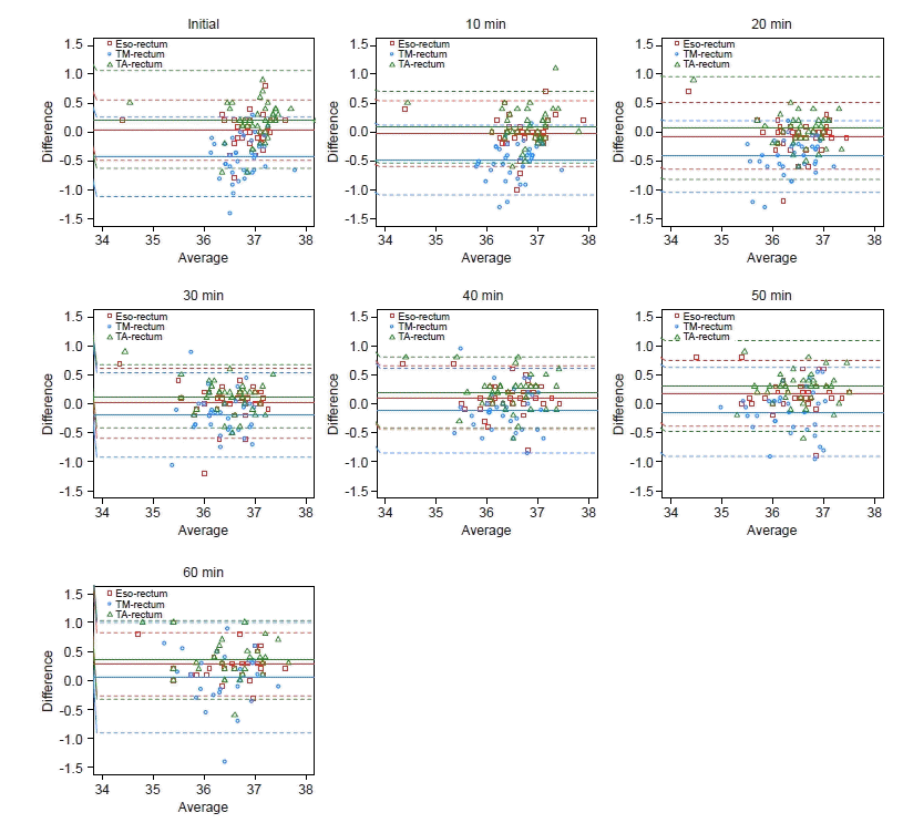
In all patients, the esophageal probe was successfully inserted through the gastric channel of the SAD. The results of the equivalence test are presented in Table 3, and the Bland–Altman plots illustrating the relationships of TE, TTM, and TZHF with TR are shown in Fig. 3. When considering the initial temperature measurements, the smallest difference was observed with TE, while the largest difference was noted with TTM, using TR as a reference. Overall, the minimal difference between temperatures was observed between measurements in the esophagus and rectum during the 0–40-min period, and between the tympanic membrane and rectum from 50–60-min period. When agreement was examined with TR as a reference, TE demonstrated equivalent results to TR (P < 0.001 at 0, 10, 20, 30, and 40-min intervals and P = 0.018 at 50-min intervals), except at the 60-min interval (P = 0.697). Compared with TR, TTM was not equivalent until 30 min after surgery (P > 0.999 at 0, 10, and 20-min intervals and P = 0.084 at the 30-min interval) and then showed equivalent results until the end of the surgery (P = 0.006, P = 0.025, P = 0.013 at the 40, 50, 60-min intervals). When TZHF was compared with TR as a reference, there was no difference from 10 min to 40 min after the surgery (P = 0.216, P < 0.001, P = 0.003, P < 0.001, P = 0.048, P > 0.999, P > 0.999 at 10-min intervals from 0 to 60 min).
Analysis of the intraclass correlation coefficients (ICCs) at each time point revealed that TE exhibited good reliability with TR as the reference throughout the intraoperative period (ICC > 0.75, Table 4). Both TZHF and TTM exhibited reliability exceeding the moderate level intraoperatively; however, TE exhibited the best reliability among the three measurement methods.
In 33 of the 41 included patients, gastric suction through the TE probe effectively reduced gastric air volume (80.5%). Gastric decompression was not clearly observed on sonography in the remaining eight patients.

Statistical analysis

Based on our clinical experience and findings from a previous study, we considered a difference of up to 0.3°C with a standard deviation (SD) of 0.5 to be accurate [12]. Sample size calculation revealed that enrolling 32 patients would result in a power of 90% at an α-level of 0.05. A final sample size of 42 was selected to account for potential dropout rates.
The Bland–Altman method, which accounts for the agreement of repeated measurements, was used to analyze the accuracy of TE measurement through the SAD and compare it with that of TTM, TZHF, and TR [13]. We performed an equivalence test to evaluate whether the TE, TZHF, and TTM values were equivalent to the TR value with a margin of 0.3°C. For reliability analysis, we calculated ICCs using a two-way random model [14]. Categorical variables were presented as numbers and percentages. Continuous variables were presented as means ± SDs with 95% confidence intervals. All analyses were performed using SAS software, version 9.4 (SAS Institute Inc.).

DISCUSSION

We observed TE measurement using a second-generation SAD to be highly accurate and reliable in pediatric patients undergoing general anesthesia, with TR as a reference, and it was feasible in all patients. Moreover, effective reduction of gastric air volume was achieved through suction via the gastric channel of the second-generation SAD.
Accurate intraoperative temperature monitoring is important, especially in children. Several guidelines exist for perioperative temperature monitoring, but none of them specify the best device or site for temperature measurement [15,16]. To ensure patient safety, an ideal temperature monitoring technique should accurately reflect the core temperature while being non-invasive [17]. The device or site used for measuring body temperature is selected based on the type of surgery, accessibility of monitoring sites, and physician’s preference. To the best of our knowledge, this is the first study investigating the most appropriate temperature measurement device and site for accurately determining the core temperature with the utilization of SAD in pediatric patients undergoing surgery.
Our study demonstrated that TE measurements obtained using a SAD exhibited higher agreement and reliability over an extended period than TTM or TZHF measurements when TR was used as a reference. This could be attributed to the fact that the distal esophagus receives sufficient blood perfusion from the core, resulting in more accurate and consistent temperature measurements than those at other sites. However, when TR serves as the reference point in an equivalent test, accuracy of TE decreases towards the end of the surgery. Conversely, TTM may be more accurate toward the end of surgery, as its accuracy is maintained throughout the later stages. The tympanic membrane, which is located near the hypothalamus, can indeed reflect core temperature [15]. However, accurately inserting the temperature probe can be challenging, particularly in pediatric patients with a small aural canal. Furthermore, concerns exist regarding the feasibility of continuous measurement, reliance on the operator’s skill for determining accuracy, and variability of measurements [18]. These limitations prevent precise core temperature measurement using this method [19]. Other monitoring devices, such as zero-heat-flux cutaneous thermometers, have an appealing theoretical basis; however, inconsistent accuracy has been noted, both in our study and in previous studies [19,20].
SADs have gained popularity as advantageous airway management tools in pediatric patients [21]. However, the absence of space for temperature probe insertion in previous SADs necessitated its insertion through the nasopharynx [6]. Consequently, accurate body temperature measurement was challenging due to the cooling effect of anesthetic gases and the potential for procedural trauma during probe insertion into the nasopharynx [22,23]. In a previous study, body temperature measurements in patients with SAD insertion primarily focused on pharyngeal measurements [24]; however, the incorporation of a gastric channel in second-generation SADs for improved esophageal access enables accurate temperature measurement in the distal esophagus and smooth insertion of temperature probes.
In contrast to tracheal intubation, utilization of SADs may lead to increased gastric insufflation in a pressure-dependent manner [25,26]. Moreover, even minor malpositioning of the SAD can result in air leakage from its seal, causing air to flow into the esophagus [27]. Gastric insufflation may increase the risk of pulmonary aspiration of gastric contents and postoperative nausea and vomiting, particularly in high-risk pediatric patients, such as those with an impaired ability to protect their airways [28,29]. The gastric channel of second-generation SADs offers the additional advantage of effectively allowing the suctioning of air that might be introduced into the stomach during mask ventilation [30].
The first limitation of our study pertains to the accuracy of the TE probe placement in routine clinical practice. The sensor is intended to be placed in the distal esophagus close to the pulmonary artery, and this is confirmed using sonography [31]. A previous study has demonstrated that the accuracy of TE measurement increases when the probe is appropriately positioned [32]. However, performing sonography in routine clinical practice is challenging, which might reduce the accuracy compared with that in our study. The second limitation is that our findings are applicable only within a limited range of temperature variations. While events such as tourniquet deflation and cardiopulmonary bypass application could induce rapid and extreme changes in body temperature, such events were not examined in this study.
In conclusion, we demonstrated that TE measurement through a second-generation SAD is feasible and accurate, making it suitable for routine use in pediatric patients with SAD insertion. Moreover, utilization of suction through the gastric channel of a second-generation SAD effectively reduces gastric air volume.

Notes

FUNDING

None.

CONFLICTS OF INTEREST

No potential conflict of interest relevant to this article was reported.

DATA AVAILABILITY STATEMENT

The datasets generated during and/or analyzed during the current study are available from the corresponding author on reasonable request.

AUTHOR CONTRIBUTIONS

Writing - original draft: Yeon-Ju Kim, Eundong Lee, Jaedo Lee, Ha-Jung Kim Writing - review & editing: Hyungtae Kim, Won Uk Koh, Young-Jin Ro, Ha-Jung Kim. Conceptualization: Hyungtae Kim, Won Uk Koh, Young-Jin Ro, Ha-Jung Kim. Data curation: Yeon-Ju Kim, Eundong Lee, Jaedo Lee, Hyungtae Kim, Won Uk Koh, Ha-Jung Kim. Formal analysis: Yeon-Ju Kim, Jaedo Lee, Won Uk Koh, Ha-Jung Kim. Methodology: Ha-Jung Kim. Investigation: Ha-Jung Kim. Resources: Yeon-Ju Kim, Ha-Jung Kim. Supervision: Won Uk Koh, Young-Jin Ro, Ha-Jung Kim.

REFERENCES

1. Pearce B, Christensen R, Voepel-Lewis T. Perioperative hypothermia in the pediatric population: prevalence, risk factors, and outcomes. J Anesth Clin Res. 2010; 1:1–4.
crossref
2. Plattner O, Semsroth M, Sessler DI, Papousek A, Klasen C, Wagner O. Lack of nonshivering thermogenesis in infants anesthetized with fentanyl and propofol. Anesthesiology. 1997; 86:772–7.
crossref
3. Martin SA, Kline AM. Can there be a standard for temperature measurement in the pediatric intensive care unit? AACN Clin Issues. 2004; 15:254–66.
crossref
4. Kimberger O, Cohen D, Illievich U, Lenhardt R. Temporal artery versus bladder thermometry during perioperative and intensive care unit monitoring. Anesth Analg. 2007; 105:1042–7.
crossref
5. Hebbar K, Fortenberry JD, Rogers K, Merritt R, Easley K. Comparison of temporal artery thermometer to standard temperature measurements in pediatric intensive care unit patients. Pediatr Crit Care Med. 2005; 6:557–61.
crossref
6. Cook T, Howes B. Supraglottic airway devices: recent advances. Cont Educ Anaesthesia, Critical Care & Pain. 2011; 11:56–61.
7. Ahn JH, Park J, Ryu kH, Jo JS, Kang RA, Ko JS, et al. Utility of ultrasound evaluation of I‐Gel® placement in children: An observational study. Paediatr Anaesth. 2021; 31:902–10.
crossref
8. Teunissen LPJ, Klewer J, De Haan A, De Koning JJ, Daanen HAM. Non-invasive continuous core temperature measurement by zero heat flux. Physiol Meas. 2011; 32:559–70.
crossref
9. Bouvet L, Albert ML, Augris C, Boselli E, Ecochard R, Rabilloud M, et al. Real-time detection of gastric insufflation related to facemask pressure–controlled ventilation using ultrasonography of the antrum and epigastric auscultation in nonparalyzed patients: a prospective, randomized, double-blind study. Anesthesiology. 2014; 120:326–34.
10. Park JH, Kim JY, Lee JM, Kim YH, Jeong HW, Kil HK. Manual vs. pressure‐controlled facemask ventilation for anaesthetic induction in paralysed children: a randomised controlled trial. Acta Anaesthesiol Scand. 2016; 60:1075–83.
crossref
11. Cubillos J, Tse C, Chan VWS, Perlas A. Bedside ultrasound assessment of gastric content: an observational study Evaluation échographique du contenu gastrique au chevet du patient: une étude observationnelle. Can J Anaesth. 2012; 59:416–23.
12. Lawson L, Bridges EJ, Ballou I, Eraker R, Greco S, Shively J, et al. Accuracy and precision of noninvasive temperature measurement in adult intensive care patients. Am J Crit Care. 2007; 16:485–96.
crossref
13. Bland JM, Altman DG. Agreement between methods of measurement with multiple observations per individual. J Biopharm Stat. 2007; 17:571–82.
crossref
14. Koo TK, Li MY. A guideline of selecting and reporting intraclass correlation coefficients for reliability research. J Chiropr Med. 2016; 15:155–63.
crossref
15. Sessler DI. Perioperative temperature monitoring. Anesthesiology. 2021; 134:111–8.
crossref
16. Hopf HW. Perioperative temperature management: time for a new standard of care? Anesthesiology. 2015; 122:229–30.
17. Cote CJ, Lerman J, Todres ID. A practice of anesthesia for infants and children: expert consult. Philadelphia: Elsevier Health Sciences;2012.
18. Bijur PE, Shah PD, Esses D. Temperature measurement in the adult emergency department: oral, tympanic membrane and temporal artery temperatures versus rectal temperature. Emerg Med J. 2016; 33:843–7.
crossref
19. Imamura M, Matsukawa T, Ozaki M, Sessler D, Nishiyama T, Kumazawa T. The accuracy and precision of four infrared aural canal thermometers during cardiac surgery. Acta Anaesthesiol Scand. 1998; 42:1222–6.
crossref
20. Langham GE, Maheshwari A, Contrera K, You J, Mascha E, Sessler DI. Noninvasive temperature monitoring in postanesthesia care units. Anesthesiology. 2009; 111:90–6.
crossref
21. Lopez‐Gil M, Brimacombe J, Alvarez M. Safety and efficacy of the laryngeal mask airway A prospective survey of 1400 children. Anaesthesia. 1996; 51:969–72.
crossref
22. Whitby JD, Dunkin LJ. Temperature differences in the oesophagus. The effects of intubation and ventilation. Br J Anaesth. 1969; 41:615–8.
crossref
23. Bissonnette B, Sessler DI, LaFlamme P. Intraoperative temperature monitoring sites in infants and children and the effect of inspired gas warming on esophageal temperature. Anesth Analg. 1989; 69:192–6.
24. Wadhwa A, Sessler DI, Sengupta P, Hanni K, Akça O. Core temperature measurements through a new airway device, perilaryngeal airway (CobraPLA). J Clin Anesth. 2005; 17:358–62.
25. Devitt JH, Wenstone R, Noel AG, O'Donnell MP. The laryngeal mask airway and positive-pressure ventilation. Anesthesiology. 1994; 80:550–5.
26. Litman RS. Complications of laryngeal masks in children: big data comes to pediatric anesthesia. Anesthesiology. 2013; 119:1239–40.
27. Graziotti PJ. Intermittent positive pressure ventilation through a laryngeal mask airway: is a nasogastric tube useful? Anaesthesia. 1992; 47:1088–90.
28. Mirzakhani H, Williams JN, Mello J, Joseph S, Meyer MJ, Waak K, et al. Muscle weakness predicts pharyngeal dysfunction and symptomatic aspiration in long-term ventilated patients. Anesthesiology. 2013; 119:389–97.
29. Lerman J. Surgical and patient factors involved in postoperative nausea and vomiting. Br J Anaesth. 1992; 69(7 Suppl 1):24S–32.
crossref
30. Lee J, Lim H, Son KG, Ko S. Optimal nasopharyngeal temperature probe placement. Anesth Analg. 2014; 119:875–9.
crossref
31. Hong SH, Lee J, Jung JY, Shim JW, Jung HS. Simple calculation of the optimal insertion depth of esophageal temperature probes in children. J Clin Monit Comput. 2020; 34:353–9.
crossref
32. Robinson JL, Seal RF, Spady DW, Joffres MR. Comparison of esophageal, rectal, axillary, bladder, tympanic, and pulmonary artery temperatures in children. J Pediatr. 1998; 133:553–6.
crossref

Fig. 1.
ST probe with 9Fr used to measure esophageal temperature. ST: standard transverse.
apm-23150f1.tif
Fig. 2.
Flowchart of the study population in accordance with STROBE guidelines. STROBE: Strengthening the Reporting of Observational Studies in Epidemiology.
apm-23150f2.tif
Fig. 3.
Results of the Bland–Altman analysis comparing the temperatures recorded in the esophagus, tympanic membrane, and forehead with those recorded in the rectum. The solid line is the mean value of the difference, and the dashed line is the mean ± 1.96 standard deviation.
apm-23150f3.tif
Table 1.
Patient Characteristics and Perioperative Variables
Variables Patients (n = 41)
Baseline characteristics
 Age (mo) 41.1 ± 22.2
 M/F 23/18 (56.1/43.9)
 Weight (kg) 18.3 ± 14.1
 Height (cm) 98.2 ± 15.4
 ASA PS 1/2 21/20 (51.2/48.8)
Intraoperative data
 Anesthetic time (min) 110.3 ± 43.9
 Operation time (min) 60.8 ± 37.8
 Type of surgery
  Hand surgery 17 (41.5)
  Arm surgery 9 (22.0)
  Knee surgery 1 (2.4)
  Foot surgery 14 (34.1)

Values are presented as mean ± SD, number only, or number (%). ASA PS: American Society of Anesthesiologists physical status.

Table 2.
Range of Body Temperature Data according to the Measurement Sites during Anesthesia
Time (min) TE TR TTM TZHF
0 36.9 ± 0.6 36.9 ± 0.6 36.5 ± 0.5 37.1 ± 0.6
10 36.7 ± 0.5 36.7 ± 0.6 36.3 ± 0.5 36.8 ± 0.5
20 36.5 ± 0.5 36.5 ± 0.6 36.2 ± 0.5 36.6 ± 0.5
30 36.4 ± 0.6 36.4 ± 0.6 36.2 ± 0.5 36.5 ± 0.5
40 36.5 ± 0.6 36.3 ± 0.6 36.3 ± 0.6 36.6 ± 0.6
50 36.5 ± 0.6 36.3 ± 0.7 36.3 ± 0.6 36.6 ± 0.6
60 36.6 ± 0.6 36.3 ± 0.7 36.8 ± 0.6 36.7 ± 0.7
70 36.8 ± 0.6 36.5 ± 0.7 36.5 ± 0.6 36.8 ± 0.6
80 36.9 ± 0.5 36.5 ± 0.7 36.7 ± 0.5 36.8 ± 0.6
90 37.0 ± 0.5 36.6 ± 0.5 36.7 ± 0.3 36.9 ± 0.6
100 37.1 ± 0.5 36.7 ± 0.6 36.8 ± 0.3 36.9 ± 0.7
110 37.1 ± 0.1 36.7 ± 0.2 36.8 ± 0.2 37.1 ± 0.2
120 37.0 ± 0.2 36.7 ± 0.2 36.7 ± 0.3 37.0 ± 0.2

Values are presented as mean ± SD. TE: core temperature measured in the esophagus, TR: core temperature measured in the rectum, TTM: core temperature measured in the tympanic membrane, TZHF: core temperature measured using a zero-heat flux thermometer on the forehead cutaneous site.

Table 3.
The Results of the Equivalent Test with TR as a Reference
TE
TTM
TZHF
Time (min) Mean difference 95% CI P value Mean difference 95% CI P value Mean difference 95% CI P value
0 0.030 –0.055 0.115 < 0.001* 0.433 0.319 0.547 > 0.999 0.215 0.077 0.353 0.219
10 –0.025 –0.117 0.067 < 0.001* 0.482 0.381 0.582 > 0.999 0.085 –0.016 0.186 < 0.001*
20 –0.068 –0.161 0.026 < 0.001* 0.418 0.313 0.522 > 0.999 0.073 –0.072 0.217 0.003*
30 0.010 –0.088 0.108 < 0.001* 0.189 0.061 0.316 0.084 0.125 0.036 0.214 < 0.001*
40 0.108 0.018 0.197 < 0.001* 0.113 –0.017 0.244 0.006* 0.200 0.101 0.299 0.048*
50 0.181 0.084 0.279 0.018* 0.139 0.001 0.278 0.025* 0.305 0.172 0.439 > 0.999
60 0.280 0.176 0.384 0.697 –0.046 –0.242 0.150 0.013* 0.357 0.227 0.486 > 0.999

CI: confidence interval, TR: core temperature measured in the rectum, TE: core temperature measured in the esophagus, TTM: core temperature measured in the tympanic membrane, TZHF: core temperature measured using a zero-heat flux thermometer on the forehead cutaneous site.

* P < 0.05, it means less than the mean difference ± 0.3.

Table 4.
Reliability in Temperature
TE & TR
TTM & TR
TZHF & TR
Time (min) ICC* 95% CI ICC* 95% CI ICC* 95% CI
0 0.890 0.812 0.937 0.593 -0.028 0.833 0.690 0.453 0.826
10 0.863 0.768 0.920 0.625 -0.080 0.868 0.825 0.706 0.898
20 0.854 0.753 0.915 0.692 -0.017 0.890 0.677 0.490 0.804
30 0.860 0.764 0.919 0.760 0.572 0.865 0.856 0.722 0.923
40 0.879 0.778 0.933 0.793 0.658 0.878 0.826 0.574 0.918
50 0.864 0.664 0.936 0.808 0.677 0.888 0.711 0.302 0.866
60 0.843 0.314 0.944 0.740 0.581 0.845 0.771 0.174 0.915

ICC: intraclass correlation coefficient, CI: confidence interval, TE: core temperature measured in the esophagus, TTM: core temperature measured in the tympanic membrane, TZHF: core temperature measured using a zero-heat flux thermometer on the forehead cutaneous site, TR: core temperature measured in the rectum.

* Values of ICC were within 0–1 and were classified as follows: less than 0.5, poor; between 0.5-0.75, moderate; between 0.75–0.9, good; greater than 0.9, excellent.

TOOLS
Similar articles