Journal List > Endocrinol Metab > v.39(5) > 1516088776

Oh, Choi, Jang, Son, Park, Song, Kim, Cho, Chai, Chung, and Park: Small Multi-Gene DNA Panel Can Aid in Reducing the Surgical Resection Rate and Predicting the Malignancy Risk of Thyroid Nodules

Abstract

Background

We explored the utility of a small multi-gene DNA panel for assessing molecular profiles of thyroid nodules and influencing clinical decisions by comparing outcomes between tested and untested nodules.

Methods

Between April 2022 and May 2023, we prospectively performed fine-needle aspiration (FNA) with gene testing via DNA panel of 11 genes (BRAF, RAS [NRAS, HRAS, KRAS], EZH1, DICER1, EIF1AX, PTEN, TP53, PIK3CA, TERT promoter) in 278 consecutive nodules (panel group). Propensity score-matching (1:1) was performed with 475 nodules that consecutively underwent FNA without gene testing between January 2021 and December 2021 (control group).

Results

In the panel group, positive call rate for mutations was 41.7% (BRAF 16.2%, RAS 12.6%, others 11.5%, double mutation 1.4%) for all nodules, and 40.0% (BRAF 4.3%, RAS 19.1%, others 15.7%, double mutation 0.9%) for indeterminate nodules. Benign call rate was 69.8% for all nodules, and 75.7% for indeterminate nodules. In four nodules, additional TP53 (in addition to BRAF or EZH1) or PIK3CA (in addition to BRAF or TERT) mutations were co-detected. Sensitivity, specificity, positive predictive value, and negative predictive value were 80.0%, 53.3%, 88.1%, 38.1% for all nodules, and 78.6%, 45.5%, 64.7%, 62.5% for indeterminate nodules, respectively. Panel group exhibited lower surgical resection rates than the control group for all nodules (27.0% vs. 52.5%, P<0.001), and indeterminate nodules (23.5% vs. 68.2%, P<0.001). Malignancy risk was significantly different between the panel and control groups (81.5% vs. 63.9%, P=0.008) for all nodules.

Conclusion

Our panel aids in managing thyroid nodules by providing information on malignancy risk based on mutations, potentially reducing unnecessary surgery in benign nodules or patients with less aggressive malignancies.

INTRODUCTION

Thyroid nodules are a common clinical occurrence, with their prevalence varying from 20% to 65% in the general population [1]. The critical distinction between benign and malignant or premalignant lesions is paramount for effective management, particularly when determining the need for surgery. The initial evaluation of these nodules often involves ultrasound imaging coupled with fine-needle aspiration (FNA) cytological analysis [2,3], but these methods have limitations, including variable diagnostic accuracy, especially in cytologically indeterminate nodules where the risk of malignancy (ROM) hovers between 13% and 34% [4]. Hence, subjecting most patients with indeterminate nodules to diagnostic surgery results in a substantial proportion undergoing unnecessary procedures [5]. The emergence of molecular testing has shown promise in overcoming diagnostic challenges and improving clinical management [6]. Multigene panels, which analyze mutations across various relevant genes altering the molecular characterization of thyroid tumors, offer a broader perspective on thyroid cancer pathogenesis [7,8]. This comprehensive approach enhances the assessment of ROM, helping to make more informed decisions for individual patients [9].
Initially, molecular tests focused on a single or a small number of genetic variants in key driver genes, such as BRAF, RAS, peroxisome proliferator-activated receptor γ (PPARγ)-paired box 8 (PAX8), and/or rearranged during transfection/papillary thyroid carcinoma (RET/PTC) 1/3 (7-gene panel), but their diagnostic efficacy was insufficient [10]. Commercial multi-gene panels, including ThyroSeq v3 (ThyroSeq, Rye Brook, NY, USA), Afirma gene expression classifier (GEC), gene sequencing classifier (GSC) (Afirma, Washington, DC, USA), and ThyGeNext/ThyraMIR (Parsippany, NJ, USA), employing nextgeneration sequencing (NGS) methods, have demonstrated significantly improved diagnostic performance by including a large number of genes [6]. Thus, the American Thyroid Association 2015 guideline [2] and recent European guidelines [11] recommend that molecular testing can be considered for cytologically indeterminate nodules, although studies on the actual clinical and surgical implications remain limited [12]. However, considering the cost of these commercial NGS tests, European guidelines state these tests should be used “if possible” and advocate the development of less expensive tests. The price of commercial NGS tests can range from US$3,000 to over US$6,000, which includes the cost of RNA or miRNA sequencing in addition to DNA tests [13,14].
On the other hand, a substantial portion of indeterminate nodules identified as follicular pattern tumors harbor mutations associated with low-risk molecular subtypes, such as RAS-like or non-BRAF-non-RAS (NBNR), with a low ROM or risk of recurrence (i.e., low-risk mutations) [2,15,16]. More than 90% of RAS-like or NBNR tumors with genetic alterations possess a point mutation in RAS, eukaryotic translation initiation factor 1A X-linked (EIF1AX), dicer 1, ribonuclease III (DICER1), phosphatase and tensin homolog (PTEN), enhancer of zeste 1 polycomb repressive complex 2 subunit (EZH1), or tumor protein p53 (TP53). Therefore, a small DNA gene panel testing these genes with the addition of BRAF gene can yield superior diagnostic performance compared to the 7-gene panel, even though it cannot detect RET and PPARγ fusions that the 7-gene panel can [16]. Moreover, although the sensitivity or negative predictive value (NPV) of this small DNA gene testing alone may be lower than that of commercial NGS tests that include RNA or miRNA testing, it could still support clinical decision-making for the management of indeterminate nodules with a cost at least five to six times lower. Additionally, this small DNA gene testing with the addition of telomerase reverse transcriptase (TERT) promoter and phosphatidylinositol-4,5-bisphosphate 3-kinase catalytic subunit alpha (PIK3CA) genes can also cover common double mutations often found in aggressive thyroid cancer [17]. Therefore, the small DNA testing of those 11 genes can aid not only in diagnosis but also in prognosis prediction. Therefore, this study investigated the clinical efficacy of molecular testing, specifically in the diagnostic performance and the ability of reducing the surgical resection rate (RR), through prospective DNA panel testing of the 11 genes. These genes included eight main driver genes of follicular patterned tumors (BRAF, NRAS, HRAS, KRAS, EIF1AX, DICER1, PTEN, and EZH1) and three genes associated with high-risk cancers (TERT promoter, PIK3CA, and TP53). We assessed its diagnostic accuracy by comparing it with three commercial gene panel tests and further analyzed the surgical RR through a comparison with propensity score-matched controls who did not undergo molecular testing.

METHODS

Subjects

A total of 281 consecutive thyroid nodules that underwent FNA and multi-gene panel testing between April 2022 and May 2023 at Seoul National University Hospital were prospectively included. We excluded those associated with syndromic conditions such as multiple endocrine neoplasia. There were no patients with a previous history of head and neck malignancy. Nodules exhibiting suspicious ultrasound features according to the 2021 Korean Thyroid Imaging Reporting and Data System (K-TIRADS) [18] and 2016 Korean Thyroid Association (KTA) guideline [19] underwent FNA and were subjected to both cytological analysis and 11-gene DNA panel testing. In brief, the KTIRADS description of the sonographic patterns of a K-TIRADS 2 (benign) nodule includes (1) iso-/hyperechoic spongiform; (2) partially cystic nodule with intracystic echogenic foci and comet-tail artifact; or (3) pure cyst. The sonographic patterns of a K-TIRADS 3 (low-suspicion) nodule include partially cystic or iso-/hyperechoic nodule without any of the three suspicious features (punctate echogenic foci, nonparallel orientation, and irregular margins). The sonographic patterns of a K-TIRADS 4 (intermediate-suspicion) nodule include (1) solid hypoechoic nodules without any of the three suspicious features; (2) a partially cystic or iso-/hyperechoic nodule with any of the three suspicious ultrasound features; or (3) entirely calcified nodules. Finally, the sonographic patterns of a K-TIRADS 5 (high-suspicion) nodule include a solid hypoechoic nodule with any of the three suspicious features [18]. In addition to the FNA performed for cytology, FNA for the gene panel was conducted with 26-gauge needles in two passes. The aspirated specimen was then preserved in Eppendorf DNA LoBind Tubes (Eppendorf, Hamburg, Germany) containing 1 mL of preservation solution to maintain nucleic acid integrity. It was refrigerated and transported on the same day with packed ice to the laboratory for testing. Subsequently, the specimen was stored in a deep freezer at –80℃ until analysis, which was performed within 3 to 5 days of storage. The cytology of the nodules was confirmed by three experienced endocrine pathologists according to the Bethesda classification (BC) [20]. Three nodules were excluded from the analysis due to inadequate samples for gene panel testing. Finally, 278 nodules including 19 Bethesda I nodules, 87 Bethesda II nodules, 115 indeterminate (102 Bethesda III and 13 IV) nodules, nine Bethesda V nodules, and 48 Bethesda VI nodules were included. The gene mutation features of the nodules were analyzed and correlated with clinical characteristics, including age, sex, nodule size, K-TIRADS category, and BC.
To assess the impact of molecular panel testing on RR, we retrospectively included 475 nodules that underwent consecutive FNA without gene testing from January 2021 to December 2021 for the control group. To mitigate confounding variables in baseline clinical characteristics, we conducted 1:1 propensity score-matching (PSM) through the nearest neighbor matching method with a caliper width of 0.2, using age, sex, nodule size, K-TIRADS score, and BC as variables for matching, resulting in a total of 278 nodules in each group. The quality of the PSM model was assessed using the Hosmer-Lemeshow test for goodness-of-fit and the C-statistic for discriminatory ability.
To guide the decision-making process between surgery and active surveillance, in accordance with the 2023 KTA guidelines [3], we engaged patients in a shared decision-making approach. Clinical decisions were made by considering molecular results alongside other factors, such as nodule size, growth pattern, ultrasound features, patient history, and patient preferences. We provided comprehensive information, including FNA or gun biopsy results, malignancy risk, and prognosis linked to identified genetic mutations. Additionally, we discussed the potential risks associated with surgery, such as those related to general anesthesia, as well as complications like scarring, functional impairment, and voice changes after surgery. For patients opting for active surveillance without surgery, we conveyed the risks associated with potentially overlooking malignancy, the importance of ongoing diagnosis, and the necessity for ultrasound monitoring every 6 to 24 months, as determined by follow-up test results.
The study received approval from the Institutional Review Board at Seoul National University Hospital (IRB no. 1608-088-785). All Panel group patients provided informed consent prior to FNA, and the need for informed consent for the patients in the control group was waived.

DNA isolation and targeted sequencing for 11-gene DNA panel of FNA samples

Molecular testing involved an 11-gene DNA panel with targeted sequencing for mutations in eight main driver genes of follicular patterned tumors (BRAF, NRAS, HRAS, KRAS, EIF1AX, DICER1, PTEN, and EZH1) and three genes associated with high-risk cancers (TERT promoter, PIK3CA, and TP53). The mutations analyzed included point mutations (missense/nonsense), insertions or deletions (indels), and regulatory mutations.
In brief, genomic DNA was isolated from the FNA samples using an automated nucleic acid purification platform, Maxwell CCS system (Promega, Madison, WI, USA) with the Maxwell CSC Blood DNA kit (Promega). The isolated DNAs were quantified and qualified using QubitTM4 Fluorometer (Invitrogen, Waltham, MA, USA), 4200 TapeStation (Agilent Technologies Inc., Santa Clara, CA, USA), and NANODROP 8000 Spectrophotometer (Thermo Scientific, Waltham, MA, USA). The xGen DNA Library Prep EZ Kit (IDT Inc., Coralville, IA, USA) was used to construct DNA into an NGS library following the manufacturer’s instructions. Target genes were captured using an 11-gene DNA panel (DCGen Co. Ltd., Seoul, Korea). Subsequently, the sequencing libraries were sequenced with NextSeq 550Dx (Illumina, San Diego, CA, USA). Targeted DNA sequencing reads were aligned to the hg38 reference genome using Burrows-Wheeler Aligner (BWA)-0.7.17. Putative duplications were marked by the GATK-4.1.8.0 module. Sites potentially harboring small insertions or deletions (indels) were recalibrated by applying the modules of GATK-4.1.8.0 with known variant sites identified from phase I of the 1000 Genomes Project (http://www.1000genomes.org/), dbSNP-151 (https://www.ncbi.nlm.nih.gov/snp/), and Mills and 1000G gold standard indels. We used MuTect-4.1.8.0 and HaplotypeCaller to detect single nucleotide variations (SNVs) and small indels. The targeted sequencing coverage was 2000x for tumors.
The mean concentration of the 281 samples was 793 ng, and 60 samples (21%) registered concentrations below 100 ng. Samples were classified as inadequate if DNA concentrations were below 50 ng, leading to the identification of three nodules deemed inadequate. In response to quality control issues, 18 samples (6.3%) underwent retesting. To remove germline-like variants, we excluded any variant with an allelic frequency greater than 0.01. Furthermore, sequencing artifacts and germline variants were filtered out using a panel of normals (PoN). Annotations for potential functional consequences and other mutation information were performed using variant effect predictor (VEP). For confident detection of somatic mutations, we applied the following criteria: (1) total reads at the mutation site ≥20 and (2) variant allele frequency ≥0.02. The specific types of detected mutations are summarized in Supplemental Table S1.

Comparison of diagnostic performance of the panel

The positive call rate (PCR) and benign call rate (BCR) were calculated. The PCR represented the percentage of nodules with positive test results, while the BCR denoted the percentage of nodules with low-risk (NBNR) or negative molecular test outcomes. Sensitivity, specificity, NPV, and positive predictive value (PPV) were analyzed using histopathology data from surgically resected nodules. These analyses were conducted for all nodules as well as for indeterminate (Bethesda III and IV) nodules exclusively. We conducted a literature review that included both validation studies and real-world studies of the diagnostic performance of other available commercial gene panels for thyroid nodules, including the 7-gene panel, Afirma GEC panel, Afirma GSC panel, and ThyroSeq v3 panel, for comparison with our multi-gene panel [7,21-36].
For the analysis, malignant nodules consisted of those diagnosed as carcinoma through surgical pathology, including papillary thyroid carcinoma (PTC), follicular thyroid carcinoma (FTC), differentiated high-grade thyroid carcinoma (DHGTC), oncocytic carcinoma (OC). Non-invasive follicular thyroid neoplasm with papillary-like nuclear feature (NIFTP), representing a premalignant entity warranting surgical intervention, was also considered malignant. Nodules diagnosed with BRAFV600E mutations that did not undergo surgery were also classified as malignant in the diagnostic performance analysis. Follicular adenoma (FA), oncocytic adenoma (OA), and follicular nodular disease (FND) were considered benign.

Comparison of resection rate and risk of malignancy

RR and ROM were compared between the panel and the control groups. RR was defined as the percentage of nodules undergoing surgery, and ROM as the percentage of malignant nodules among those undergoing surgery. Nodules harboring BRAFV600E mutations that had not yet undergone surgery were considered malignant and included in the ROM analysis.

Statistical analysis

Clinical characteristics were presented as frequencies with percentages for categorical variables; mean±standard deviation was used for continuous variables. Prior to conducting tests for comparison, the normal distribution of continuous variables was assessed using the Shapiro-Wilk test. Continuous variables were compared using the t test or Mann-Whitney U test, whereas categorical variables were compared using the chi-square or Fisher’s exact test for two groups. PSM was performed using the MatchIt package of the R software. All statistical analyses were performed using R version 4.3.1 (R foundation for Statistical Computing, Vienna, Austria), and all P values less than 0.05 were considered statistically significant.

RESULTS

Characteristics of subjects

Table 1 summarizes the characteristics of the subjects before and after PSM. Baseline clinical characteristics of the 278 nodules in the panel group and 475 nodules in the control group, before PSM, showed significant differences. However, PSM successfully balanced all baseline clinical characteristics, including sex, age, nodule size, K-TIRADS category, and BC (P>0.050). The C-statistic value of the propensity score model was 0.811, and the Hosmer-Lemeshow statistic showed the following results: χ2=10.545, degrees of freedom=8, and P=0.229. In the panel group, there were 218 (78.4%) females, with a mean age of 54 years. The mean nodule size was 1.7 cm. K-TIRADS categories II, III, IV, and V were observed in one (0.4%), 73 (26.3%), 103 (37.1%), and 101 (36.3%) nodules, respectively. Cytology results revealed BC I, II, III–IV, and V–VI categories in 19 (6.8%), 87 (31.3%), 115 (41.4%), and 57 (20.5%) nodules, respectively. Of the 58 (20.9%) nodules in the panel group with available histopathological diagnosis, 42 (72.4%) were malignant, one (1.7%) was NIFTP, and 16 (27.6%) were benign. In the control group, consisting of 111 (39.9%) nodules with available histopathological diagnosis, 66 (59.5%) were malignant, five (4.5%) were NIFTP, and 40 (36.0%) were benign (Table 1).

Diagnostic performance of the panel

Table 2 summarizes the diagnostic performance of the 11-gene DNA panel in comparison with other available commercial panels. The PCR was 116/278 (41.7%) for all nodules, and 46/115 (40.0%) for indeterminate (Bethesda III or IV) nodules. The BCR was 194/278 (69.8%) for all nodules and 87/115 (75.7%) for indeterminate nodules. The sensitivity, specificity, PPV, and NPV were 80.0%, 53.3%, 88.1%, and 38.1%, respectively, for all nodules in the panel group, and 78.6%, 45.5%, 64.7%, and 62.5%, respectively, for indeterminate nodules.
Comparing our results to previous reports on the diagnostic performance of the commercial multi-gene panels, we found our PCR to be consistent with those panels, while the BCR was slightly higher in our study. The diagnostic performance of our 11-gene DNA panel for indeterminate nodules generally demonstrated lower performance compared to other panels. However, the specificity of real-world studies of the Afirma GEC, the PPV of a validation study for the Afirma GSC, and the NPV of both validation and real-world studies of the Afirma GEC were comparable to the corresponding values of the 11-gene DNA panel (Table 2).

Comparison of resection rate and risk of malignancy

The RR and ROM for each BC are summarized in Table 2. The panel group maintained a lower RR (75/278, 27.0%) than the control group (146/278, 52.5%) (P <0.001) for all nodules, and for the indeterminate Bethesda III–IV group (23.5% vs. 68.2%, P<0.001) and the Bethesda V–VI group (70.2% vs. 88.3%, P=0.015). The RR was lower in the panel group for Bethesda II nodules, but without statistical significance (8.0% vs. 17.0%, P=0.073).
The ROM was calculated in 192 (89 indeterminate) nodules, including 169 resected and 16 BRAFV600E-positive unresected nodules (84 and five in indeterminate nodules, respectively). The ROM was significantly different between the two groups (81.5% vs. 63.9%, P=0.008) for all nodules, but not significantly different for each subgroup (Bethesda II, II–IV indeterminate, or V–VI) (Table 3).
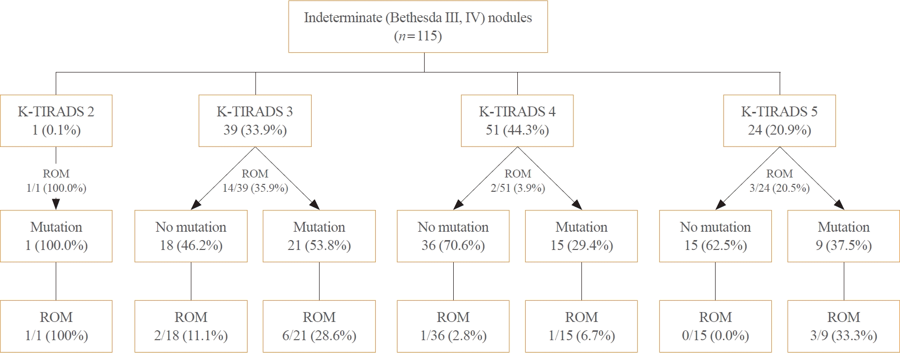
Subsequently, the ROM was evaluated according to the results from the 11-gene DNA panel and sonographic characteristics. The ROM in indeterminate nodules with mutations was higher compared to those without mutations across all K-TIRADS categories. The ROM of indeterminate nodules without mutations was 11.1%, 2.8%, and 0.0% for K-TIRADS 3, 4, and 5, while it was 28.6%, 6.7%, or 22.2% in nodules harboring mutations, respectively (P=0.184, P=0.159, or P=0.019, respectively) (Fig. 1). The ROM according to K-TIRADS category in Bethesda II and Bethesda V–VI nodules is shown in Supplemental Figs. 1, 2, respectively.

Molecular profiles of nodules by 11-gene DNA panel testing

The detected mutations included 45 BRAFV600E, 35 RAS, 12 EZH1, 11 DICER1, seven EIF1AX, and two PTEN mutations. Four nodules had additional TP53 (in addition to BRAFV600E or EZH1) or PIK3CA (in addition to BRAFV600E or TERT) mutations, and three showed additional mutation in the same gene (BRAF, KRAS, and DICER1). The RR was high in the BRAFV600E mutation (62.2%) and double mutation groups (100%), intermediate in the RAS mutation group (25.7%), and low in the no mutation (18.5%) group. Of the 20 nodules with no mutations that underwent surgery and had available histopathology results, 12 were malignant and eight were benign. Nodules with EZH1, DICER1 yielded one FA each, and two nodules with EIF1AX mutations yielded one FTC and one FA. Of the eight nodules with NRAS mutations, four were malignant (two PTCs, one FTC, and one NIFTP) and four were benign (one FA, one OA, and one FND). One nodule with HRAS mutation was diagnosed as FTC. All nodules with BRAFV600E mutations that had available histopathology were found to be malignant (21 PTCs and one DHGTC) (Table 4).
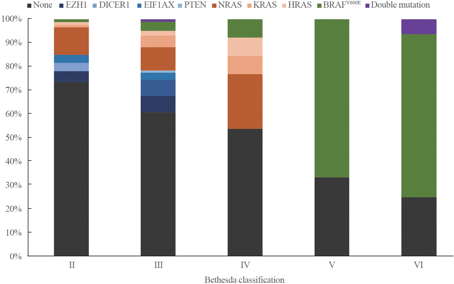
The molecular profiles of the panel group nodules were different according to the Bethesda groups (Fig. 2). Of the Bethesda II nodules, 73.6% had no mutations, 13.8% had RAS mutations, 11.5% had NBNR mutations, and 1.1% had BRAFV600E mutations. Of the intermediate (Bethesda III–IV) nodules, 60% had no mutations, 19% had RAS mutations, 16% had NBNR mutations, 4% had BRAFV600E mutations, and 1% had double mutations. Of the Bethesda V–VI nodules, 26% had no mutations, 69% had BRAFV600E mutations, and 5% had double mutations (Supplemental Table S2, Supplemental Fig. S3).

Clinical characteristics of molecular groups by the 11-gene DNA panel testing

Table 5 summarizes the comparison of clinical characteristics between nodules managed with active surveillance or surgery for each molecular group. In all mutation subtype groups, patients who underwent surgery had larger nodules than those under active surveillance, but without statistical significance. No significant differences were seen in any of the subtype groups for other clinical characteristics, including sex, age, and K-TIRADS category.
In four nodules, dual mutations (BRAFV600E/PIK3CA, BRAFV600E/TP53, PIK3CA/TERT, and EZH1/TP53) were detected, and all underwent surgery. Three of the nodules were diagnosed as PTC with extrathyroidal extension and lymphatic invasion, whereas the nodule with EZH1/TP53 mutation was diagnosed as OA (Table 6).

DISCUSSION

This study demonstrated that a small 11-gene DNA panel, composed of genes prevalent in indeterminate nodules or associated with prognosis, could result in a clinically significant decrease in the RR and an increase in active surveillance for low-risk cancers. Although the diagnostic accuracy of this panel test was lower than that of other existing commercial tests with a larger number of genetic mutations, it still yielded reasonably good results.
Our findings demonstrate that the utilization of the small DNA panel can significantly reduce the RR of thyroid nodules. In particular, the RR of both all nodules and indeterminate nodules was notably lower in the panel group compared to the control group (27.0% vs. 52.5% and 23.5% vs. 68.2%, respectively). The RR in this study was lower than the PCR rates (41.7% and 40.0%, respectively). Most commercial panel tests recommend surgery in cases with positive results, but they suggest surveillance as an alternative approach if the molecular results suggest a low risk of cancer progression [37]. In this study, only 25.7% of patients with RAS mutations, 12.5% with NBNR mutations, or 18.5% without mutations in 11 genes of our panel underwent surgery, contrasting with 68.2% of control patients with indeterminate nodules. After the panel test, in Bethesda III nodules, 47.1% of RAS tumors were resected, while only 16.7% of NBNR tumors, similar to the 17.7% rate in the no mutation group, underwent surgery. This suggests that a larger proportion of patients with no or low-risk mutations preferred surveillance or delayed surgery instead of immediate surgery in our subjects, leading to a 50% (from 52.5% to 27%) reduction in surgery in all nodules and a 65% (from 68.2% to 23.5%) reduction in indeterminate nodules by 11-gene DNA panel testing. A gene mutation detected by our panel does not automatically categorize the nodule as malignant; each mutation’s significance is assessed individually. BRAF mutations typically indicate nearly a 100% ROM, and double mutations are also associated with a high ROM with aggressive behavior [38]. RAS mutations generally signify potential malignancy or premalignant lesions, with a ROM ranging 44% to 66% [38,39], often favoring surgical intervention in consideration with other clinical factors. Other single mutations (NBNR), such as EZH1 mutation, are generally considered low-risk mutations more likely associated with adenomas, with a ROM ranging from 6% to 47.6% [39-41]. It is crucial to note that despite the improvement in molecular testing, current clinical decision-making for surgical procedures involves a myriad of considerations beyond cytopathological findings, including nodule size, growth pattern, ultrasound features, patient history, and patient preferences.
There are risks associated with the reduced surgical RR and increased reliance on active surveillance, particularly in the context of cancer patients. These risks include the potential progression of premalignant lesions into cancer, the risk of cancer advancement and metastasis, as well as the potentially increased surgical complexity and associated complications. These are crucial aspects that deserve careful consideration. However, it is noteworthy that the risks associated with active surveillance of thyroid nodules or even thyroid cancer without mutations or with single mutations, particularly RAS-like or NBNR mutations, may not be extensively significant, as highlighted in previous studies [42,43]. Nevertheless, our small 11-gene panel does not detect copy number alterations common in OAs or low-risk fusions found in FAs or FTCs, which are detectable by larger commercial panels. Thus, while our panel includes aggressive mutations prevalent in thyroid carcinomas, a broader genetic assessment is necessary to fully evaluate the potential risks associated with active surveillance of thyroid nodules or cancers. Furthermore, gene panel testing results can offer predictions regarding the ROM, potentially making it easier to accept the decision to pursue active surveillance for lower-risk patients. On the other hand, for patients inclined towards surgical resection, the typical recommendation is diagnostic lobectomy. However, the use of the panel introduces the option of considering an initial total thyroidectomy instead of lobectomy, dependent on the number and type of mutations identified. Additionally, the genetic information provided by the gene panel can guide long-term patient management, allowing for more active and careful follow-up for those at higher risk and less intensive follow-up for those at lower risk. As a result, the panel emerges as a vital clinical tool that may assist in determining the optimal surgical extent and guide personalized patient care, and thus, integrating the panel into routine clinical practice may be beneficial.
Although the ROM was significantly higher in the panel group than in the control group for all nodules (81.5% vs. 63.9%), suggesting that the panel’s molecular information could potentially contribute to sparing patients from unnecessary surgery, the difference was not very large, and there were no significant differences in the ROM among specific BC cytologies. This lack of significance may be attributed to the relatively small sample size, but it is also important to consider that RAS mutations and some other single gene mutations are not exclusive to carcinomas and can also occur in adenomas [44]. For certain genes, such as EZH1, the likelihood of a nodule being an adenoma with these mutations is relatively higher [45,46]. Thus, if the surgical resection of all nodules with a positive mutation of any gene is considered for malignant or premalignant lesions, it can heighten the sensitivity in cancer diagnosis. However, this lowers the specificity, impacting the ROM associated with nodules containing such mutations. Moreover, in cases where a gene mutation is present and the nodule is actually malignant, the decision to opt for active surveillance due to it being categorized as low-risk cancer can also impact ROM. Given that the ROM is influenced by these paradoxical decisions stemming from mutation results, it is imperative to interpret the outcomes of molecular tests with caution.
Unlike some other commercial panels, our multi-gene panel exhibited lower diagnostic performance, especially in terms of sensitivity or NPV, compared with Afirma GSC and ThyroSeq v3, which was anticipated due to the smaller number of genes covered in our panel. In theory, an increase in the number of genes associated with thyroid cancers covered in the panel would lead to higher NPV. However, it is important to note that this would also result in a greater inclusion of mutations associated with adenomas, making it challenging for PPV to improve. Consequently, even established commercial panels have faced difficulties in achieving optimal diagnostic accuracy [13]. Given these inherent challenges, our panel was not expected to attain perfect accuracy. While our panel exhibited a lower NPV than other commercial gene panels, the PPV was similar.
Hence, if we solely evaluate the NPV of our small DNA panel, it may be challenging to regard it as a viable alternative to larger commercial panels. Nevertheless, it is crucial to emphasize that guidelines for thyroid nodule management recommend active surveillance, as an alternative to surgery, for low-risk PTCs and indeterminate nodules, including follicular neoplasms. This consideration depends on various factors such as nodular size, growth rate, imaging characteristics, and patient preference, in addition to molecular findings. Even when dealing with nodules that could potentially be FTC or follicular variant PTC, active surveillance becomes a viable option if they are relatively small, exhibit a low TIRADS score, show no significant changes on ultrasonography, and notably lack high-risk mutations. Although the small multi-gene panel may not cover all known mutations associated with thyroid cancers, as demonstrated by the reduction in RR in this study, it can effectively reduce diagnostic surgery and increase active surveillance of thyroid cancer with low risk at an economical price, provided that it covers prevalent mutations associated with the risk of cancer development or progression.
The cost-effectiveness of multi-gene panels remains a subject of debate. It has been postulated that molecular testing can help avoid unnecessary surgery and reduce costs [47], partially based on the fact that, despite the high costs of large-scale molecular tests themselves (estimated costs for tests like ThyroSeq v3, Afirma GEC, Afirma GSC, and ThyGeNext/ThyraMIR ranging from US$3,000 to over US$6,000) [14], these molecular tests are cheaper than cost estimate for a diagnostic lobectomy [48]. However, this assertion holds particularly true in countries or regions where surgical expenses are notably high, such as in the United States. In contrast, in locations like Korea, an open thyroid lobectomy, inclusive of preoperative assessments and immediate postoperative visits, costs approximately US$712 [49], and this cost is even lower when a substantial part is covered by National Health Insurance in patients diagnosed with cancer. Therefore, it is crucial to consider the application of appropriate molecular tests, taking into account varying cost-effectiveness across different countries. To assess the cost-effectiveness, we indirectly compared our 11-gene panel to the established ThyroSeq3 commercial panel for thyroid nodules. ThyroSeq3, priced at $3,200 [6], demonstrated a reduction in RRs from 54% to 24% [50]. Given the United States diagnostic lobectomy cost of approximately $9,602 [48], the cost per avoided lobectomy was approximately –$21,340. In contrast, our 11-gene panel, priced at $550, showcased a decline in RRs from 53% to 25%. Considering the Korean diagnostic lobectomy cost of $712 [49], the cost per avoided lobectomy was approximately –$580. A negative cost per avoided lobectomy suggests cost savings, indicating our smaller panel may offer a more cost-effective option for thyroid nodule evaluation. However, further comprehensive studies with larger samples are needed to validate these findings.
Clinicians should carefully evaluate clinical characteristics during follow-up, even in mutation-negative nodules, which may turn out to be a cancer showing progression [51]. Additionally, proper education on molecular testing is crucial for clinicians, considering the lack of specificity in existing guidelines regarding the appropriate usage and interpretation of these tests. Given the significant added costs and potential challenges in interpretation, clinicians must exercise caution when ordering and interpreting molecular tests [12]. Recognizing the inherent limitations of molecular testing and staying updated with evolving research are essential for optimizing the application of molecular markers in clinical settings [52].
Although probing only the surface of underlying tumor biology, molecular testing offers accessible prognostic information. Multi-gene panels can identify mutations highly specific for malignancy, such as BRAFV600E or double mutations with TERT or TP53 [53]. Preoperatively identifying these higher-risk mutations empowers clinicians to identify potentially high-risk thyroid nodules and tailor management accordingly, including the appropriate surgical extent for those undergoing surgery [54]. Our study detected four nodules with double mutations, indicating that the discovery of high-risk mutations is possible even with a panel focused on a limited number of essential genes.
This study has some limitations. First, in cases where surgery was not performed, we could not definitively determine whether the nodules were truly malignant or benign, leading to uncertainty regarding the actual ROM. Second, being conducted by a single clinician at a single center increases the risk of bias and limits the depth of research compared to multicenter studies. However, for our investigation using a small 11-gene panel, we deliberately chose a single-center approach to minimize result interpretation variations among clinicians, given the panel’s novelty and specific focus. Although we acknowledge the evolving utility of this 11-gene panel, future research is encouraged to leverage more extensive multicenter studies with larger sample sizes and a broader spectrum of clinicians for comprehensive insights. Third, our findings may not be generalizable to other populations or settings. While expensive commercial panels may be suitable in countries like the USA, where the higher cost is justified by slightly higher diagnostic rates, they are not as suitable for Korea. Thus, we developed this 11-gene panel originally for the Korean setting, considering the local need for a cost-effective and genetically appropriate panel. However, with satisfactory results, the 11-gene panel might also prove useful in other populations, although further research is needed to confirm its effectiveness and cost-efficiency in different contexts. Fourth, the decision to conduct molecular testing was not performed in all consecutive nodules because of patient preferences, introducing a potential selection bias. We anticipated that some patients might opt out of the study if they were required to undergo an additional FNA for gene testing after receiving cytology results. To mitigate this risk, we streamlined the process by performing both the cytological analysis and multi-gene panel testing during the same FNA procedure, although this approach deviates from the standard clinical practice. Furthermore, the study periods differed between the prospective panel and retrospective control groups, potentially introducing bias. Given the non-randomized nature, we saw that identical time frames would likely introduce more patient selection bias. To address this, we intentionally set distinct study periods to minimize unwarranted influence on patient selection and outcomes. Specifically, the control group had a minimal 1-year difference from the prospective panel, chosen strategically to reduce variations in practices impacting outcomes. Importantly, no significant changes in guidelines, diagnostic procedures, or surgical practices occurred within the 1 to 2 years of the study period. Lastly, the inherent limitations of the panel itself have implications for its diagnostic performance. The panel exclusively tests for point mutations in 11 genes from DNA samples, lacking the capacity to detect other types of genetic alterations such as fusions and copy number variations. Therefore, further research is required to develop a multi-gene panel that encompasses the most essential mutations for the molecular diagnosis of thyroid cancer while remaining cost-effective.
In conclusion, our small 11-gene DNA panel is not only an economical test, but it also is capable of aiding in the clinical management of thyroid nodules by providing information on the ROM or aggressiveness of thyroid cancer based on accompanying mutations, complementing standard clinical, radiological, and cytological evaluations. This approach holds the potential to reduce unnecessary surgery, particularly for patients seeking active surveillance for non-aggressive malignant nodules. By integrating molecular information into risk assessment and management strategies, multi-gene panels contribute to refining thyroid nodule management practices and advancing patient care. Further research is necessary to identify the optimal diagnostic molecular tool for managing indeterminate thyroid nodules, focusing on cost-effective gene panels with an appropriate number and combination of genes.

Supplementary Material

Supplemental Table S1.

Specific Types of Detected Mutations
enm-2024-2034-Supplemental-Table-S1.pdf

Supplemental Table S2.

Molecular Profile according to Bethesda Classification
enm-2024-2034-Supplemental-Table-S2.pdf

Supplemental Fig. S1.

Rate of malignancy of Bethesda classification II nodules by Korean Thyroid Imaging Reporting and Data System (K-TIRADS) category and mutation characteristics. ROM, risk of malignancy.
enm-2024-2034-Supplemental-Fig-S1.pdf

Supplemental Fig. S2.

Rate of malignancy of Bethesda classification V–VI nodules by Korean Thyroid Imaging Reporting and Data System (K-TIRADS) category and mutation characteristics. ROM, risk of malignancy.
enm-2024-2034-Supplemental-Fig-S2.pdf

Supplemental Fig. S3.

Percentage distribution of detected mutations of nodules by Bethesda classification groups. NBNR, non-BRAF-non-RAS; EZH1, enhancer of zeste 1 polycomb repressive complex 2 subunit; DICER1, dicer 1, ribonuclease III; EIF1AX, eukaryotic translation initiation factor 1A X-linked; PTEN, phosphatase and tensin homolog.
enm-2024-2034-Supplemental-Fig-S3.pdf

Notes

CONFLICTS OF INTEREST

Young Joo Park is an editor-in-chief and Sun Wook Cho is a deputy editor of the journal. But they were not involved in the peer reviewer selection, evaluation, or decision process of this article. No other potential conflicts of interest relevant to this article were reported.

Young Joo Park received fundings from DCGen Co. Ltd. (No. SNUH 0620204943 and 0620234950), but has no other conflicts of interest relevant to this article. Hye-Mi Choi, Woosung Chung, and Young Jun Chai, who are affiliated with DCGen Co. Ltd., had no influence on the outcomes of this study.

AUTHOR CONTRIBUTIONS

Conception or design: M.Y.O., H.M.C., W.C., Y.J.C., Y.J.P. Acquisition, analysis, or interpretation of data: M.Y.O., H.M.C. Drafting the work or revising: M.Y.O., H.M.C., W.C., Y.J.P. Final approval of the manuscript: M.Y.O., H.M.C., W.C., J.J., H.S., S.S.P., M.S., Y.H.K., S.W.C., Y.J.C., Y.J.P.

ACKNOWLEDGMENTS

This study was supported by the ICT & Future Planning of the Basic Science Research Program through the National Research Foundation of Korea (NRF) funded by the Ministry of Science (NRF-2019R1A2C2084332), the BK21FOUR Program of NRF funded by the Ministry of Education (5120200513755), the Research Grant (0620204930) from the Seoul National University Hospital, and the grant from the Technology development Program (grant number: RS-2023-00224272) funded by the Ministry of SMEs and Startups (MSS, Korea). The Biospecimens and data used in this study were provided by the Biobank of Seoul National University Hospital, a member of Korea Biobank Network.

REFERENCES

1. Mu C, Ming X, Tian Y, Liu Y, Yao M, Ni Y, et al. Mapping global epidemiology of thyroid nodules among general population: a systematic review and meta-analysis. Front Oncol. 2022; 12:1029926.
crossref
2. Haugen BR, Alexander EK, Bible KC, Doherty GM, Mandel SJ, Nikiforov YE, et al. 2015 American Thyroid Association management guidelines for adult patients with thyroid nodules and differentiated thyroid cancer: the American Thyroid Association Guidelines task force on thyroid nodules and differentiated thyroid cancer. Thyroid. 2016; 26:1–133.
crossref
3. Park YJ, Lee EK, Song YS, Kang SH, Koo BS, Kim SW, et al. 2023 Korean Thyroid Association management guidelines for patients with thyroid nodules. Int J Thyroidol. 2023; 16:1–31.
crossref
4. Ali SZ, Baloch ZW, Cochand-Priollet B, Schmitt FC, Vielh P, VanderLaan PA. The 2023 Bethesda system for reporting thyroid cytopathology. Thyroid. 2023; 33:1039–44.
crossref
5. Hundahl SA, Cady B, Cunningham MP, Mazzaferri E, McKee RF, Rosai J, et al. Initial results from a prospective cohort study of 5583 cases of thyroid carcinoma treated in the United States during 1996. U.S. and German Thyroid Cancer Study Group. An American College of Surgeons Commission on Cancer Patient Care Evaluation study. Cancer. 2000; 89:202–17.
crossref
6. Khan TM, Zeiger MA. Thyroid nodule molecular testing: is it ready for prime time? Front Endocrinol (Lausanne). 2020; 11:590128.
crossref
7. Patel KN, Angell TE, Babiarz J, Barth NM, Blevins T, Duh QY, et al. Performance of a genomic sequencing classifier for the preoperative diagnosis of cytologically indeterminate thyroid nodules. JAMA Surg. 2018; 153:817–24.
crossref
8. Steward DL, Carty SE, Sippel RS, Yang SP, Sosa JA, Sipos JA, et al. Performance of a multigene genomic classifier in thyroid nodules with indeterminate cytology: a prospective blinded multicenter study. JAMA Oncol. 2019; 5:204–12.
crossref
9. Nylen C, Mechera R, Marechal-Ross I, Tsang V, Chou A, Gill AJ, et al. Molecular markers guiding thyroid cancer management. Cancers (Basel). 2020; 12:2164.
crossref
10. Bardet S, Goardon N, Lequesne J, Vaur D, Ciappuccini R, Leconte A, et al. Diagnostic and prognostic value of a 7-panel mutation testing in thyroid nodules with indeterminate cytology: the SWEETMAC study. Endocrine. 2021; 71:407–17.
crossref
11. Durante C, Hegedus L, Czarniecka A, Paschke R, Russ G, Schmitt F, et al. 2023 European Thyroid Association clinical practice guidelines for thyroid nodule management. Eur Thyroid J. 2023; 12:e230067.
crossref
12. Aragon Han P, Olson MT, Fazeli R, Prescott JD, Pai SI, Schneider EB, et al. The impact of molecular testing on the surgical management of patients with thyroid nodules. Ann Surg Oncol. 2014; 21:1862–9.
crossref
13. Stewardson P, Eszlinger M, Paschke R. Diagnosis of endocrine disease: usefulness of genetic testing of fine-needle aspirations for diagnosis of thyroid cancer. Eur J Endocrinol. 2022; 187:R41–52.
crossref
14. Lasolle H, Lopez J, Pattou F, Borson-Chazot F, Bardet S, Groussin L, et al. SFE-AFCE-SFMN 2022 consensus on the management of thyroid nodules: role of molecular tests for cytologically indeterminate thyroid nodules. Ann Endocrinol (Paris). 2022; 83:395–400.
crossref
15. Jung CK, Bychkov A, Song DE, Kim JH, Zhu Y, Liu Z, et al. Molecular correlates and nuclear features of encapsulated follicular-patterned thyroid neoplasms. Endocrinol Metab (Seoul). 2021; 36:123–33.
crossref
16. Yoo SK, Lee S, Kim SJ, Jee HG, Kim BA, Cho H, et al. Comprehensive analysis of the transcriptional and mutational landscape of follicular and papillary thyroid cancers. PLoS Genet. 2016; 12:e1006239.
crossref
17. Pinto N, Ruicci KM, Khan MI, Shaikh MH, Zeng YF, Yoo J, et al. Introduction and expression of PIK3CAE545K in a papillary thyroid cancer BRAFV600E cell line leads to a dedifferentiated aggressive phenotype. J Otolaryngol Head Neck Surg. 2022; 51:7.
18. Ha EJ, Chung SR, Na DG, Ahn HS, Chung J, Lee JY, et al. 2021 Korean thyroid imaging reporting and data system and imaging-based management of thyroid nodules: Korean Society of Thyroid Radiology consensus statement and recommendations. Korean J Radiol. 2021; 22:2094–123.
crossref
19. Yi KH. The revised 2016 Korean Thyroid Association guidelines for thyroid nodules and cancers: differences from the 2015 American Thyroid Association guidelines. Endocrinol Metab (Seoul). 2016; 31:373–8.
crossref
20. Cibas ES, Ali SZ. The 2017 Bethesda system for reporting thyroid cytopathology. Thyroid. 2017; 27:1341–6.
crossref
21. Cantara S, Capezzone M, Marchisotta S, Capuano S, Busonero G, Toti P, et al. Impact of proto-oncogene mutation detection in cytological specimens from thyroid nodules improves the diagnostic accuracy of cytology. J Clin Endocrinol Metab. 2010; 95:1365–9.
crossref
22. Nikiforov YE, Ohori NP, Hodak SP, Carty SE, LeBeau SO, Ferris RL, et al. Impact of mutational testing on the diagnosis and management of patients with cytologically indeterminate thyroid nodules: a prospective analysis of 1056 FNA samples. J Clin Endocrinol Metab. 2011; 96:3390–7.
crossref
23. Beaudenon-Huibregtse S, Alexander EK, Guttler RB, Hershman JM, Babu V, Blevins TC, et al. Centralized molecular testing for oncogenic gene mutations complements the local cytopathologic diagnosis of thyroid nodules. Thyroid. 2014; 24:1479–87.
crossref
24. Eszlinger M, Krogdahl A, Munz S, Rehfeld C, Precht Jensen EM, Ferraz C, et al. Impact of molecular screening for point mutations and rearrangements in routine air-dried fineneedle aspiration samples of thyroid nodules. Thyroid. 2014; 24:305–13.
crossref
25. Eszlinger M, Piana S, Moll A, Bosenberg E, Bisagni A, Ciarrocchi A, et al. Molecular testing of thyroid fine-needle aspirations improves presurgical diagnosis and supports the histologic identification of minimally invasive follicular thyroid carcinomas. Thyroid. 2015; 25:401–9.
crossref
26. Labourier E, Shifrin A, Busseniers AE, Lupo MA, Manganelli ML, Andruss B, et al. Molecular testing for miRNA, mRNA, and DNA on fine-needle aspiration improves the preoperative diagnosis of thyroid nodules with indeterminate cytology. J Clin Endocrinol Metab. 2015; 100:2743–50.
crossref
27. Alexander EK, Kennedy GC, Baloch ZW, Cibas ES, Chudova D, Diggans J, et al. Preoperative diagnosis of benign thyroid nodules with indeterminate cytology. N Engl J Med. 2012; 367:705–15.
crossref
28. Alexander EK, Schorr M, Klopper J, Kim C, Sipos J, Nabhan F, et al. Multicenter clinical experience with the Afirma gene expression classifier. J Clin Endocrinol Metab. 2014; 99:119–25.
crossref
29. Harrell RM, Bimston DN. Surgical utility of Afirma: effects of high cancer prevalence and oncocytic cell types in patients with indeterminate thyroid cytology. Endocr Pract. 2014; 20:364–9.
crossref
30. Lastra RR, Pramick MR, Crammer CJ, LiVolsi VA, Baloch ZW. Implications of a suspicious afirma test result in thyroid fine-needle aspiration cytology: an institutional experience. Cancer Cytopathol. 2014; 122:737–44.
crossref
31. McIver B, Castro MR, Morris JC, Bernet V, Smallridge R, Henry M, et al. An independent study of a gene expression classifier (Afirma) in the evaluation of cytologically indeterminate thyroid nodules. J Clin Endocrinol Metab. 2014; 99:4069–77.
crossref
32. Brauner E, Holmes BJ, Krane JF, Nishino M, Zurakowski D, Hennessey JV, et al. Performance of the Afirma gene expression classifier in Hurthle cell thyroid nodules differs from other indeterminate thyroid nodules. Thyroid. 2015; 25:789–96.
crossref
33. Marti JL, Avadhani V, Donatelli LA, Niyogi S, Wang B, Wong RJ, et al. Wide inter-institutional variation in performance of a molecular classifier for indeterminate thyroid nodules. Ann Surg Oncol. 2015; 22:3996–4001.
crossref
34. Nasr CE, Andrioli M, Endo M, Harrell RM, Livhits MJ, Osakwe I, et al. Real-world performance of the Afirma genomic sequencing classifier (GSC): a meta-analysis. J Clin Endocrinol Metab. 2023; 108:1526–32.
35. Nikiforov YE, Baloch ZW. Clinical validation of the ThyroSeq v3 genomic classifier in thyroid nodules with indeterminate FNA cytology. Cancer Cytopathol. 2019; 127:225–30.
crossref
36. Lee E, Terhaar S, McDaniel L, Gorelik D, Gerhard E, Chen C, et al. Diagnostic performance of the second-generation molecular tests in the assessment of indeterminate thyroid nodules: a systematic review and meta-analysis. Am J Otolaryngol. 2022; 43:103394.
crossref
37. Livhits MJ, Zhu CY, Kuo EJ, Nguyen DT, Kim J, Tseng CH, et al. Effectiveness of molecular testing techniques for diagnosis of indeterminate thyroid nodules: a randomized clinical trial. JAMA Oncol. 2021; 7:70–7.
crossref
38. Goldner WS, Angell TE, McAdoo SL, Babiarz J, Sadow PM, Nabhan FA, et al. Molecular variants and their risks for malignancy in cytologically indeterminate thyroid nodules. Thyroid. 2019; 29:1594–605.
crossref
39. Hlozek J, Pekova B, Rotnagl J, Holy R, Astl J. Genetic changes in thyroid cancers and the importance of their preoperative detection in relation to the general treatment and determination of the extent of surgical intervention: a review. Biomedicines. 2022; 10:1515.
crossref
40. Bandargal S, Chen T, Pusztaszeri MP, Forest VI, da Silva SD, Payne RJ. Prognostic indicators of EIF1AX-mutated thyroid tumor malignancy and cancer aggressiveness. Cancers (Basel). 2022; 14:6097.
crossref
41. Bandargal S, Rajab M, Forest VI, Pusztaszeri MP, Hier MP, da Silva SD, et al. Characteristics of PTEN mutation in thyroid tumours: a retrospective chart review. Cancers (Basel). 2023; 15:1575.
crossref
42. Lee CR. A review of active surveillance of papillary thyroid microcarcinoma. J Endocr Surg. 2022; 22:1–9.
crossref
43. Miyauchi A, Ito Y, Fujishima M, Miya A, Onoda N, Kihara M, et al. Long-term outcomes of active surveillance and immediate surgery for adult patients with low-risk papillary thyroid microcarcinoma: 30-year experience. Thyroid. 2023; 33:817–25.
crossref
44. Marotta V, Bifulco M, Vitale M. Significance of RAS mutations in thyroid benign nodules and non-medullary thyroid cancer. Cancers (Basel). 2021; 13:3785.
crossref
45. Jung CK, Kim Y, Jeon S, Jo K, Lee S, Bae JS. Clinical utility of EZH1 mutations in the diagnosis of follicular-patterned thyroid tumors. Hum Pathol. 2018; 81:9–17.
crossref
46. Song YS, Park YJ. Genomic characterization of differentiated thyroid carcinoma. Endocrinol Metab (Seoul). 2019; 34:1–10.
crossref
47. Labourier E. Utility and cost-effectiveness of molecular testing in thyroid nodules with indeterminate cytology. Clin Endocrinol (Oxf). 2016; 85:624–31.
crossref
48. Nicholson KJ, Roberts MS, McCoy KL, Carty SE, Yip L. Molecular testing versus diagnostic lobectomy in Bethesda III/IV Thyroid Nodules: a cost-effectiveness analysis. Thyroid. 2019; 29:1237–43.
crossref
49. Kim K, Choi JY, Kim SJ, Lee EK, Lee YK, Ryu JS, et al. Active surveillance versus immediate surgery for low-risk papillary thyroid microcarcinoma patients in South Korea: a cost-minimization analysis from the MAeSTro Study. Thyroid. 2022; 32:648–56.
crossref
50. Li W, Justice-Clark T, Cohen MB. The utility of ThyroSeq® in the management of indeterminate thyroid nodules by fine-needle aspiration. Cytopathology. 2021; 32:505–12.
crossref
51. Zhu CY, Donangelo I, Gupta D, Nguyen DT, Ochoa JE, Yeh MW, et al. Outcomes of indeterminate thyroid nodules managed nonoperatively after molecular testing. J Clin Endocrinol Metab. 2021; 106:e1240. –7.
crossref
52. Noureldine SI, Olson MT, Agrawal N, Prescott JD, Zeiger MA, Tufano RP. Effect of gene expression classifier molecular testing on the surgical decision-making process for patients with thyroid nodules. JAMA Otolaryngol Head Neck Surg. 2015; 141:1082–8.
crossref
53. Salameh S, Rajab M, Forest VI, Pusztaszeri M, Payne RJ. Surgical outcomes of thyroid nodules positive for gene expression alterations using ThyroSeq V3 genomic classifier. Cancers (Basel). 2022; 15:49.
crossref
54. Yang H, Park H, Ryu HJ, Heo J, Kim JS, Oh YL, et al. Frequency of TERT promoter mutations in real-world analysis of 2,092 thyroid carcinoma patients. Endocrinol Metab (Seoul). 2022; 37:652–63.

Fig. 1.
Rate of malignancy of indeterminate (classification III and IV) nodules by Korean Thyroid Imaging Reporting and Data System (KTIRADS) category and mutation characteristics. ROM, risk of malignancy.
enm-2024-2034f1.tif
Fig. 2.
Percentage distribution of detected mutations of nodules by Bethesda classfication. EZH1, enhancer of zeste 1 polycomb repressive complex 2 subunit; DICER1, dicer 1, ribonuclease III; EIF1AX, eukaryotic translation initiation factor 1A X-linked; PTEN, phosphatase and tensin homolog.
enm-2024-2034f2.tif
Table 1.
Characteristics of Subjects before and after Propensity Score-Matching
Characteristic Before propensity score-matching
After propensity score-matching
Control group (n=475) Panel group (n=278) SMD before matching P value Control group (n=278) Panel group (n=278) SMD after matching P value
Female sex 401 (84.4) 218 (78.4) 0.155 0.038 223 (80.2) 218 (78.4) 0.044 0.601
Age, yr 54.6±14.3 54.0±13.4 0.044 0.565 53.6±14.4 54.0±13.4 0.036 0.733
Nodule size, cm 1.8±1.3 1.7±1.1 0.106 0.150 1.7±1.1 1.7±1.1 0.103 0.903
K-TIRADS 0.434 –1.014
 2 16 (3.4) 1 (0.4) <0.001 1 (0.4) 1 (0.4) 0.962
 3 192 (40.4) 73 (26.3) 76 (27.3) 73 (26.3)
 4 166 (34.9) 103 (37.1) 106 (38.1) 103 (37.1)
 5 101 (21.3) 101 (36.3) 95 (34.2) 101 (36.3)
Bethesda classification 0.105 –0.045
 I 27 (5.7) 19 (6.8) 0.085 20 (7.2) 19 (6.8) 0.859
 II 192 (40.4) 87 (31.3) 88 (31.7) 87 (31.3)
 III 143 (30.1) 102 (36.7) 103 (37.1) 102 (36.7)
 IV 32 (6.7) 13 (4.7) 7 (2.5) 13 (4.7)
 V 17 (3.6) 9 (3.2) 9 (3.2) 9 (3.2)
 VI 64 (13.5) 48 (17.3) 51 (18.3) 48 (17.3)
Management
 Surgery 221 (46.5) 75 (27.0) <0.001 146 (52.5) 75 (27.0) <0.001
 Surveillance 254 (53.5) 204 (73.4) 132 (47.5) 204 (73.4)
Definite histopathologya
 Malignant 103 (61.3) 42 (72.4) 0.175 74 (66.7) 42 (72.4) 0.588
  PTC 83 34 60 34
  DHGTC - 1 - 1
  FTC 10 5 8 5
  OCA - 2 - 2
  FT-UMP 10 - 6 -
 NIFTP 8 (4.8) 1 (1.7) 6 (5.4) 1 (1.7)
 Benign 57 (33.9) 15 (25.9) 31 (27.9) 15 (25.9)
  FA 36 7 17 7
  OA 6 3 4 3
  FND 15 5 10 5

Values are expressed as number (%) or mean±standard deviation. One-to-one propensity score-matching of sex, age, nodule size, K-TIRADS criteria, and Bethesda classification.

SMD, standardized mean difference; K-TIRADS, Korean Thyroid Imaging Reporting and Data System; PTC, papillary thyroid carcinoma; DHGTC, differentiated high-grade thyroid carcinoma; FTC, follicular thyroid carcinoma; OCA, oncocytic carcinoma of the thyroid; FT-UMP, follicular tumor of uncertain malignant potential; NIFTP, non-invasive follicular thyroid neoplasm with papillary-like nuclear features; FA, follicular adenoma; OA, oncocytic adenoma of the thyroid; FND, follicular nodular disease.

a Available in 168 patients in the control group and 58 in the panel group before propensity score-matching, and available in 111 patients in the control group and 58 in the panel group after propensity score-matching.

Table 2.
Diagnostic Performance of the Multi-Gene Panel Testing
Study Present study
Commercial panels
11-Gene DNA panel 7-Gene panel
Afirma GEC
Afirma GSC
ThyroSeq v3
Real world Validation Real world Validation Real world Validation Real world
Included nodules (included studies) 278 115 956 (7) [20-25] 265 (1) [26] 793 (6) [27-32] 190 (1) [7] 1,976 (13 of 1 meta-analysis) [33] 257 (1) [34] 530 (6 of 1 meta-analysis) [35]
Bethesda category All III–IV III–IV III–IV III–IV III–IV III–IV III–IV III–IV
Panel results
 PCR, % 41.7 40.0 10–39a 59 45–74a 46 32.9 41 29–39a
 BCR, % 69.8 75.7 61–90a 41 26–55a 54 66.9 61 61–71a
Diagnostic performance
No. of resected nodules 75 27 956 265 360 190 582 257 530
 Sensitivity, % 80.0 (68.2–88.9)b 78.6 (49.2–95.3)b 18–86a 92 83–100a 91 97 (91–100)c 94 95 (59–86)c
 Specificity, % 53.3 (26.6–78.7)b 45.5 (16.8–76.6)b 82–99a 52 8–24a 68 88 (76–100)c 82 50 (5–75)c
 PPV, % 88.1 (81.0–92.8)b 64.7 (50.0–77.1)b 19–88a 47 75–100a 47 65 (35–100)c 66 70 (17–83)c
 NPV, % 38.1 (23.8–54.8)b 62.5 (33.6–84.6)b 56–97a 93 14–57a 96 99 (96–100)c 97 92 (21–93)c

GEC, gene expression classifier; GSC, gene sequencing classifier; PCR, positive call rate; BCR, benign call rate (no mutation+low-risk mutation); PPV, positive predictive value; NPV, negative predictive value.

a Range;

b 95% confidence interval;

c Pooled data % (range) from meta-analysis.

Table 3.
Resection Rate and Risk of Malignancy of the Nodules
Groups All II III VI V VI
RR Panel group 75/278 (27.0) 7/87 (8.0) 23/102 (22.5) 4/13 (30.8) 4/9 (44.4) 36/48 (75.0)
Control group 146/278 (52.5) 15/88 (17.0) 69/103 (67.0) 6/7 (85.7) 8/9 (88.9) 45/51 (88.2)
P value <0.001 0.073 <0.001 0.022 0.052 0.090
ROM Panel group 66/81 (81.5) 2/6 (33.3) 11/21 (52.4) 2/4 (50.0) 6/6 (100.0) 37/37 (100.0)
Control group 71/111 (63.9) 4/10 (40.0) 33/58 (56.9) 4/6 (66.7) 3/3 (100.0) 33/33 (100.0)
P value 0.008 0.796 0.723 0.617 >0.999 >0.999

Values are expressed as number/total number (%).

RR, resection rate; ROM, risk of malignancy.

Table 4.
Molecular Profile and Outcomes of Panel-Tested Thyroid Nodules
Molecular group
Mutated gene
Pathology of resected nodules
Group Total (n=278) Resected (n=75, 27.0%) Gene Total (n=278) Resected (n=75, 27.0%) Malignant (n=43, 57.3%) Benign (n=16, 21.3%) Unknowna (n=16, 21.3%)
No mutation 162 (58.3) 30 (18.5) Noneb 162 (58.3) 30 (18.5) 12 (8 PTC, 2 FTC, 2 OCA) 8 (3 FA, 1 OA, 4 FND) 10
NBNR 32 (12.6) 4 (12.5) EZH1 12 (4.3) 1 (8.3) - 1 (FA) -
DICERc 11 (4.0) 1 (9.1) - 1 (FA) -
EIF1AX 7 (2.5) 2 (28.6) 1 (FTC) 1 (FA) -
PTEN 2 (0.7) - - - -
RAS 35 (12.6) 9 (25.7) NRAS 24 (8.6) 8 (33.3) 4 (2 PTC, 1 FTC, 1 NIFTP) 4 (1 FA, 1 OA, 1 FND) 1
KRASc 7 (2.5) - - - -
HRAS 4 (1.4) 1 (25.0) 1 (FTC) - -
BRAFV600E 45 (16.2) 28 (62.2) BRAFV600Ec 45 (16.2) 28 (62.2) 22 (21 PTC, 1 DHGTC) - 6
Double mutation 4 (1.4) 4 (100.0) BRAFV600E/PIK3CA 1 (0.4) 1 (100.0) 1 (PTC) - -
BRAFV600E/TP53 1 (0.4) 1 (100.0) 1 (PTC) -
PIK3CA/TERT 1 (0.4) 1 (100.0) 1 (PTC) - -
EZH1/TP53 1 (0.4) 1 (100.0) - 1 (OA)

Values are expressed as number (%), unless otherwise specified.

NBNR, non-BRAF-non-RAS; EZH1, enhancer of zeste 1 polycomb repressive complex 2 subunit; DICER1, dicer 1, ribonuclease III; EIF1AX, eukaryotic translation initiation factor 1A X-linked; PTEN, phosphatase and tensin homolog; PIK3CA, phosphatidylinositol-4,5-bisphosphate 3-kinase catalytic subunit alpha; TP53, tumor protein p53; TERT, telomerase reverse transcriptase; PTC, papillary thyroid carcinoma; FTC, follicular thyroid carcinoma; OCA, oncocytic carcinoma of the thyroid; FA, follicular adenoma; OA, oncocytic adenoma of the thyroid; FND, follicular nodular disease; NIFTP, noninvasive follicular thyroid neoplasm with papillary-like nuclear features; DHGTC, differentiated high-grade thyroid carcinoma.

a Unknown pathology results because surgery was performed at different centers;

b Currently negative by the 11-gene DNA panel test;

c One nodule with additional mutation in the same gene: DICER1E1813K/DICER1D1810V (variant allele frequency [VAF] 0.19/0.05) KRASG12V/KRASG12C (VAF 0.22/0.22), BRAFV600E/BRAFG469A (VAF 0.19/0.03).

Table 5.
Characteristics by Molecular Groups of 11-Gene DNA Panel-Tested Thyroid Nodules
Characteristic None (n=162)
NBNR (n=32)
RAS (n=35)
BRAFV600E (n=45)
Double (n=4)
Surveillance Surgery P value Surveillance Surgery P value Surveillance Surgery P value Surveillance Surgery P value Surgery
Number 132 (81.5) 30 (18.5) 28 (87.5) 4 (12.5) 26 (74.3) 9 (25.7) 17 (37.8) 28 (62.2) 4 (100.0)
Female sex 105 (78.4) 25 (83.3) 0.578 24 (85.7) 4 (100.0) 0.426 22 (84.6) 9 (100.0) 0.218 9 (52.9) 20 (71.4) 0.214 1 (25.0)
Age, yr 55.6±12.4 51.0±15.3 0.136 54.5±14.2 58.8±12.0 0.578 57.0±13.7 54.3±16.5 0.642 51.6±16.5 46.3±9.9 0.238 61.8±10.9
Nodule size, cm 1.7±1.0 2.3±1.7 0.077 1.9±0.7 2.6±1.9 0.469 1.7±1.0 2.2±1.0 0.197 1.0±0.5 1.3±0.8 0.287 2.0±1.3
K-TIRADS category
 2 - - 0.489 - - 0.536 - - 0.108 1 (5.9) - 0.112 -
 3 38 (28.8) 6 (20.0) 11 (39.3) 2 (50.0) 7 (26.9) 6 (66.7) 2 (11.8) - 1 (25.0)
 4 58 (43.9) 13 (43.3) 10 (35.7) 2 (50.0) 11 (42.3) 2 (22.2) 1 (5.9) 5 (17.9) 1 (25.0)
 5 36 (27.3) 11 (36.7) 7 (25.0) 0 8 (30.8) 1 (11.1) 13 (76.5) 23 (82.1) 2 (50.0)
Bethesda classification
 I 13 (7.3) 1 (3.3) <0.001 4 (14.3) 0 0.642 1 (3.8) - 0.039 - - 0.024 -
 II 58 (43.9) 6 (20.0) 9 (32.1) 1 (25.0) 12 (46.2) - 1 (5.9) - -
 III 51 (38.6) 11 (36.7) 17.7 15 (53.6) 3 (75.0) 16.7 9 (34.6) 8 (88.9) 47.1 4 (23.5) - 1 (25.0)
 IV 4 (3.0) 3 (10.0) - - 4 (15.4) 1 (11.1) 1 (5.9) - -
 V 3 (2.3) - - - - - 2 (11.8) 4 (14.3) -
 VI 3 (2.3) 9 (30.0) - - - - 9 (52.9) 24 (85.7) 3 (75.0)

Values are expressed as number (%) or mean±standard deviation.

NBNR, non-BRAF-non-RAS; K-TIRADS, Korean Thyroid Imaging Reporting and Data System.

Table 6.
Clinical Characteristics of Four Nodules with Multiple Mutations
Double mutation Age, yr Sex Nodule size, cm K-TIRADS Bethesda Surgical extent Pathology Multifocality ETE LN metastasis (metastasis/harvested) Lymphatic/vascular invasion
BRAFV600E/PIK3CAE542K (VAF 0.29/0.2) 47 Male 1.10 5 VI Right lobectomy+RCND PTC Unifocal Micro 1/2 Positive/Negative
BRAFV600E/TP53R110P (VAF 0.3/0.02) 70 Female 1.68 4 VI Total thyroidectomy+BCND PTC Multifocal Gross 4/23 Positive/Negative
PIK3CAK111E/TERTpC228T (VAF 0.08/0.11) 60 Male 1.30 5 VI Total thyroidectomy+LCND PTC Unifocal Gross 0/4 Positive/Negative
EZH1Y642F/TP53R248Q (VAF 0.11/0.02) 70 Male 3.91 3 III Left lobectomy OA Unifocal No NA Negative/Negative

K-TIRADS, Korean Thyroid Imaging Reporting and Data System; ETE, extrathyroidal extension; LN, lymph node; PIK3CA, phosphatidylinositol-4,5-bisphosphate 3-kinase catalytic subunit alpha; VAF, variant allele frequency; RCND, right central node dissection; PTC, papillary thyroid carcinoma; TP53, tumor protein p53; BCND, bilateral central node dissection; TERT, telomerase reverse transcriptase; LCND, left central node dissection; EZH1, enhancer of zeste 1 polycomb repressive complex 2 subunit; OA, oncocytic adenoma; NA, not available.

TOOLS
Similar articles