Journal List > Endocrinol Metab > v.39(5) > 1516088774

Jang, Moon, Jung, Han, Rhee, and Lee: Impact of Diabetes on COVID-19 Susceptibility: A Nationwide Propensity Score Matching Study

Abstract

Prior research has highlighted poor clinical outcomes in coronavirus disease 2019 (COVID-19)-infected patients with diabetes; however, susceptibility to COVID-19 infection in patients with diabetes has not been extensively studied. Participants aged ≥30 years who underwent COVID-19 testing from December 2019 to April 2020 were analyzed using the National Health Insurance Service data in South Korea. In a cohort comprising 29,433 1:1 propensity score-matched participants, COVID-19 positivity was significantly higher in participants with diabetes than in those without diabetes (512 [3.5%] vs. 395 [2.7%], P<0.001). Logistic regression analysis indicated that diabetes significantly increased the risk of COVID-19 test positivity (odds ratio, 1.307; 95% confidence interval, 1.144 to 1.493; P<0.001). Patients with diabetes exhibited heightened COVID-19 infection rates compared to individuals without diabetes, and diabetes increased the susceptibility to COVID-19, reinforcing the need for heightened preventive measures, particularly considering the poor clinical outcomes in this group.

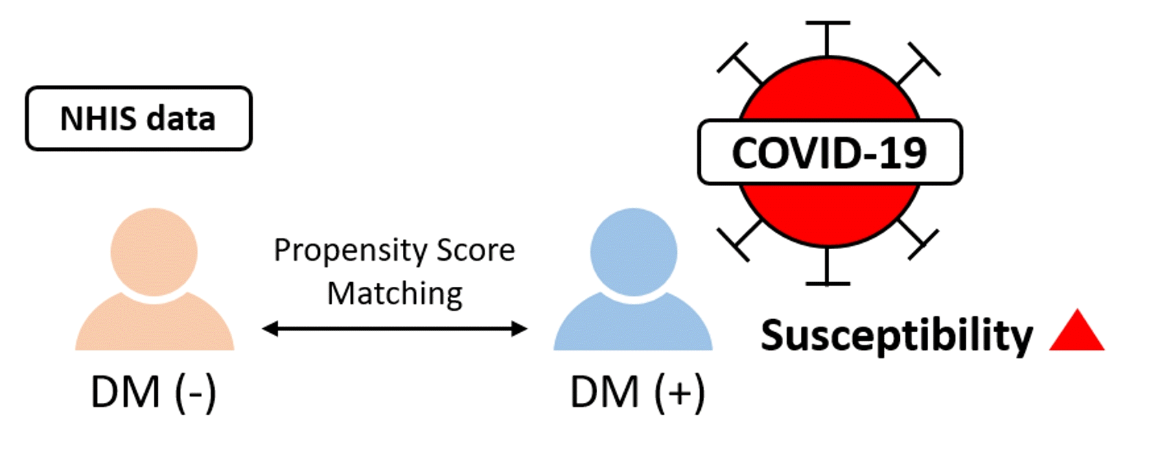
GRAPHICAL ABSTRACT

INTRODUCTION

Since 2019, coronavirus disease 2019 (COVID-19) has resulted in more than 765 million infections and 6.9 million deaths worldwide as of May 2023 [1,2]. It is widely recognized that diabetes exacerbates disease severity and mortality among individuals with COVID-19 [3,4]. The prevalence of COVID-19 among individuals with diabetes has displayed considerable variation (1.7% to 39.7%) owing to discrepancies in diabetes definitions and patient ages, thereby complicating the evaluation of COVID-19 susceptibility [5,6]. Moreover, there is a paucity of studies directly comparing susceptibility to COVID-19 between individuals with and without diabetes. This indicates the need for further research on the susceptibility of patients with diabetes to COVID-19. Therefore, this study aimed to investigate the susceptibility of patients with diabetes to COVID-19 by comparing the infection rates of COVID-19 between individuals with and without diabetes using nationwide population-based data from the National Health Insurance Service (NHIS) of South Korea.

METHODS

Participants who underwent COVID-19 testing between December 2019 and April 2020 were analyzed using data from the NHIS of South Korea. Participants taking antidiabetic drugs or insulin within one year before COVID-19 testing were classified as having diabetes. Considering that the incidence of COVID-19 was high in certain regions during the study period [7], the region was categorized into Seoul, Daegu, Gyeonggi, Gyeongbuk, and others. Comorbidities were defined using diagnostic codes, whereas blood test results were based on the most recent data from 2017. COVID-19 tests were conducted using diagnostic kits or real-time polymerase chain reaction with nasopharyngeal swabs or sputum samples.
Groups with and without diabetes were compared using 1:1 propensity score matching (PSM) with variables including sex, age, comorbidities, medications, etc. Sensitivity analyses included 1:2 and 1:3 PSM. The relationship between the presence of diabetes and the risk of COVID-19 was examined using logistic regression analysis. Statistical significance was set at P<0.05. Statistical analyses were performed using SAS version 9.4 (SAS Institute, Cary, NC, USA). This study was approved by the Institutional Review Board (IRB) of the Kangbuk Samsung Hospital (IRB no. KBSMC 2020-04-040). Patient data were de-identified, and consent from the participants was waived according to the Bioethics and Safety Act of South Korea.

RESULTS

After excluding participants with missing claim data, data from 115,235 participants revealed that 97,965 did not have diabetes, while 17,270 had diabetes. Following 1:1 PSM, the groups with and without diabetes, each comprising 14,661 participants, were compared (Table 1, Supplemental Fig. S1). Both groups exhibited a similar proportions of male sex. Age distribution displayed no significant difference between the two groups, with the highest proportion aged 60 to 79 years. Body mass index averaged approximately 25.0 kg/m² in both groups. In the group with diabetes, glucose levels were significantly higher at 132.6±50.9 mg/dL compared to that in the group without diabetes at 98.1±14.6 mg/dL (P<0.001). Furthermore, the group without diabetes had a significantly higher proportion with dyslipidemia, whereas the group with diabetes had a significantly higher Charlson Comorbidity Index score.
Following 1:1 PSM, in the group without diabetes, 395 (2.7%) tested positive for COVID-19, whereas in the group with diabetes, the proportion was significantly higher (512 patients, 3.5%) (P<0.001). This distinction persisted in the 1:2 and 1:3 PSM analyses, with the group with diabetes consistently presenting a significantly greater proportion of COVID-19 positive cases compared to that in the group without diabetes (Supplemental Table S1).
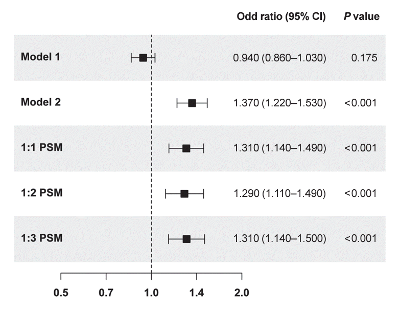
The risk of COVID-19 in relation to the presence of diabetes was analyzed (Fig. 1). In univariate analyses, diabetes did not increase the susceptibility to COVID-19. However, after adjusting for factors such as sex, age, comorbidities, and medication, diabetes significantly increased the risk of COVID-19 (odds ratio [OR], 1.366; 95% confidence interval [CI], 1.217 to 1.534; P<0.001). This increased risk remained significant even after 1:1 PSM, with patients with diabetes showing an increase in susceptibility to COVID-19 (OR, 1.307; 95% CI, 1.144 to 1.493; P<0.001). Sensitivity analyses in the 1:2 and 1:3 PSM cohorts also demonstrated that diabetes significantly increased the risk of COVID-19 (1:2 PSM: OR, 1.310; 95% CI, 1.140 to 1.504; P<0.001; 1:3 PSM: OR, 1.288; 95% CI, 1.114 to 1.491; P<0.001).

DISCUSSION

In this study, we conducted a nationwide population-based propensity score-matched cohort analysis to evaluate the influence of diabetes on COVID-19 susceptibility. Among individuals who underwent COVID-19 testing, those with diabetes had a 1.3-fold higher infection rate than those without diabetes.
In a previous study using NHIS data, there was no significant difference in susceptibility to COVID-19 between individuals without diabetes and those using only oral hypoglycemic agents (OHAs) [8]. However, patients using both insulin and OHA, similar to this study, exhibited a 1.25 times higher susceptibility to COVID-19 compared to individuals without diabetes. However, the previous study targeted a smaller number of diabetic patients (4,246 individuals) compared to the present study. In addition, it used only sex, age, and region as matching variables and included sex, age, region, diabetic medication, comorbidities, and socioeconomic status as adjustment variables. In contrast, the present study targeted a larger number of patients and used multiple variables as both PSM matching variables and adjustment variables. Considering that the results may vary depending on the adjustment, the present study, which utilized multiple adjustment variables, is considered demonstrating statistically more significant results.
When the immune system malfunctions, pathogens such as bacteria, viruses, and fungi breach the body’s defense mechanisms. Diabetes, characterized by insulin deficiency and hyperglycemia, has a notable effect on the immune system [9]. Monocytes isolated from diabetic patients exhibited reduced secretion of interleukin-1β upon lipopolysaccharide stimulation compared to controls [10]. Furthermore, hyperglycemia impairs cytokine production, which is critical for protection against pathogens and adaptive immune responses through antibodies and effector T cell production [11]. Moreover, hyperglycemia is reported to disrupt leukocyte recruitment [12] and trigger dysfunction of neutrophils, macrophages, and natural killer cells [13,14].
Angiotensin-converting enzyme 2 (ACE-2) is known as a cellular entry receptor for severe acute respiratory syndrome coronavirus 2 [15]. ACE-2 expression increases in patients with diabetes and in response to hyperglycemia [16]. Moreover, antidiabetic drugs, such as glucagon-like peptide-1 receptor agonists and thiazolidinediones, have been linked to elevated ACE-2 expression [17,18], whereas medications commonly used in patients with diabetes, such as angiotensin-converting enzyme inhibitors and angiotensin receptor blockers, induce ACE-2 overexpression [19]. As a result, hyperglycemia in diabetes can trigger immune system impairment, and both hyperglycemia and medication usage can amplify ACE-2 expression, potentially heightening the vulnerability of patients with diabetes to COVID-19. As a reflection of these mechanisms, a systematic review reported that patients with diabetes were overrepresented among COVID-19 cases compared to population averages [20].
The current study has several limitations. Because this study categorized participants receiving antidiabetic drugs as patients with diabetes, it did not include those with diabetes who were not taking medication. And this study has the limitation that it did not consider the participants’ glycemic control status, type of antidiabetic drugs, and whether they were admitted to nursing homes or long-term care facilities. Nonetheless, the strength of this study lies in its use of NHIS data to analyze the impact of diabetes on COVID-19 susceptibility across a substantial population that underwent testing. Furthermore, our PSM analyses address a wide range of confounding variables and used diverse adjustment variables, thereby substantially enhancing the accuracy of the results.
Patients with diabetes exhibit a higher COVID-19 infection rate than non-diabetic individuals, and diabetes increases their susceptibility to COVID-19. Given the poor clinical outcomes observed in patients with diabetes and COVID-19, it is imperative to adopt preventive measures against COVID-19 for patients with diabetes.

Supplementary Material

Supplemental Table S1.

Baseline Characteristics and Infection Rate of COVID-19 according to Diabetes Status
enm-2024-2014-Supplemental-Table-S1.pdf

Supplemental Fig. S1.

Study subjects. COVID-19, coronavirus disease 2019; NHIS, National Health Insurance Service.
enm-2024-2014-Supplemental-Fig-S1.pdf

Notes

CONFLICTS OF INTEREST

Won-Young Lee is an editor-in-chief and Eun-Jung Rhee is a deputy editor of the journal. But they were not involved in the peer reviewer selection, evaluation, or decision process of this article. No other potential conflicts of interest relevant to this article were reported.

AUTHOR CONTRIBUTIONS

Conception or design: E.J.R., W.Y.L. Acquisition, analysis, or interpretation of data: S.J.M., J.H.J., K.D.H. Drafting the work or revising: H.N.J., S.J.M., E.J.R., W.Y.L. Final approval of the manuscript: H.N.J., S.J.M., J.H.J., K.D.H., E.J.R., W.Y.L.

ACKNOWLEDGMENTS

This work was supported by the EnM Research Award from the Korean Endocrinology Society to Sun Joon Moon in 2020.

REFERENCES

1. Lu R, Zhao X, Li J, Niu P, Yang B, Wu H, et al. Genomic characterisation and epidemiology of 2019 novel coronavirus: implications for virus origins and receptor binding. Lancet. 2020; 395:565–74.
crossref
2. World Health Organization. COVID-19 weekly epidemiological update, edition 141, 4 May 2023 [Internet]. Geneva: WHO;2023. [cited 2024 Jun 17]. Available from: https://iris.who.int/handle/10665/367666.
3. Hartantri Y, Debora J, Widyatmoko L, Giwangkancana G, Suryadinata H, Susandi E, et al. Clinical and treatment factors associated with the mortality of COVID-19 patients admitted to a referral hospital in Indonesia. Lancet Reg Health Southeast Asia. 2023; 11:100167.
crossref
4. Moon SJ, Rhee EJ, Jung JH, Han KD, Kim SR, Lee WY, et al. Independent impact of diabetes on the severity of coronavirus disease 2019 in 5,307 patients in South Korea: a nationwide cohort study. Diabetes Metab J. 2020; 44:737–46.
crossref
5. Lechien JR, Chiesa-Estomba CM, Place S, Van Laethem Y, Cabaraux P, Mat Q, et al. Clinical and epidemiological characteristics of 1420 European patients with mild-to-moderate coronavirus disease 2019. J Intern Med. 2020; 288:335–44.
crossref
6. Gold JA, Wong KK, Szablewski CM, Patel PR, Rossow J, da Silva J, et al. Characteristics and clinical outcomes of adult patients hospitalized with COVID-19: Georgia, March 2020. MMWR Morb Mortal Wkly Rep. 2020; 69:545–50.
7. Kang D, Choi J, Kim Y, Kwon D. An analysis of the dynamic spatial spread of COVID-19 across South Korea. Sci Rep. 2022; 12:9364.
crossref
8. Chun SY, Kim DW, Lee SA, Lee SJ, Chang JH, Choi YJ, et al. Does diabetes increase the risk of contracting COVID-19?: a population-based study in Korea. Diabetes Metab J. 2020; 44:897–907.
crossref
9. Berbudi A, Rahmadika N, Tjahjadi AI, Ruslami R. Type 2 diabetes and its impact on the immune system. Curr Diabetes Rev. 2020; 16:442–9.
crossref
10. Mooradian AD, Reed RL, Meredith KE, Scuderi P. Serum levels of tumor necrosis factor and IL-1 alpha and IL-1 beta in diabetic patients. Diabetes Care. 1991; 14:63–5.
11. Tanaka T, Narazaki M, Kishimoto T. IL-6 in inflammation, immunity, and disease. Cold Spring Harb Perspect Biol. 2014; 6:a016295.
crossref
12. Martinez N, Ketheesan N, Martens GW, West K, Lien E, Kornfeld H. Defects in early cell recruitment contribute to the increased susceptibility to respiratory Klebsiella pneumoniae infection in diabetic mice. Microbes Infect. 2016; 18:649–55.
crossref
13. Stegenga ME, van der Crabben SN, Blumer RM, Levi M, Meijers JC, Serlie MJ, et al. Hyperglycemia enhances coagulation and reduces neutrophil degranulation, whereas hyperinsulinemia inhibits fibrinolysis during human endotoxemia. Blood. 2008; 112:82–9.
crossref
14. Berrou J, Fougeray S, Venot M, Chardiny V, Gautier JF, Dulphy N, et al. Natural killer cell function, an important target for infection and tumor protection, is impaired in type 2 diabetes. PLoS One. 2013; 8:e62418.
crossref
15. Li W, Moore MJ, Vasilieva N, Sui J, Wong SK, Berne MA, et al. Angiotensin-converting enzyme 2 is a functional receptor for the SARS coronavirus. Nature. 2003; 426:450–4.
crossref
16. Roca-Ho H, Riera M, Palau V, Pascual J, Soler MJ. Characterization of ACE and ACE2 expression within different organs of the NOD mouse. Int J Mol Sci. 2017; 18:563.
crossref
17. Romani-Perez M, Outeirino-Iglesias V, Moya CM, Santisteban P, Gonzalez-Matias LC, Vigo E, et al. Activation of the GLP-1 receptor by liraglutide increases ACE2 expression, reversing right ventricle hypertrophy, and improving the production of SP-A and SP-B in the lungs of type 1 diabetes rats. Endocrinology. 2015; 156:3559–69.
crossref
18. Zhang W, Li C, Liu B, Wu R, Zou N, Xu YZ, et al. Pioglitazone upregulates hepatic angiotensin converting enzyme 2 expression in rats with steatohepatitis. Ann Hepatol. 2013; 12:892–900.
crossref
19. Kriszta G, Kriszta Z, Vancsa S, Hegyi PJ, Frim L, Eross B, et al. Effects of angiotensin-converting enzyme inhibitors and angiotensin receptor blockers on angiotensin-converting enzyme 2 levels: a comprehensive analysis based on animal studies. Front Pharmacol. 2021; 12:619524.
crossref
20. Hartmann-Boyce J, Rees K, Perring JC, Kerneis SA, Morris EM, Goyder C, et al. Risks of and from SARS-CoV-2 infection and COVID-19 in people with diabetes: a systematic review of reviews. Diabetes Care. 2021; 44:2790–811.
crossref

Fig. 1.
Logistic regression analyses for susceptibility of coronavirus disease 2019 (COVID-19) according to diabetes status. Statistical analyses were performed using logistic regression. Model 1: unadjusted; Model 2: adjusted for sex, age, region, low income, body mass index, smoking, systolic blood pressure, aspartate aminotransferase, alanine aminotransferase, gamma-glutamyltransferase, hypertension, dyslipidemia, ischemic heart disease, stroke, asthma or chronic obstructive pulmonary disease, cancer, end-stage renal disease, Charlson Comorbidity Index score, angiotensin-converting enzyme inhibitor, angiotensin II receptor blocker, steroids, and immunosuppressants; propensity score matching (PSM): adjusted for the same variables as in model 2. CI, confidence interval.
enm-2024-2014f1.tif
enm-2024-2014f2.tif
Table 1.
Baseline Characteristics and Infection Rate of COVID-19 according to Diabetes Status
Characteristic Pre PSM
Post PSM
Non-DM DM P value ASD Non-DM DM P value ASD
Number 97,965 17,270 14,661 14,661
Male sex 44,544 (45.5) 9,862 (57.1) <0.001 0.234 8,204 (56.0) 8,114 (55.3) 0.290 0.012
Age, yr <0.001 0.822
 20–29 8,073 (8.2) 83 (0.5) 76 (0.5) 81 (0.6)
 30–39 22,257 (22.7) 457 (2.7) 0.633 416 (2.8) 450 (3.1) 0.014
 40–49 21,028 (21.5) 1,281 (7.4) 0.408 1,196 (8.2) 1,236 (8.4) 0.010
 50–59 18,553 (18.9) 2,860 (16.6) 0.062 2,607 (17.8) 2,622 (17.9) 0.003
 60–69 13,848 (14.1) 4,710 (27.3) 0.329 3,971 (27.1) 3,967 (27.1) 0.001
 70–79 8,820 (9.0) 5,042 (29.2) 0.532 3,992 (27.2) 3,949 (26.9) 0.007
 ≥80 5,386 (5.5) 2,837 (16.4) 0.355 2,403 (16.4) 2,356 (16.1) 0.009
BMI, kg/m2 23.6±3.6 25.1±3.8 <0.001 0.415 25.0±3.7 24.9±3.8 0.427 0.009
Region <0.001 0.551
 Seoul 20,635 (21.1) 3,315 (19.2) 0.047 2,762 (18.8) 2,820 (19.2) 0.010
 Daegu 13,041 (13.3) 1,983 (11.5) 0.029 1,731 (11.8) 1,709 (11.7) 0.014
 Gyeonggi 23,370 (23.9) 3,910 (22.6) 0.056 3,200 (21.8) 3,284 (22.4) 0.005
 Gyeongbuk 6,084 (6.2) 1,499 (8.7) 0.094 1,310 (8.9) 1,269 (8.7) 0.010
 Other 34,835 (35.6) 6,563 (38.0) 0.051 5,658 (38.6) 5,579 (38.1) 0.011
SBP, mm Hg 120.4±14.5 128.7±15.8 <0.001 0.543 128.0±15.5 127.9±15.6 0.652 0.005
Glucose, mg/dL 94.3±13.3 133.0±51.2 <0.001 1.035 98.1±14.6 132.6±50.9 <0.001 0.921
AST, IU/L 24.8±16.0 29.8±22.6 <0.001 0.255 29.4±23.1 29.4±21.4 0.877 0.002
ALT, IU/L 23.7±21.0 29.2±26.0 <0.001 0.236 28.6±30.8 28.7±22.8 0.946 0.001
γ-GTP, IU/L 33.1±48.6 50.2±74.0 <0.001 0.273 48.1±78.8 48.6±69.7 0.613 0.006
Low income 14,156 (14.5) 3,645 (21.1) <0.001 0.175 2,993 (20.4) 3,033 (20.7) 0.563 0.007
Smoking 17,114 (17.5) 3,347 (19.4) <0.001 0.049 2,778 (19.0) 2,770 (18.9) 0.905 0.001
Comorbidities
 Hypertension 36,131 (36.9) 14,753 (85.4) <0.001 1.149 12,269 (83.7) 12,170 (83.0) 0.121 0.018
 Dyslipidemia 43,811 (44.7) 15,508 (89.8) <0.001 1.095 13,080 (89.2) 12,936 (88.2) 0.008 0.031
 IHD 10,333 (10.6) 5,854 (33.9) <0.001 0.585 4,448 (30.3) 4,498 (30.7) 0.526 0.007
 Stroke 3,671 (3.8) 2,638 (15.3) <0.001 0.401 1,923 (13.1) 1,947 (13.3) 0.679 0.005
 Asthma or COPD 20,036 (20.5) 5,940 (34.4) <0.001 0.316 4,953 (33.8) 4,917 (33.5) 0.656 0.005
 Cancer 8,249 (8.4) 3,794 (22.0) <0.001 0.384 3,154 (21.5) 3,105 (21.2) 0.485 0.008
 ESRD 602 (0.6) 1,061 (6.1) <0.001 0.310 472 (3.2) 508 (3.5) 0.242 0.014
 CCI score 2.3±2.4 6.2±3.1 <0.001 1.429 5.5±2.6 5.5±2.7 0.035 0.025
Medication
 ACEi 389 (0.4) 306 (1.8) <0.001 0.133 224 (1.5) 211 (1.4) 0.530 0.007
 ARB 8,225 (8.4) 5,223 (30.2) <0.001 0.576 3,897 (26.6) 3,958 (27.0) 0.421 0.009
 Steroid 14,609 (14.9) 4,338 (25.1) <0.001 0.257 3,662 (25.0) 3,618 (24.7) 0.552 0.007
 Immunosuppressant 895 (0.9) 518 (3.0) <0.001 0.151 378 (2.6) 379 (2.6) 0.971 <0.001
COVID-19 3,353 (3.4) 556 (3.2) 0.174 0.011 395 (2.7) 512 (3.5) <0.001 0.046

Values are expressed as number (%) or mean±standard deviation. Statistical analyses were performed using t test or chi-squared test. Groups with and without diabetes were compared using 1:1 propensity score matching with variables including sex, age, region, BMI, low income, smoking, SBP, AST, ALT, γ-GTP, hypertension, dyslipidemia, IHD, stroke, asthma or COPD, cancer, ESRD, CCI score, ACEi, ARB, steroids, and immunosuppressants.

COVID-19, coronavirus disease 2019; PSM, propensity score matching; DM, diabetes mellitus; ASD, absolute standardized mean difference; BMI, body mass index; SBP, systolic blood pressure; AST, aspartate aminotransferase; ALT, alanine aminotransferase; γ-GTP, gamma-glutamyltransferase; IHD, ischemic heart disease; COPD, chronic obstructive pulmonary disease; ESRD, end-stage renal disease; CCI, Charlson Comorbidity Index; ACEi, angiotensin-converting enzyme inhibitor; ARB, angiotensin II receptor blocker.

TOOLS
Similar articles