

 PDF
PDF Citation
 Print
Print


INTRODUCTION
Chronic subdural hematomas (SDHs) are a common neurosurgical disease. The treatment strategy is usually divided based on whether the patients are symptomatic or not. Symptomatic SDHs are generally evacuated through either a twist-drill craniotomy, burr hole craniotomy, or a craniotomy, while asymptomatic SDHs are managed conservatively. Recently, 3 randomized, prospective trials have shown efficacy in preventing SDH recurrence after middle meningeal artery embolization (MMAE): Embolise, the squid trial for embolization of the middle meningeal artery and managing non-acute SDH using liquid materials: a Chinese randomized trial of middle meningeal artery treatment [1,2]. This endovascular approach was a response to a growing consensus that the pathophysiology of chronic SDH formation is an initially inflammatory response to trauma with subsequent angiogenesis and formation of inflammatory neovascular membranes in the subdural space that receives its vascular supply from the MMA branches [3].
Since Mandai et al. [4] first reported MMAE for chronic SDH, multiple studies have found its utility for SDH. For these cases, MMAE typically begins by obtaining vascular access through the femoral artery and, more recently, radial artery [5]. Though direct access of the MMA has been reported for treatment of dural arteriovenous fistulas (dAVFs), to the best of our knowledge, there is no literature on direct and retrograde MMAE for SDH [6,7]. Here, we present the first case of direct MMA access with retrograde embolization for the treatment of a SDH as a novel approach. Direct MMA access with retrograde embolization during mini-craniotomy for chronic or subacute SDH evacuation obviates the need for 2 separate procedures in hospitals that do not have a hybrid operating room/angiographic suite.
Go to : 
CASE REPORT
Here we present a technical note on treatment of a subacute SDH through evacuation of the hematoma through a mini-craniotomy and concurrent MMAE through catheterization of a frontal branch. A patient with a complicated past medical history including congestive heart failure, chronic kidney disease on dialysis through a right arm arteriovenous fistula, and pulmonary hypertension who presented as a transfer from an outside facility after being found down at his home during a wellness check by a family member. Upon further investigation, his close family member notified our team of multiple recent motor-vehicle collisions where the patient would get lost coming home from his dialysis sessions, most recently about 1 week prior to presentation. The patient was not formally evaluated after the car accidents. The imaging was significant for an acute holohemischepric right SDH that was non-operative at the time.
The patient was oriented to self and place and did not have any focal neurological deficits. He did require supplemental oxygen and was noted to have evidence of fluid overload. Further work up revealed multiple electrolyte abnormalities, pulmonary edema, elevated basic natriuretic peptide, and troponin. The patient was also taking aspirin daily, which was confirmed by an abnormal platelet function assay. Due to multiple active medical problems that carry an increased risk of anesthesia and after discussion with family, a decision was made to hold off on any surgical intervention until he was medically optimized and to wait for the acute component of the hematoma to liquify, which would allow a smaller surgical intervention to further limit risks of prolonged anesthesia.
The patient was admitted to the intensive care unit for dialysis and cardiac and neuro monitoring. His hospitalization was complicated by focal status epilepticus that required intubation and a propofol infusion. Surgical evacuation of the SDH was planned in a delayed fashion at 10 days after admission after a head computed tomography (CT) had shown liquefaction of a large component of the hematoma (Fig. 1A, B). Due to the medical fragility of the patient and to reduce anesthesia/intubation time, we decided to perform a MMAE in the operating room since no hybrid operating room angiography suite was available at our hospital.
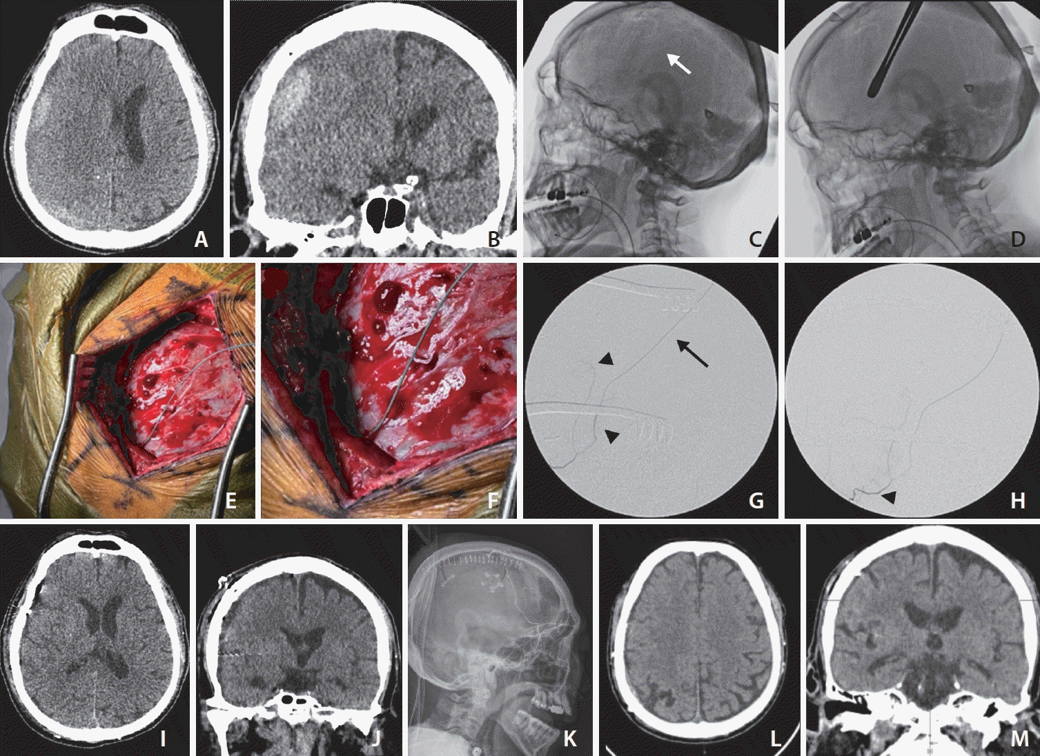 | Fig. 1.(A) Axial and (B) coronal views of computed tomography (CT) images of the head demonstrate a subacute subdural hematoma (SDH) that has partially liquefied 10 days after admission of the patient. (C) Intraoperativelateral skull X-ray. The white arrow points towards a prominent calvarial groove in which an middle meningeal artery (MMA) branch is expected to be found. (D) Lateral skull X-ray with sponge stick over the calvarial groove used to mark out the incision, note the pins of the radiolucent Mayfield head holder. (E, F) Direct catheterization of the frontal MMA branch on the dura at the craniotomy site. (G) Image of the anterior and posterior frontal MMA branches (black arrowheads). The black arrow points at the microcatheter. (H) Intraoperative digital subtraction angiography of the right middle meningeal artery. The black arrowhead points to the point from which the embolization was carried out. (I) Axial and (J) coronal views of CT images of the head demonstrate near resolution of the subacute on chronic SDH along the right cerebral convexity with improvement in midline shift on postoperative imaging. Onyx 18 cast can be seen in the main trunk of the right MMA. (K) Robust penetration of Onyx 18 embolic agent into right frontal branches of the MMA on the lateral skull X-ray. (L) Axial and (M) coronal views of a CT brain show near complete resolution of the right SDH 1.5 months after surgery.
|
Procedure
To facilitate intraoperative fluoroscopy, we placed the patient in a radiolucent Mayfield head holder. Fluoroscopy was used to localize the calvarial grooves of the MMA with a sponge stick (Fig. 1C). The incision was planned above the superior temporalis line and across the skin projection of a prominent calvarial groove. This landmark correlated well with the thickest part of the SDH collection. After we completed the craniotomy, we noted a large posterior frontal branch of the right middle meningeal on the dura that was cannulated using a 1.3 French Headway duo microcatheter (Terumo Neuro) and a 0.014 inch Synchro 2 soft microwire (Stryker) under direct visualization without difficulty (Fig. 1D). A super-selective digital subtraction angiogram of the MMA was performed to confirm intraluminal location of the microcatheter followed by a roadmap using Omnipaque 300 (GE Healthcare) (Fig. 1E, F). Under roadmap visualization, the catheter was navigated more proximally over the microwire using a Bayonet and advanced towards the greater sphenoid wing (Fig. 1G). At this point, a repeat digital subtraction angiogram of the right MMA was performed. No anastomotic connections to the eye were noted. The catheter was then loaded with dimethylsulfoxide (DMSO, Medtronic) over 90 seconds, and under direct fluoroscopic visualization Onyx 18 (Medtronic) was injected. Several frontal branches of the MMA were embolized, and no embolic material traveled towards the eye (Fig. 1H). We slightly retracted the catheter towards the end of the injection to embolize the catheterized branch of the right MMA. We glued the catheter in by injecting Onyx along the catheter into a more distal posterior frontal branch. We approximately used a total of 0.3 mL of Onyx. The catheter was cut with Metzenbaum scissors at its entry site into the MMA (Supplementary Video 1). We then proceeded with the evacuation and irrigation of the SDH in standard fashion. Most of the collection was liquified except for an area of clot over the craniotomy site that was removed using copious irrigation and a Penfield #1 instrument. Of note, we encountered no bleeding from the dural edges. A 12 French Red Robinson subdural drain was left in place, and the craniotomy was closed in standard fashion.
The operative time from placement of pins to skin closure was 100 minutes with a total intubation time of 185 minutes. Total fluoroscopy time was 4 minutes and 50 seconds, and 2 mL of Omnipaque contrast was used. The patient recovered well without visual changes or facial nerve palsy. The subdural drain was discontinued after 48 hours. Post-operative CT brain imaging revealed good evacuation of the hematoma and robust Onyx cast in the MMA (Fig. 1I-K). At the time of discharge, he was fully oriented and had no focal neurological deficits. After 1.5 months, a repeat CT brain showed resolution of the SDH (Fig. 1L, M). At the time, he was in a skilled nursing facility and had recovered to a modified Rankin scale score of 3. Retrograde MMAE during surgery has not previously been described for the treatment of SDH according to our literature search.
Go to :
DISCUSSION
As the number of patients with chronic SDHs continues to increase, it is imperative to continue ongoing efforts to optimize the workflow. Recently, 3 randomized studies have shown efficacy of MMAE in reducing hematoma recurrence [1]. Here we present a novel surgical approach of a combined mini-craniotomy and direct MMA catheterization followed by retrograde embolization for treatment of a subacute on chronic SDH in a medically complex patient to improve workflow.
Medline, PubMed, and Embase were searched since 2000 to June 2024 with the following search terms: “chronic subdural hematoma” and “middle meningeal artery.” We reviewed the abstracts of all papers that were available as full text and for which English translations were available. Those publications which included MMAE for chronic SDH (703) were reviewed for the type of access obtained for the embolization procedure. In cases for which the exact technique was not specified, conclusions were able to be drawn from either provided angiogram figures or assumed to be performed through access proximal to MMA if surgery was not performed.
Based on our literature review, this is the first reported case of a retrograde embolization of the MMA through direct catheterization during surgical treatment for SDH evacuation. MMAE is most commonly undertaken through either a transfemoral or a transradial approach. A recent study published by Qiao et al. [8] described catheterization of the superficial temporal artery in a patient with external carotid artery occlusion with further retrograde access to the internal maxillary artery followed by MMA catheterization leading to successful embolization and total resorption without a surgical adjunct. One case of direct MMA catherization was described by Oh et al. [7] for the treatment of a dAVF after the MMA branch supplying the dAVF could not be embolized successfully using a transfemoral approach. After proximal MMAE, the patient was brought to the operating room for a decortication of the cranium over a frontal branch of the MMA which was then successfully embolized using Onyx [7]. In this case, an antegrade angiogram was performed to better understand MMA branch anatomy and to plan a precise location for decortication. Another case reported by Lin et al. [6] also described direct access to an MMA branch during a temporal craniotomy for treatment of a dAVF after other conventional methods had failed. Both of these cases highlight the technical feasibility of direct catheterization of the MMA, but in both cases the MMA was embolized in an antegrade fashion. In addition, the pathology of the dAVF may have led to enlargement of the MMA, which makes catheterization easier.
Some important considerations for MMAE include obtaining an adequate super-selective MMA angiogram to ensure the absence of an orbital choroidal blush to rule out anastomoses to the eye. Despite injection against the direction of the flow of blood, the medial sphenoidal branch of MMA was easily visualized, and the contrast bolus almost traveled down to the foramen spinosum. No collaterals to the eye were noted in our patient. The systolic blood pressure at the time of embolization was only around 100 mmHg which helped with the retrograde penetration of the contrast bolus and Onyx material, and the patient had a history of congestive heart failure.
We robustly embolized the frontal branches of the MMA. A true parieto-occipital branch was not visualized during angiography, so it is possible that the frontal branch was dominant. Khorasanizadeh et al. [9] reported that treatment of more than 1 MMA branch is not associated with improved treatment efficacy.
This case is nuanced in that the patient required a mini-craniotomy as opposed to burr hole drainage due to the component of a solid clot, which is not the case for all patients. However, some patients, even in the absence of a solid clot, may benefit from resection of subdural membranes through a mini-craniotomy. At this time, studies report mixed results on the comparison of burr hole drainage versus craniotomy except that drain placement is associated with reduced recurrence as opposed to no drainage [10,11]. Additionally, more studies comparing the outcomes between different surgical approaches in combination with MMAE are needed.
We performed an Onyx embolization with DMSO administration, which is better tolerated under general anesthesia. However, the MMAE could also have been carried out under conscious sedation with n-butyl cyanoacrylate or particles. From a workflow perspective though, the intraoperative embolization only added approximately 30 minutes to the case which is difficult to beat with a separate procedure if transport and positioning times are also considered.
The direct intraoperative MMAE procedure may also introduce unwanted events or complications. The intraoperative fluoroscopy equipment may not have the same resolution as a dedicated neuro-interventional suite. The microcatheter may get glued in place and need to be cut, a procedure for which the catheter is not U.S. Food and Drug Administration-approved. A retained catheter could cause a surgical site infection or foreign body inflammatory reaction.
We do not advocate for direct MMA catheterization and retrograde embolization for all patients but do believe that this approach can save time under anesthesia for high-risk patients and reduce total combined operative and procedural time for the health care system in hospitals that do not have a hybrid operating room/angiography suite. We were able to carry out this combined procedure in a standard operating room with a single-plane C-arm. Further studies are needed to determine how often the retrograde MMAE can be carried out successfully in a larger patient cohort.
Go to :

 XML Download