Abstract
Purpose
Intracranial mycotic aneurysms (IMAs), rare and often life-threatening, result from arterial wall infections typically caused by bacteria such as Staphylococcus and Streptococcus. The standard treatment for ruptured aneurysms is not well-defined and often individualized. This study investigates the efficacy of endovascular glue embolization in managing ruptured IMAs, based on our center’s experience.
Materials and Methods
A retrospective analysis was conducted for ruptured IMAs treated with glue embolization between January 2016 and December 2023. The procedure involved aneurysm sac and parent vessel occlusion with glue delivery. Data included patient demographics, clinical presentations, and neuroimaging. Clinical outcomes were assessed using the modified Rankin scale (mRS) at 3 months, and angiographic follow-up was conducted at 6 months.
Results
The study included 28 patients, predominantly male (64.3%), with a mean age of 48 years. Headache was the primary symptom in 92.9% of cases, and positive blood/cerebro spinal fluid cultures were found in 82.14% of cases. All aneurysms were located in the distal circulation, primarily in the anterior circulation system. Glue embolization was successfully performed in all cases, achieving complete aneurysm sac and parent vessel obliteration. Follow-up at 3 months indicated mRS scores of 0 or 1 in 96.5% of cases. Six-month angiographic follow-up showed no aneurysm regrowth or new formations.
Conclusion
Endovascular glue embolization demonstrated high efficacy and safety in treating ruptured IMAs, with a 100% obliteration rate and favorable clinical outcomes in this single-center experience. Despite limitations such as its retrospective design and small sample size, the study supports glue embolization as a viable, less invasive alternative to traditional surgery. Further comparative studies are needed to confirm these findings and refine treatment approaches.
Intracranial mycotic aneurysms (IMAs) are rare, accounting for 2–3% of intracranial aneurysms [1,2]. These lesions result from infections of the arterial wall, most commonly caused by staphylococcus and streptococcus [2]. Risk factors include infective endocarditis, intravenous drug abuse, sepsis, meningitis, and immunosuppression [2,3]. Pathogenesis involves septic emboli inhabiting the arterial wall, leading to inflammation and necrotic degradation of the tunica media and adventitia while leaving intima intact [3]. This results in pseudoaneurysm formation due to pulsatile pressure [3-5]. IMAs are typically located in the distal branches of major cerebral arteries and are often fusiform, but saccular or fusi-saccular (exhibits both fusiform form and a localized sac) forms are also seen on angiograms [4,5]. These IMA commonly present as intraparenchymal or subarachnoid hemorrhage [1,6]. Unruptured IMA can often be managed with antibiotics alone, but the ruptured IMA carry a high mortality rate, with worsens prognosis if aneurysm rebleeds [7,8]. Therefore, urgent intervention either endovascular or surgical, is required for ruptured IMA to prevent rebleeding [1,4,8,9]. Endovascular treatment of IMA is favored due to challenges in identifying and managing multiple, distally located aneurysms via open surgery [9]. The literature offers limited data on the efficacy of using glue (n-butyl cyanoacrylate [NBCA]) for embolizing ruptured IMA. This study aims to explore the efficacy and outcomes of glue embolization in managing ruptured IMA, based on our center’s past experiences.
After obtaining approval from the Ethics Committee of the Institute, we conducted a retrospective analysis of the Neurointervention database at the Institute from January 2016 to December 2023. Our study included 28 cases of ruptured IMA treated endovascularly with only glue embolization. This study focused exclusively on ruptured aneurysms, excluding any unruptured cases. Included patients were aged between 15 and 80 years and had positive findings on blood/cerebro spinal fluid (CSF) culture or vegetations on echocardiography. Data collected comprised patient demographics profiles, clinical presentations, blood/CSF culture or echocardiography reports and neuroimaging findings from computed tomography (CT) and/or magnetic resonance imaging (MRI). Additionally, we reviewed angiographic findings including aneurysm location, size and shape, endovascular treatment characteristics, clinical outcome at 3 months, and follow-up angiography at 6 months.
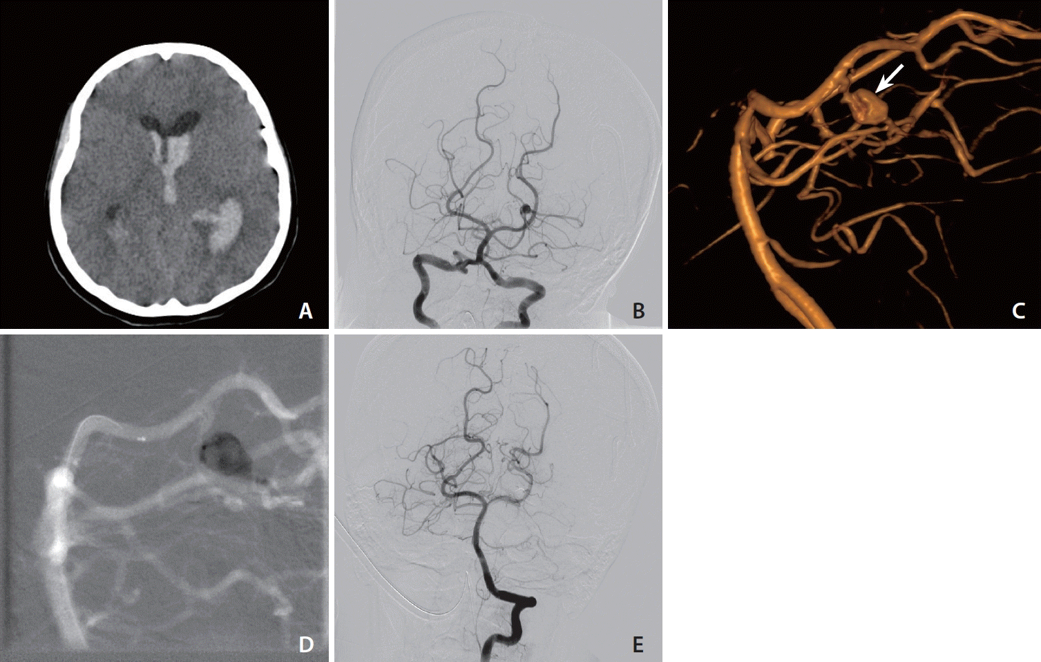
After obtaining informed consent, all procedures were performed by a single senior neurointerventionist using a uniplane angiographic system (Siemens Artis Q). Procedures were conducted under general anesthesia with femoral access. Typically, the procedure began with the placement of a long guiding sheath (Ballast 0.088) in the main vessel followed by the placement of a distal access catheter (AXS Catalyst 5) of length 115 cm for support. Subsequently, a microcatheter of appropriate size (Marathon-165 cm) was used to access the aneurysm with a suitable microwire (Traxcess or Synchro 14). The microcatheter was carefully navigated to the aneurysm sac under real-time imaging. Ensuring stable microcatheter positioning was critical for controlled glue injection. Under fluoroscopic guidance, a liquid embolic agent (glue) was delivered into the aneurysm to obliterate the sac and parent vessel (Fig. 1). The glue concentrations used ranged from 10–60%. Injection was performed slowly, with constant monitoring through fluoroscopy. The operator must be vigilant to stop the injection immediately if the glue begins to migrate beyond the target area. Immediate post-procedural angiography was performed to ensure complete aneurysm occlusion and to rule out non-target embolization. Culture-guided appropriate antibiotics were administered for 8 weeks in all cases. Clinical follow-up was conducted at 3 months, using the modified Rankin scale (mRS) to assess clinical outcomes. Follow-up radiological assessment was performed at 6 months with digital subtraction angiography (DSA) of the brain to assess for any residual or new lesions. The end point of the study was obliteration of the aneurysm.
Within the specified time frame, our study encompassed a total of 28 cases. The mean age of the patients was 48.0 years, with ages ranging from 15–73 years. Among them, 53.6% (n=15) were under 50 years of age. Of the total, 64.3% (n=18) were male while 35.7% (n=10) were females. The most prevalent clinical presentation was headache, observed in 92.9% (n=26) of cases. Additionally, fever was associated with headache in 46.4% (n=13) of cases, while 7.1% (n=2) presented with neurological deficits such as hemiparesis. At admission, cranial imaging (CT/MR scan) revealed parenchymal hematoma in 89.3% cases (n=25). Only 2 cases exhibited intraventricular hemorrhage, and 1 case presented with subdural hematoma in addition to parenchymal hematoma. Positive blood culture for bacterial agents were reported in 64.3% (n=18) of cases, while CSF cultures were positive in 17.9% (n=5) of cases. Additionally, valvular vegetations (aortic or mitral) indicative of infective endocarditis were observed in the 35.7% (n=10) of cases. All the IMA were situated in the distal circulation system. Among the 28 cases, anterior circulation aneurysms were identified in 71.4% (n=20) while posterior circulation system aneurysms were detected in 28.6% (n=8) of cases. Morphologically, fusiform IMA were observed in 57.1% (n=16) of cases and fusi-saccular in 42.9% (n=12). No saccular aneurysm was observed in our study. The mean diameter of the mycotic aneurysms was 3.7 mm (Table 1).
All cases underwent endovascular treatment with glue embolization on the same day as the detection of the mycotic aneurysm. Aneurysm sac obliteration along with parent vessel occlusion was performed in all cases (n=28). The percentage of glue used in treatment ranged from 10–60%. One case needed a decompressive craniotomy for mass effect, while another required external ventricular drainage for intraventricular hemorrhage.
Clinical outcome at 3 months, assessed using the mRS, showed scores of 0 and 1 in 78.6% (n=22) and 17.9% (n=5) of cases, respectively. One (3.6%) case had a mRS of 3. Angiographic follow-up with DSA at 6 months revealed no regrowth or newly formed aneurysm in any of the cases (Table 1).
The clinical presentation of a ruptured IMA varies depending on the severity of infection, associated comorbidities, and the aneurysm’s location. Patients with ruptured mycotic aneurysms usually manifest with persistent localized or diffuse headache, a subacute history of fever, or neurological deficits [1,3,4,7]. Accordingly, in our study, the most common presenting symptom was headache (92.9%), followed by fever (46.4%). Intraparenchymal hemorrhage is more commonly associated with mycotic aneurysm than with their saccular counterparts. Interestingly, the size of the mycotic aneurysm does not predict the likelihood of rupture [7,8]. In our study group, we found that parenchymal hemorrhage (89.2%) was the most common presentation on radiographic imaging.
The diagnosis of a mycotic aneurysm is established by detecting the aneurysm on imaging alongside concurrent predisposing factors, like endocarditis, sepsis, meningitis or immunosuppression [7,10]. Despite advancements in noninvasive neuroimaging and concerns over risks associated with DSA, conventional angiography remains the gold standard for detecting mycotic aneurysms [4,5,8,11]. Classic descriptive features on an angiogram include a distal location with predilection of middle cerebral artery (MCA) territories, fusiform shape, and changes in appearance on follow-up angiograms [6,12]. In this series, DSA was performed in all cases. Anterior circulation system mycotic aneurysms were found in the anterior cerebral artery distal to the A2 segment in 17.9% of cases, and MCA aneurysms were located in the M3 or M4 segment in 53.6% of cases. Posterior circulation aneurysms involved the distal segment of the posterior cerebral artery in 28.57% of cases. In addition to imaging, confirmation of the diagnosis was supported by positive cultures from blood or CSF or the presence of vegetations on echocardiography. Literature indicates that infective pathogens can be isolated in 50–85% of the cases, with common organisms including staphylococcus and streptococcus followed by salmonella, pseudomonas, and others [11-14]. Aortic or mitral valve vegetations are observed on echocardiography in cases of infective endocarditis. Our study demonstrates the presence of infective pathogens and valvular vegetations on cardiac imaging in 82.14% and 35.7% of cases, respectively.
The re-rupture risk is much higher during the acute phase of the disease, and life-threatening bleeding may occur during this period. Therefore, early detection and treatment is mandatory given the high mortality of 30–80% secondary to rebleed associated with ruptured IMA. Both surgical and endovascular treatments are safe and effective for treating mycotic aneurysms [1]. Due to the friable wall and fusiform nature of these aneurysms, clipping is technically challenging. Traditional surgical approach for fusiform mycotic aneurysm involves vessel sacrifice by trapping and resection of the aneurysm, proximal vessel ligation or microvascular anastomosis in reasonable cases. Difficult to access surgically due to distal and deep location, morbidity associated with surgical dissection makes minimal invasive endovascular embolization of an aneurysm with parent artery occlusion a favorable approach over surgery [3,4,6,7]. Obvious advantages over surgery is the less invasive nature, easier to access distal aneurysms, minimal procedure associated trauma to the already compromised cardiac status patients, less mortality, and shorter delay to subsequent cardiac surgery if needed [6,7,15].
Fusiform or fusi-saccular mycotic aneurysm sac obliteration and parent vessel occlusion by embolization with an endovascular approach can be a reasonable alternative to open surgery. Parent artery occlusion in fusiform morphology is safe to perform on distal vessels that supply non-eloquent areas, given there is sufficient collateral circulation. If the vessel supplies an eloquent territory, a balloon occlusion test can be useful in assessing tolerance in a cooperative patient [3,4,15]. Nevertheless, a substantial number of patients demonstrate tolerance to parent artery occlusion due to the pathophysiology of mycotic aneurysm formation. Initially, septic embolization and subsequent vessel necrosis lead to ischemia, but recanalization may induce mycotic aneurysm. If the initial event of ischemia is tolerated, endovascular embolization is likely to be well-tolerated as well [13,16]. Morphologically, a saccular mycotic aneurysm demands special attention and needs to be embolized alone without sacrificing the parent vessel [7]. Endovascular coil embolization of the mycotic aneurysm enables controlled deployment; however, it carries risks such as coil compaction, aneurysm recanalization, and rebleeding [3-5,15]. In numerous cases, the parent artery is often small, posing challenges in advancing the microcatheter to the lesion [4,5,15].
Glue embolization is known for its outstanding durability and rapid setting, making it highly effective for immediate aneurysm occlusion. In this study, the glue concentration varied from 10% to 60%, with the selection based on specific aneurysm size and flow dynamics characteristics. Larger size and high flow rates aneurysms necessitated higher concentrations to achieve rapid polymerization and minimize the risk of glue migration. Ultimately, the concentration was determined by the operator’s experience, considering intra-procedural factors, catheter stability, and real-time imaging feedback. Precise control is essential to avoid non-target embolization, and prompt catheter withdrawal is necessary to prevent adhesion to the vessel wall. A systematic review by Desai et al. [16] on mycotic aneurysm treatments found coiling (50.4%) as the most used technique, followed by glue (28.2%) and Onyx (16.1%) [6-8]. The review indicated comparable obliteration rates across different techniques: coiling (99.1%), NBCA (100%), and Onyx (100%) with similar complication rates: coiling (4.3%), NBCA (15.2%), and Onyx (8.1%). In this series, we observed a 100% obliteration rate on follow-up angiograms at 6 months. Clinical outcomes were favorable, with 96.5% of cases having a mRS score of 0-1. Rebleeding is a potential complication that can occur before, during, or after the procedure. In our study, the mycotic aneurysm was treated promptly following diagnosis, with no instances of intraoperative or postoperative rupture [16]. Vasospasm, both clinical and radiological, was noted in 2 cases and was successfully managed with intra-arterial nimodipine. Introducing foreign materials into infected areas may potentially worsen local infection or trigger abscess formation but no such complications were reported in the literature or observed in our study. Administration of antibiotics before and after the intervention may mitigate these infectious risks.
Limitations of this study include its retrospective single-center design, small sample size of 28 cases, and absence of comparative analysis with other treatment modalities.
In our single-center experience, endovascular glue embolization demonstrates promising efficacy in the treatment of ruptured IMAs. With a 100% obliteration rate and favorable clinical outcomes, glue embolization has demonstrated comparable obliteration rates with fewer complications, making it a viable option for managing ruptured IMAs. Further comparative studies are warranted to validate these findings and elucidate the optimal treatment approach for managing this challenging condition.
ACKNOWLEDGMENTS
We would like to express our sincere gratitude to everyone who supported this study. Special thanks to Palak whose insights were invaluable throughout this project.
Notes
REFERENCES
1. Bhattacharyya A, Mittal S, Yadav RR, Jain K, Gupta B, Parihar A, et al. Endovascular management of infective intracranial aneurysms with acrylic glue. A report of two cases. Interv Neuroradiol. 2009; 15:443–447.
2. Sirakov A, Elhasan HA, Aguilar Pérez M, Serna Candel C, Bäzner H, Henkes H. Middle cerebral artery (M3) aneurysm: two “mycotic” aneurysms of the middle cerebral artery due to bacterial endocarditis; endovascular treatment of one aneurysm with glue (nBCA) injection during adenosine-induced asystole; spontaneous resolution of the second aneurysm. In: Henkes H, Lylyk P, Ganslandt O. The Aneurysm Casebook. Springer, Cham, 2020;1027-1038.
3. Asai T, Usui A, Miyachi S, Ueda Y. Endovascular treatment for intracranial mycotic aneurysms prior to cardiac surgery. Eur J Cardiothorac Surg. 2002; 21:948–950.
4. Kong KH, Chan KF. Ruptured intracranial mycotic aneurysm: a rare cause of intracranial hemorrhage. Arch Phys Med Rehabil. 1995; 76:287–289.
5. La Barge DV 3rd, Ng PP, Stevens EA, Friedline NK, Kestle JR, Schmidt RH. Extended intracranial applications for ethylene vinyl alcohol copolymer (Onyx): mycotic and dissecting aneurysms. Technical note. J Neurosurg. 2009; 111:114–118.
6. Yuan SM, Wang FG. Cerebral mycotic aneurysm as a consequence of infective endocarditis: a literature review. Cor et Vasa. 2017; 59:e257–e265.
7. Lucas JTM, Elhamdani S, Jeong SW, Yu A. Mycotic aneurysm presenting as subdural empyema: illustrative case. J Neurosurg Case Lessons. 2022; 3:CASE21507.
8. Korkmazer B, Karaman AK, Üstündağ A, Süleyman K, Arslan S, Kızılkılıç O, et al. Outcomes of endovascular treatment for intracranial mycotic aneurysms: a retrospective data analysis of a tertiary center. Cerrahpaşa Med J. 2024; 48:1–7.
9. Cheng-Ching E, John S, Bain M, Toth G, Masaryk T, Hui F, et al. Endovascular embolization of intracranial infectious aneurysms in patients undergoing open heart surgery using n-butyl cyanoacrylate. Interv Neurol. 2017; 6:82–89.
10. Esenkaya A, Duzgun F, Cinar C, Bozkaya H, Eraslan C, Ozgiray E, et al. Endovascular treatment of intracranial infectious aneurysms. Neuroradiology. 2016; 58:277–284.
11. Ducruet AF, Hickman ZL, Zacharia BE, Narula R, Grobelny BT, Gorski J, et al. Intracranial infectious aneurysms: a comprehensive review. Neurosurg Rev. 2010; 33:37–46.
12. Alawieh AM, Dimisko L, Newman S, Grossberg JA, Cawley CM, Pradilla G, et al. Management and long-term outcomes of patients with infectious intracranial aneurysms. Neurosurgery. 2023; 92:515–523.
13. Matsubara N, Miyachi S, Izumi T, Yamanouchi T, Asai T, Ota K, et al. Results and current trends of multimodality treatment for infectious intracranial aneurysms. Neurol Med Chir (Tokyo). 2015; 55:155–162.
14. Kiwan R, Son M, Mayich M, Boulton M, Pandey S, Sharma M. Ruptured intracranial infectious aneurysms: single Canadian center experience. Surg Neurol Int. 2022; 13:185.
15. Bele K, Ullal S, Mahale A, Rani S. Unusual Intracranial Manifestation of Infective Endocarditis. Open Neuroimaging J. 2021; 14:28–32.
16. Desai B, Soldozy S, Desai H, Kumar J, Shah S, Raper DM, et al. Evaluating the safety and efficacy of various endovascular approaches for treatment of infectious intracranial aneurysms: a systematic review. World Neurosurg. 2020; 144:293–298.e15.