Journal List > Cardiovasc Prev Pharmacother > v.6(4) > 1516088710

Kim: Use of dual-energy x-ray absorptiometry for body composition in chronic disease management

Abstract

As individuals age or contend with chronic diseases, shifts in body composition often emerge, characterized by a loss of muscle mass and an increase in fat mass, even among those with stable body weight. Both obesity and sarcopenia are key drivers of frailty, disability, and heightened morbidity and mortality. The simultaneous decline in skeletal muscle and accumulation of visceral fat can work synergistically, magnifying their detrimental effects on physical function and metabolic health. Today, dual-energy x-ray absorptiometry (DEXA) is widely recognized as one of the most versatile imaging techniques for assessing not only osteoporosis but also sarcopenia and obesity. Whole-body DEXA facilitates comprehensive analysis, offering detailed insights into fat mass, non-bone lean mass, and bone mineral content at both total and regional levels. DEXA is highly valued for its accuracy, reproducibility, speed, affordability, and low radiation exposure. Furthermore, advancements in DEXA technology and software now allow for precise estimation of visceral adipose tissue. This review underscores the clinical applications of whole-body DEXA, focusing on the use of muscle and fat mass indices in diagnosing low muscle mass, sarcopenia, and sarcopenic obesity, aligned with the latest research and guidelines.

INTRODUCTION

The issues of obesity and an aging population are global concerns in the 21st century, not just limited to Korea. Both aging, particularly from middle age onwards, and obesity significantly influence changes in body composition, which in turn contribute to the onset and exacerbation of chronic diseases [13]. Over recent decades, the prevalence of obesity has surged globally. In Korea, the rate of obesity, defined as a body mass index (BMI) of 25.0 kg/m2 or higher, rose by 18%, from 32.6% in 2009 to 38.5% in 2018 [4]. This increase in obesity prevalence was noted across all age groups, with the most significant rise observed in those aged 80 years and older [5]. BMI is commonly used as an indicator of obesity and is strongly correlated with body fat percentage. However, relying solely on BMI to assess obesity in older adults who typically experience a decrease in lean body mass and an increase in fat mass may lead to an underestimation of their actual fat mass.
Obesity is defined as the excessive accumulation of fat to a degree that is harmful to health [6]. However, recent studies suggest that using BMI as a diagnostic criterion for obesity in the general adult population is not suitable for older adults. This has led to the introduction of the concept of sarcopenic obesity [79]. Sarcopenic obesity is characterized by an excessive accumulation of fat coupled with a concurrent decrease in lean body mass [10]. Research indicates that both the prevalence and risk of sarcopenic obesity increase with age [8,11]. In older adults, obesity exacerbates sarcopenia by increasing fat infiltration within the muscles, reducing the capacity for physical activity, and elevating the risk of mortality [1215]. With the proportion of the population aged 65 years and older rapidly increasing, and the prevalence of obesity within this group continuing to rise, the prevention and treatment of obesity necessitate management at a national level, not merely as an individual concern. Particularly for older adults, accurate diagnosis is crucial for the effective management of obesity.
When evaluating the reliability and validity of methods to assess obesity, the concept of a body compartment model is utilized. This model divides the body's structural framework into sections [16]. The two-compartment model separates the body into fat mass and fat-free mass. The latter is further broken down chemically into water, protein, and minerals, which allows for the expansion of the two-compartment model into a chemical four-compartment model [17].
To diagnose obesity, various physical measurement methods such as BMI, waist circumference, and skinfold thickness are employed, along with instrumental methods including bioelectrical impedance analysis, dual-energy x-ray absorptiometry (DEXA), magnetic resonance imaging (MRI), computed tomography (CT), and air displacement plethysmography (Table 1). Physical measurement methods and bioelectrical impedance analysis are relatively simple and can be used to diagnose obesity or overweight in large populations. However, these physical measurements have limitations in accurately calculating muscle mass and fat mass [18,19]. CT and MRI are instrumental in accurately measuring body composition and determining the distribution of fat and muscle mass by body segment. However, CT involves a high risk of radiation exposure and is costly [20,21]. MRI, while also providing clear differentiation between visceral and subcutaneous fat, does not involve radiation exposure. Nevertheless, it is limited by high costs and restricted accessibility. DEXA has been actively used in clinical research as a standard method for measuring body fat and shows a high correlation with muscle mass measurements obtained by CT or MRI. It offers the added benefit of lower radiation exposure and is recognized as a standard test for measuring limb muscle mass. Recently, in Korea, the use of DEXA for body composition analysis in sarcopenia was endorsed by the New Health Technology Assessment Committee in 2019 as a safe and effective technology for diagnosing and monitoring treatment outcomes in suspected and confirmed cases of sarcopenia, facilitating its widespread clinical use.

BASIC PRINCIPLES OF DEXA

X-ray imaging operates by emitting photons from an x-ray source at a specific energy level. These photons are either absorbed or scattered by obstacles, with the resulting image brightness reflecting the number of photons that reach the detector [22]. DEXA is considered the gold standard for body composition analysis, utilizing a three-compartment model to measure fat mass, fat-free mass, and bone mineral content (Table 2). The fundamental principle of DEXA involves assessing the rate of radiation absorption by different materials in the body as low-dose radiation at two distinct energy levels passes through it. As the x-ray beam travels through tissues, its energy is attenuated, a process influenced by the beam's energy intensity and the thickness and density of the tissues. Essentially, higher photon energy results in lower x-ray attenuation, allowing photons to pass more easily through low-density materials, such as soft tissue, compared to high-density materials like bone. The attenuation difference at the two energy peaks of the x-ray varies according to the type of tissue encountered. Consequently, DEXA calculates the attenuation coefficient ratio (R-value) at these two energy peaks. The R-value remains consistent for bone and fat across all subjects. However, the R-value for soft tissue varies with an individual's tissue composition, where a lower R-value indicates a higher fat percentage.
While DEXA provides measurements of total or regional fat mass, fat-free mass, and bone mineral content, it does not directly measure all three components simultaneously. In whole-body scans, approximately 40% to 45% of the pixels contain bone, potentially including adjacent soft tissues. In DEXA images, pixels are categorized as either soft tissue or bone pixels to facilitate the calculation of bone mineral density and fat mass. It is assumed that bone pixels represent a mix of soft tissue mass and bone mass. Soft tissue pixels are presumed to consist of fat-free tissue, excluding both fat and bone, and the fat mass of these pixels is calculated first. The soft tissue mass in bone pixels is assumed to contain the same fat mass as that in soft tissue pixels. Subsequently, the bone mass is determined using the specific attenuation value from the soft tissue within the bone pixels, which also enables the calculation of soft tissue mass.

USES OF DEXA

Accurate measurements of body composition are useful for understanding energy metabolism in various physiological or clinical contexts. Chronic diseases and their progression can impact body composition, affecting bones and muscles, and these changes may potentially occur in all diseases. Therefore, comprehensive body composition analysis using whole-body DEXA scans offers a valuable approach that can be utilized in a range of clinical studies and practical clinical settings. Applications of body composition analysis span physiological and pathophysiological conditions, including athletes, growth, and aging processes (e.g., sarcopenia), as well as diverse populations and diseases (e.g., obesity, eating disorders, endocrine, gastrointestinal, renal, and infectious diseases) [2325].
The 2013 official position guidelines of the International Society for Clinical Densitometry (ISCD) endorsed the broader application of DEXA for body composition analysis, specifically recommending its use in three pathological conditions.
  • (1) For HIV-positive patients, it is important to evaluate fat distribution when using antiretroviral therapy due to the associated risk of lipoatrophy.

  • (2) For patients undergoing bariatric surgery, or those on medications, diets, or weight loss therapies anticipated to result in significant weight loss, it is advisable to evaluate both fat and fat-free mass when weight loss exceeds 10%.

  • (3) For patients with reduced strength or poor physical function, it is essential to evaluate both fat and fat-free mass.

According to the official ISCD guidelines, studies and reports on body composition analysis should include key variables such as bone density, bone mineral density, bone mineral content, total mass, total fat-free mass, total fat mass, and body fat percentage [26,27].
In recent years, more specific DEXA metrics and measurements have been established for assessing muscle mass and obesity, though their clinical utility remains uncertain. DEXA metrics for fat and muscle mass include the fat mass index (FMI; total fat mass / height2), visceral adipose tissue (VAT), subcutaneous adipose tissue (SAT), the android (A) to gynoid (G) fat ratio, trunk to leg fat mass, lean mass index (total lean mass / height2), appendicular lean mass (ALM; arm lean mass + leg lean mass), ALM adjusted for height2 (ALM/height2), ALM adjusted for weight (ALM/weight), and ALM adjusted for BMI (ALM/BMI) [27,28]. The lean mass index, ALM/height2, ALM/weight, and ALM/BMI are used as diagnostic indicators of "low muscle mass" in cases of sarcopenia. Since the European Working Group on Sarcopenia in Older People (EWGSOP) defined sarcopenia in 2010 as a condition characterized by a gradual and overall decline in muscle mass, along with muscle strength or physical performance, both muscle mass and strength or physical performance must be evaluated to diagnose sarcopenia [29]. ALM/height2 has been shown to be a good predictor of physical disability and mortality risk. Recent guidelines from the revised 2018 EWGSOP [30] and the 2019 Asian Working Group for Sarcopenia [31] recommend using appendicular muscle mass divided by height2 as the primary indicator of muscle mass reduction. Some reports suggest that indicators adjusted for BMI or weight may be more closely related to metabolic indicators or better at predicting muscle function decline and disability than ALM/height2 [32,33]; however, further research is needed in this area. Recent DEXA programs allow for the assessment of muscle mass and sarcopenia status by inputting separate measurements of muscle strength and walking speed (Table 3).
Obesity indicators, such as total fat mass, FMI, body fat percentage, and the total fat to muscle ratio, show strong associations with an increased risk of cardiovascular and metabolic diseases, regardless of BMI [34]. Additionally, the distribution of fat within the body may be more clinically significant than the total fat mass itself [35,36]. For this reason, DEXA variables that reflect central and peripheral fat distribution—such as android and gynoid body fat mass, the A/G fat ratio, trunk to leg fat ratio, and VAT, SAT, and VAT/SAT—can be clinically useful. DEXA devices calculate the A/G fat ratio as the ratio of fat mass between the android and gynoid regions of interest (ROI). The android ROI is typically defined as the area between the last rib and the iliac crest. The upper boundary of the gynoid ROI is located at a distance of 1.5 times the height of the android ROI below it, and the lower boundary, which includes the thigh-buttock region, is determined by a line extending twice the height of the android ROI downward from the upper boundary [37]. DEXA variables that represent waist circumference and waist to hip ratio, such as android fat mass and the A/G fat ratio, are strongly associated with insulin resistance and dyslipidemia [38]. The trunk to leg fat ratio can serve as an indicator to assess fat redistribution in patients experiencing lipoatrophy or lipodystrophy during treatment [27]. FMI has been explored as an alternative to BMI for obesity classification, focusing on excess fat mass rather than excess weight [27]. FMI utilizes sex- and race-specific reference ranges and, although its clinical utility has not yet been established, it may offer a more accurate assessment of obesity in individuals with high muscle mass. Several obesity-related organizations and institutions recommend against using BMI alone to assess obesity in individuals with high muscle mass and suggest that body composition analysis can provide a more accurate determination of obesity levels [27,39].
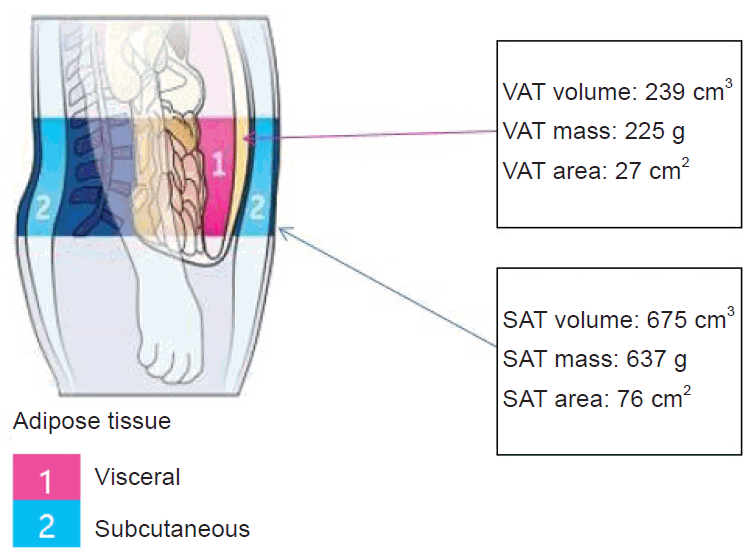
One of the recent advancements in whole-body DEXA technology is its capability to assess VAT (Fig. 1). The latest whole-body DEXA programs initially detect intra-abdominal and external fat within the android ROI to estimate the amount of SAT. The estimated SAT is then subtracted from the total fat mass in the android ROI to calculate the final amount of VAT. In terms of diagnostic criteria, VAT is traditionally measured in square centimeters (cm2) in CT scans, whereas DEXA provides VAT volume, weight, and cross-sectional area. Additionally, DEXA-derived VAT measurements have demonstrated a high correlation with values obtained from CT scans (r=0.93) [40]. DEXA-derived VAT measurements offer several advantages over CT, most notably improved accuracy with lower radiation doses. As the clinical significance of VAT has been recognized, several studies have demonstrated that VAT predicts mortality and cardiovascular disease risk more accurately than SAT or the A/G fat ratio [27,41]. Even with a normal BMI, an individual can exhibit a high degree of visceral obesity, which complicates the detection of metabolically obese normal weight using traditional anthropometric methods alone. Particularly when evaluating sarcopenia or obesity in older or chronically ill patients, the simultaneous measurement of muscle mass, fat mass, and visceral fat through a DEXA scan can minimize diagnostic errors.
ALM measured by DEXA (DEXA-ALM) serves as a crucial indicator of low muscle mass, while VAT assessed by CT (CT-VAT) is the standard for evaluating visceral obesity. Sarcopenia and visceral obesity can interact metabolically, leading to significant adverse effects on metabolic diseases and atherosclerosis. However, most existing studies have either examined the roles of sarcopenia and visceral obesity separately using DEXA-ALM and CT-VAT, or have utilized the ratio of DEXA-ALM to CT-VAT as a marker of sarcopenic obesity [9,42]. With advancements in DEXA technology, it is now feasible to measure both appendicular muscle mass and VAT simultaneously. This capability not only offers convenience but also provides a range of body composition analysis results, which is expected to further broaden the applications of DEXA.

CONCLUSIONS

Body composition analysis using DEXA achieves accuracy comparable to that of CT and MRI, and its use is growing due to several advantages over other diagnostic tools. Initially, whole-body DEXA was primarily employed to determine body fat percentage. However, recent technological advancements have enhanced its precision in analyzing regional body composition. This improvement allows for the differentiation and assessment of VAT and SAT in the abdominal area. Notably, in 2019, DEXA was recognized as a new medical technology for diagnosing and monitoring the effects of sarcopenia treatment. It is now also employed to evaluate muscle mass in patients diagnosed with or suspected of having sarcopenia. Furthermore, DEXA is less invasive and more cost-effective in terms of both equipment and examination expenses compared to CT and MRI, while still providing similar accuracy and utilizing a lower radiation dose. DEXA is versatile and useful for diagnosing conditions like frailty, sarcopenia, or obesity in older individuals, and for assessing the impact of various interventions. It is also valuable for monitoring body composition changes or evaluating the prognosis in patients with chronic diseases, regardless of age.

Notes

Conflicts of interest

The author has no conflicts of interest to declare.

Funding

The author received no financial support for this study.

REFERENCES

1. Roubenoff R. Sarcopenic obesity: the confluence of two epidemics. Obes Res. 2004; 12:887–8.
crossref
2. Mokdad AH, Bowman BA, Ford ES, Vinicor F, Marks JS, Koplan JP. The continuing epidemics of obesity and diabetes in the United States. JAMA. 2001; 286:1195–200.
crossref
3. Kim TN, Choi KM. The implications of sarcopenia and sarcopenic obesity on cardiometabolic disease. J Cell Biochem. 2015; 116:1171–8.
crossref
4. Nam GE, Kim YH, Han K, Jung JH, Rhee EJ, Lee WY, et al. Obesity fact sheet in Korea, 2020: prevalence of obesity by obesity class from 2009 to 2018. J Obes Metab Syndr. 2021; 30:141–8.
crossref
5. Nam GE, Kim YH, Han K, Jung JH, Rhee EJ, Lee SS, et al. Obesity fact sheet in Korea, 2019: prevalence of obesity and abdominal obesity from 2009 to 2018 and social factors. J Obes Metab Syndr. 2020; 29:124–32.
crossref
6. Zamboni M, Mazzali G, Zoico E, Harris TB, Meigs JB, Di Francesco V, et al. Health consequences of obesity in the elderly: a review of four unresolved questions. Int J Obes (Lond). 2005; 29:1011–29.
crossref
7. Zamboni M, Mazzali G, Fantin F, Rossi A, Di Francesco V. Sarcopenic obesity: a new category of obesity in the elderly. Nutr Metab Cardiovasc Dis. 2008; 18:388–95.
crossref
8. Kim TN, Yang SJ, Yoo HJ, Lim KI, Kang HJ, Song W, et al. Prevalence of sarcopenia and sarcopenic obesity in Korean adults: the Korean sarcopenic obesity study. Int J Obes (Lond). 2009; 33:885–92.
crossref
9. Kim TN, Park MS, Lim KI, Yang SJ, Yoo HJ, Kang HJ, et al. Skeletal muscle mass to visceral fat area ratio is associated with metabolic syndrome and arterial stiffness: the Korean Sarcopenic Obesity Study (KSOS). Diabetes Res Clin Pract. 2011; 93:285–91.
crossref
10. Prado CM, Wells JC, Smith SR, Stephan BC, Siervo M. Sarcopenic obesity: a critical appraisal of the current evidence. Clin Nutr. 2012; 31:583–601.
crossref
11. Johnson Stoklossa CA, Sharma AM, Forhan M, Siervo M, Padwal RS, Prado CM. Prevalence of sarcopenic obesity in adults with class II/III obesity using different diagnostic criteria. J Nutr Metab. 2017; 2017:7307618.
12. Kalinkovich A, Livshits G. Sarcopenic obesity or obese sarcopenia: a cross talk between age-associated adipose tissue and skeletal muscle inflammation as a main mechanism of the pathogenesis. Ageing Res Rev. 2017; 35:200–21.
crossref
13. Barbat-Artigas S, Pion CH, Leduc-Gaudet JP, Rolland Y, Aubertin-Leheudre M. Exploring the role of muscle mass, obesity, and age in the relationship between muscle quality and physical function. J Am Med Dir Assoc. 2014; 15:303.
crossref
14. Tian S, Xu Y. Association of sarcopenic obesity with the risk of all-cause mortality: a meta-analysis of prospective cohort studies. Geriatr Gerontol Int. 2016; 16:155–66.
crossref
15. Newman AB, Haggerty CL, Goodpaster B, Harris T, Kritchevsky S, Nevitt M, et al. Strength and muscle quality in a well-functioning cohort of older adults: the Health, Aging and Body Composition Study. J Am Geriatr Soc. 2003; 51:323–30.
crossref
16. Wang ZM, Pierson RN Jr, Heymsfield SB. The five-level model: a new approach to organizing body-composition research. Am J Clin Nutr. 1992; 56:19–28.
crossref
17. Rico H, Revilla M, Villa LF, Alvarez de Buergo M. Age-related differences in total and regional bone mass: a cross-sectional study with DXA in 429 normal women. Osteoporos Int. 1993; 3:154–9.
crossref
18. Buckley DC, Kudsk KA, Rose BS, Fatzinger P, Koetting CA, Schlatter M. Anthropometric and computerized tomographic measurements of lower extremity lean body mass. J Am Diet Assoc. 1987; 87:196–9.
crossref
19. Heymsfield SB, McManus C, Smith J, Stevens V, Nixon DW. Anthropometric measurement of muscle mass: revised equations for calculating bone-free arm muscle area. Am J Clin Nutr. 1982; 36:680–90.
crossref
20. van der Kooy K, Seidell JC. Techniques for the measurement of visceral fat: a practical guide. Int J Obes Relat Metab Disord. 1993; 17:187–96.
21. Plourde G. The role of radiologic methods in assessing body composition and related metabolic parameters. Nutr Rev. 1997; 55:289–96.
crossref
22. Adams JE. Single and dual energy X-ray absorptiometry. Eur Radiol. 1997; 7:20–31.
crossref
23. Andreoli A, Scalzo G, Masala S, Tarantino U, Guglielmi G. Body composition assessment by dual-energy X-ray absorptiometry (DXA). Radiol Med. 2009; 114:286–300.
crossref
24. Albanese CV, Diessel E, Genant HK. Clinical applications of body composition measurements using DXA. J Clin Densitom. 2003; 6:75–85.
crossref
25. Cawthon PM. Assessment of lean mass and physical performance in sarcopenia. J Clin Densitom. 2015; 18:467–71.
crossref
26. Kendler DL, Borges JL, Fielding RA, Itabashi A, Krueger D, Mulligan K, et al. The official positions of the International Society for Clinical Densitometry: indications of use and reporting of DXA for body composition. J Clin Densitom. 2013; 16:496–507.
crossref
27. Petak S, Barbu CG, Yu EW, Fielding R, Mulligan K, Sabowitz B, et al. The official positions of the International Society for Clinical Densitometry: body composition analysis reporting. J Clin Densitom. 2013; 16:508–19.
crossref
28. Kim KM, Jang HC, Lim S. Differences among skeletal muscle mass indices derived from height-, weight-, and body mass index-adjusted models in assessing sarcopenia. Korean J Intern Med. 2016; 31:643–50.
crossref
29. Cruz-Jentoft AJ, Baeyens JP, Bauer JM, Boirie Y, Cederholm T, Landi F, et al. Sarcopenia: European consensus on definition and diagnosis: report of the European Working Group on Sarcopenia in Older People. Age Ageing. 2010; 39:412–23.
30. Cruz-Jentoft AJ, Bahat G, Bauer J, Boirie Y, Bruyere O, Cederholm T, et al. Sarcopenia: revised European consensus on definition and diagnosis. Age Ageing. 2019; 48:601.
crossref
31. Chen LK, Woo J, Assantachai P, Auyeung TW, Chou MY, Iijima K, et al. Asian Working Group for Sarcopenia: 2019 consensus update on sarcopenia diagnosis and treatment. J Am Med Dir Assoc. 2020; 21:300–7.
crossref
32. Baker JF, Long J, Leonard MB, Harris T, Delmonico MJ, Santanasto A, et al. Estimation of skeletal muscle mass relative to adiposity improves prediction of physical performance and incident disability. J Gerontol A Biol Sci Med Sci. 2018; 73:946–52.
crossref
33. Kim TN, Park MS, Kim YJ, Lee EJ, Kim MK, Kim JM, et al. Association of low muscle mass and combined low muscle mass and visceral obesity with low cardiorespiratory fitness. PLoS One. 2014; 9:e100118.
crossref
34. Shea JL, King MT, Yi Y, Gulliver W, Sun G. Body fat percentage is associated with cardiometabolic dysregulation in BMI-defined normal weight subjects. Nutr Metab Cardiovasc Dis. 2012; 22:741–7.
crossref
35. Carey DG, Jenkins AB, Campbell LV, Freund J, Chisholm DJ. Abdominal fat and insulin resistance in normal and overweight women: direct measurements reveal a strong relationship in subjects at both low and high risk of NIDDM. Diabetes. 1996; 45:633–8.
crossref
36. Brochu M, Starling RD, Tchernof A, Matthews DE, Garcia-Rubi E, Poehlman ET. Visceral adipose tissue is an independent correlate of glucose disposal in older obese postmenopausal women. J Clin Endocrinol Metab. 2000; 85:2378–84.
crossref
37. Bazzocchi A, Ponti F, Albisinni U, Battista G, Guglielmi G. DXA: technical aspects and application. Eur J Radiol. 2016; 85:1481–92.
crossref
38. Karpe F, Pinnick KE. Biology of upper-body and lower-body adipose tissue: link to whole-body phenotypes. Nat Rev Endocrinol. 2015; 11:90–100.
crossref
39. Muller MJ, Lagerpusch M, Enderle J, Schautz B, Heller M, Bosy-Westphal A. Beyond the body mass index: tracking body composition in the pathogenesis of obesity and the metabolic syndrome. Obes Rev. 2012; 13 Suppl 2:6–13.
40. Micklesfield LK, Goedecke JH, Punyanitya M, Wilson KE, Kelly TL. Dual-energy x-ray performs as well as clinical computed tomography for the measurement of visceral fat. Obesity (Silver Spring). 2012; 20:1109–14.
crossref
41. Kuk JL, Katzmarzyk PT, Nichaman MZ, Church TS, Blair SN, Ross R. Visceral fat is an independent predictor of all-cause mortality in men. Obesity (Silver Spring). 2006; 14:336–41.
crossref
42. Kim TN, Park MS, Ryu JY, Choi HY, Hong HC, Yoo HJ, et al. Impact of visceral fat on skeletal muscle mass and vice versa in a prospective cohort study: the Korean Sarcopenic Obesity Study (KSOS). PLoS One. 2014; 9:e115407.
crossref

Fig. 1.
Abdominal visceral adipose tissue (VAT) and subcutaneous adipose tissue (SAT) results are obtained with details including volume, mass, and area for each by dual-energy x-ray absorptiometry.
cpp-2024-6-e13f1.tif
Table 1.
Measurement methods for evaluating obesity
Anthropometry Mechanical anthropometry
Body fat Body mass index Air displacement plethysmography
Skin fold thickness Bioelectrical impedance analysis
Dual-energy x-ray absorptiometry
Isotope dilution
Whole-body potassium count
Body fat distribution Waist circumference Computed tomography
Waist to height ratio Magnetic resonance imaging
Waist to hip ratio Ultrasonography
Table 2.
Body composition analysis software facilitates detailed evaluation of total and regional distribution of fat mass, lean mass, and BMC
Region Tissue (%fat) Region (%fat) Tissue (g) Fat (g) Lean (g) BMC (g) Total mass (kg)
Arms 36.9 35.5 6,420 2,369 4,051 255 6.7
 Right 36.8 35.4 3,309 1,218 2,090 133 3.4
 Left 37.0 35.6 3,111 1,150 1,961 122 3.2
 Difference –0.1 –0.2 197 68 129 12 0.2
Legs 34.6 33.3 22,815 7,888 14,928 867 23.7
 Right 34.7 33.4 11,547 4,003 7,544 422 12.0
 Left 34.5 33.2 11,268 3,885 7,384 446 11.7
 Difference 0.2 0.3 279 119 161 –24 0.3
Trunk 33.9 33.1 32,422 10,995 21,427 787 33.2
 Right 34.1 33.3 16,057 5,480 10,577 401 16.5
 Left 33.7 32.9 16,365 5,515 10,850 385 16.8
 Difference 0.4 0.4 –308 –34 –274 16 –0.3
Android 32.0 31.7 4,327 1,386 2,942 44 4.4
Gynoid 39.3 38.5 11,874 4,671 7,203 259 12.1
Total 33.7 32.5 65,610 22,110 43,500 2,488 68.1
 Right 33.8 32.6 32,970 11,150 21,820 1,256 34.2
 Left 33.6 32.4 32,639 10,960 21,680 1,233 33.9
 Difference 0.2 0.2 331 191 140 23 0.4

BMC, bone mineral content.

Table 3.
Diagnosis of sarcopenia through skeletal muscle mass evaluation via dual-energy x-ray absorptiometry
Measured date Age (yr) ALM/height2 (kg/m2) Gait speed (m/sec) Grip strength (kg) Classification
2019-12-24 69 6.71 1.14 20 Not Sarcopenic

ALM, appendicular lean mass.

TOOLS
Similar articles