Journal List > Cancer Res Treat > v.56(4) > 1516088706

Oh, Jang, Kim, Jeong, Kim, Kwak, Kong, Park, Lee, Cho, Kim, and Yang: Identification of New Pathogenic Variants of Hereditary Diffuse Gastric Cancer

Abstract

Purpose

Hereditary diffuse gastric cancer (HDGC) presents a significant genetic predisposition, notably linked to mutations in the CDH1 and CTNNA1. However, the genetic basis for over half of HDGC cases remains unidentified. The aim of this study is to identify novel pathogenic variants in HDGC and evaluate their protein expression.

Materials and Methods

Among 20 qualifying families, two were selected based on available pedigree and DNA. Whole genome sequencing (WGS) on DNA extracted from blood and whole exome sequencing on DNA from formalin-fixed paraffin-embedded tissues were performed to find potential pathogenic variants in HDGC. After selection of a candidate variant, functional validation, and enrichment analysis were performed.

Results

As a result of WGS, three candidate germline mutations (EPHA5, MCOA2, and RHOA) were identified in one family. After literature review and in-silico analyses, the RHOA mutation (R129W) was selected as a candidate. This mutation was found in two gastric cancer patients within the family. In functional validation, it showed RhoA overexpression and a higher GTP-bound state in the RhoaR129W mutant. Decreased phosphorylation at Ser127/397 suggested altered YAP1 regulation in the Rho-ROCK pathway. Gene Ontology (GO) and Kyoto Encyclopedia of Genes and Genomes (KEGG) analyses linked RhoaR129W overexpression to changed migration/adhesion in MKN1 cell line. However, this RHOA mutation (R129W) was not found in index patients in other families.

Conclusion

The RHOA mutation (R129W) emerges as a potential causative gene for HDGC, but only in one family, indicating a need for further studies to understand its role in HDGC pathogenesis fully.

Introduction

Gastric cancer is the third leading cause of cancer-associated mortality worldwide [1]. Most gastric cancers are sporadic. Atrophic gastritis, Helicobacter pylori infection, smoking, consumption of salted food, and a history of gastric surgery are known risk factors for gastric cancer [2-6]. Familial aggregation has been found in approximately 5%-10% of patients with gastric cancer. Twin studies have revealed a higher concordance rate for gastric cancer in monozygotic twins than that in dizygotic twins, suggesting a genetic component in the development of gastric cancer [7,8]. Ethnic differences in the incidence of gastric cancer also suggest a genetic influence [9]. However, not all familial cancers have identified genetic etiologies. Only 1%-3% of all gastric cancer cases correspond to hereditary gastric cancer for which a genetic cause has been identified [10].
Hereditary diffuse gastric cancer (HDGC) is one of the representative syndromes whose genetic causes have been identified. The first reported causative gene for HDGC was CDH1, which was discovered in 1998 by Guilford et al. [11] in a study conducted in New Zealand and Australia. CDH1 is located on chromosome 16q22, and encodes the transmembrane protein E-cadherin. E-cadherin mediates intercellular adhesion and establishes and maintains the integrity and structure of epithelial tissue. The adherens junctions formed in this manner are critical for proper tissue organization [12]. Decreased E-cadherin expression impairs cell adhesion and increases cell motility. This allows cancer cells to detach from the primary tumor and invade surrounding tissues, leading to metastasis [13].
CTNNA1 is another causative gene of HDGC. In 2013, Majewski et al. [14] conducted a study on a HDGC family without CDH1 mutations and revealed CTNNA1 to be another causative gene in HDGC. CTNNA1 is located on chromosome 5q31 and is responsible for coding the cytoplasmic protein, α-E-catenin. Similar to E-cadherin, the main function of α-E-catenin was to link the cadherin-based adhesion complexes at the cell membrane to the actin filaments in the cytoskeleton. It acts as a bridge between the intercellular domain of E-cadherin and the actin cytoskeleton, facilitating the transmission of mechanical forces and stabilizing cell-cell adhesion [15]. Impairment of α-E-catenin function caused by CTNNA1 mutations can disrupt intercellular adhesion and compromise tissue organization. This disruption contributes to cancer progression and metastasis [16].
CDH1 or CTNNA1 mutations have been identified in only 40% of HDGC families, and for the remaining families the factors driving susceptibility remain unknown. This study aimed to identify new pathogenic variants of HDGC and validate their effects on protein expression in new pathogenic mutants.

Materials and Methods

1. Study participants

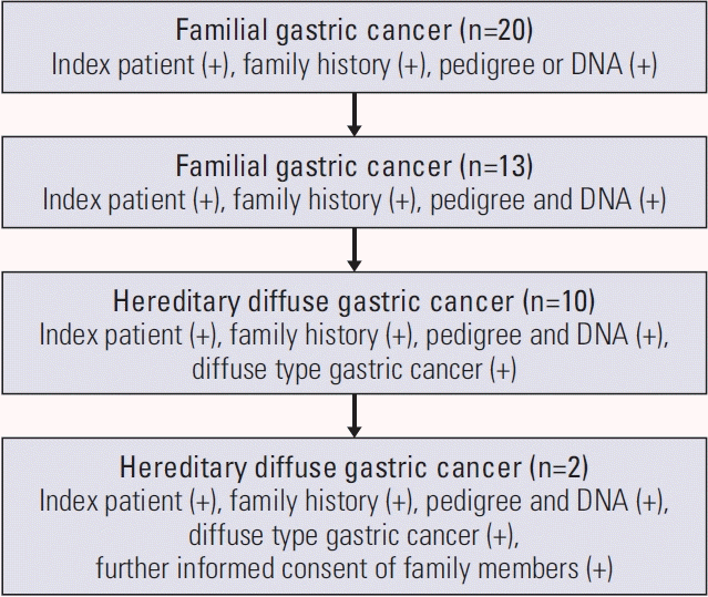
Since 2006, we collected pedigree data of patients who underwent surgery for gastric cancer at Seoul National University Hospital and whose family history met the diagnostic criteria for familial gastric cancer; two first- or second-degree relatives with gastric cancer before the age of 50 years or three first- or second-degree relatives with gastric cancer independent of age. DNA was also extracted from whole blood samples of patients and their relatives. Among these 20 families, both the pedigree and the DNA extracted at that time were available for 13 families, and 10 families met the diagnostic criteria for HDGC. Informed consent for further genetic analysis was obtained from two families included in this study (Fig. 1).

2. Whole genome sequencing

To identify candidates for new pathogenic germline mutations in HDGC, nine blood samples from the index patients and their family members with or without gastric cancer were sequenced using whole genome sequencing (WGS) (Fig. 2). Sequencing reads were aligned to the reference genome GRCh37 using Burrows-Wheeler Aligner (BWA). Variants were called using HaplotypeCaller from the Genome Analysis Toolkit (GATK) and all processes followed the GATK best-practice guidelines. Using WGS data, we detected three candidate genes with germline mutations, EPHA5, MCOA2, and RHOA. Among these, RHOA was selected based on its high frequency of mutations reported in gastric cancer.

3. In-silico analysis

We conducted extensive in-silico analysis to explore the potential impact of the newly identified final candidate on protein function and pathogenicity. Tools such as Sorting Intolerant From Tolerant (SIFT), Polymorphism Phenotyping v2 (PolyPhen-2), Mutation Taster, Combined Annotation Dependent Depletion (CADD) score, Protein Variation Effect Analyzer (PROVEAN), and I-Mutant Suite were used.

4. Whole exome sequencing

To identify RHOA mutations in cancer tissue, we extracted DNA from the formalin-fixed paraffin-embedded tissue block of the two patients whose whole blood samples were previously used for WGS and whose tissue was stored in our institution. Whole exome sequencing (WES) of the extracted DNA was performed.

5. Validation of candidate variant and functional enrichment analysis

To determine whether RHOA mutations could lead to cancer, we designed a functional validation study. We chose cell lines without CDH1 mutations and with low RhoA expression levels. MKN1, SNU216, and SNU668 cells were grown in Roswell Park Memorial Institute 1640 (RPMI 1640, Hyclone, Logan, UT) medium supplemented with 15% fetal bovine serum (Invitrogen, Carlsbad, CA) and 1% penicillin-streptomycin (Invitrogen). All cells were maintained in a humidified incubator at 37°C and 5% CO2. Using these cell lines, we performed site-directed mutagenesis targeting RHOA and overexpressed RhoA. After confirming the RhoA expression pattern in the mutant, we analyzed the change in Rho-ROCK signaling pathways in which the RhoA protein is involved using a RhoA pull-down assay to explore the potential role of RHOA mutation in regulating cellular function.

6. Candidate variant in other families

After functional validation of the germline mutation in the RHOA, we performed WES on the DNA of the index patients from 12 other families who met the diagnostic criteria for familial gastric cancer to determine whether germline mutations in the RHOA were present. None of these patients had mutations in CDH1 or CTNNA1, and the causative gene was not identified.

Results

The mean age at gastric cancer diagnosis among the index patients from 13 families, for which both DNA extracted from whole blood samples and pedigree were available, was 36.8 years, and 76.9% of these patients presented with diffuse-type gastric cancer (Table 1).
There were no CDH1 and CTNNA1 mutations in the two families who provided blood samples of the patient and family members for DNA extraction and WGS (Fig. 2), and the same germline mutation was confirmed among family members in only one family (Fig. 2B, right). Among the three candidate germline mutations of HDGC, EPHA5, MCOA2, and RHOA, RHOA with the most reports related to gastric cancer was selected as the final candidate. This mutation was identified at position 49399952 on chromosome 3, according to the hg19 genomic build, where the reference allele is G, and variant allele is A (chr3:49399952G>A). The variant was R129W, which is present in the insert domain of the RhoA protein [17].
The results from the in-silico analysis to predict the pathogenic effects of the variant suggest that the RHOA mutation (R129W) would induces changes in protein function (Table 2).
To identify the RHOA mutation (R129W) in the cancer tissue, we extracted DNA from the formalin-fixed paraffin-embedded tissue blocks of two patients whose (1) germline mutation of RHOA (R129W) was confirmed in the result of WGSs using DNA extracted from blood samples and (2) cancer tissue was stored at our institution. The same RHOA mutation (R129W) was identified in both DNA samples from tissue blocks of the two patients.
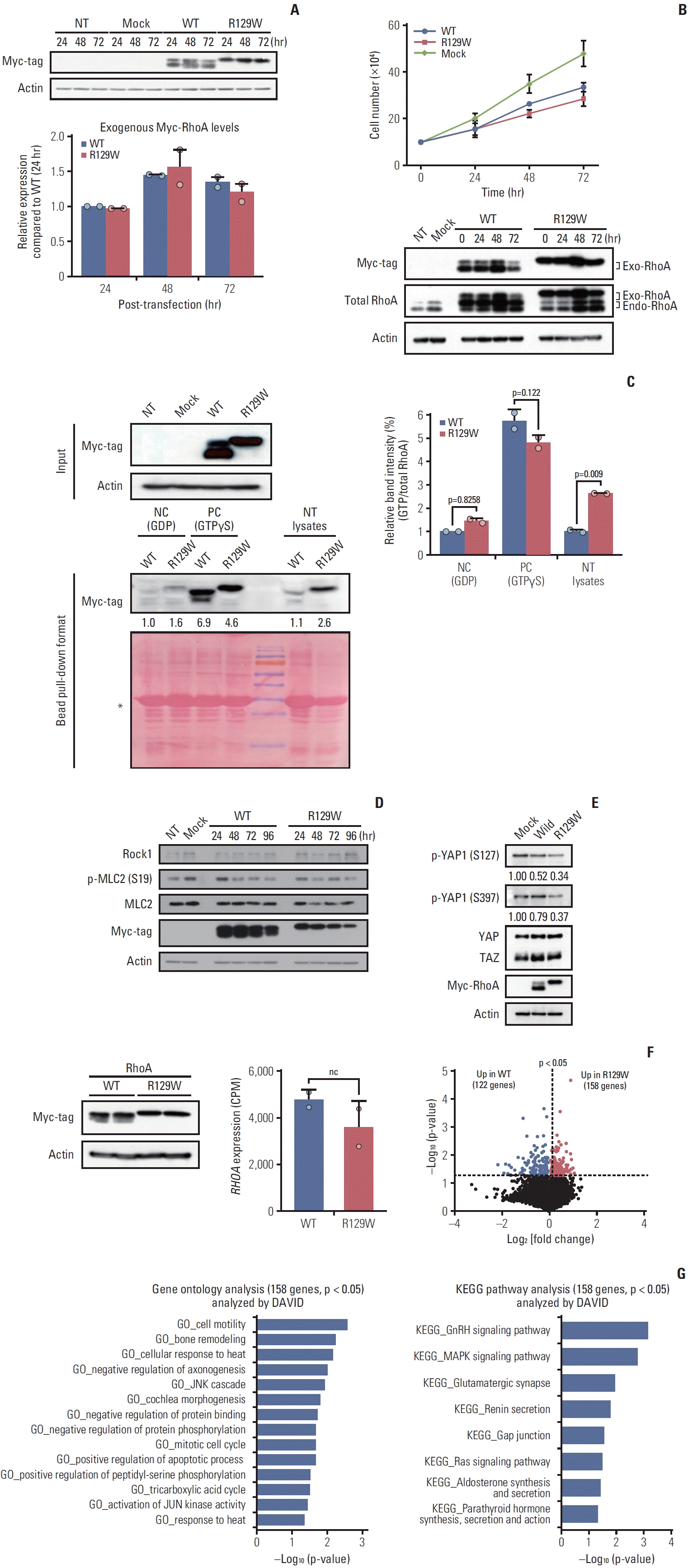
To investigate the cellular implications of the RHOA mutation (R129W) specifically independent of CDH1 mutations, we selected the MKN1, SNU216, and SNU668 gastric cancer cell lines based on genomic data analysis using cBioPortal (https://www.cbioportal.org/). These cell lines were identified to have wild-type CDH1, which excluded the effect of mutant CDH1, a well-known gene that causes gastric cancer [10,11,18,19], and wild-type RHOA genes. After performing immunoblotting analysis, the MKN1 gastric cancer cell line was selected for further experiments as it exhibited the lowest level of RhoA expression compared to the SNU216 and SNU668 cell lines (Fig. 3). Hence, we expected to observe the overexpression effect of mutant RhoA (R129W) with minimal contribution from wild-type RhoA in MKN1 cells. To generate RHOA mutation (R129W) in the Rho insert domain, a wild-type RHOA cloned plasmid (pRK5 myc-RhoA WT, #15899, Addgene) was used for site-directed mutagenesis. Next, we examined the levels of exogenous Myc-tagged wild-type RhoA (myc-RhoA) and Myc-tagged mutant RhoA (myc-RhoAR129W) by immunoblotting following transient transfection. While myc-RhoA showed two bands, myc-RhoAR129W was detected as one band, and although they have the same size (kDa), the movement of myc-RhoAR129W was confirmed to be relatively slow (Fig. 4A, upper). Under our experimental conditions, the expression levels of both proteins (mycRhoA and myc-RhoAR129W) in MKN1 cells peaked 48 hours after transfection. In addition, myc-RhoAR129W showed no significant difference in expression levels compared to myc-RhoA within 72 hours of transfection, as shown in Fig. 4A (bottom).
Based on the comparable expression levels of myc-RhoA and myc-RhoAR129W, their effects on cell growth were evaluated. However, when both proteins were overexpressed, a common phenomenon of inhibited cell growth was observed compared with the control group (mock, empty vector only). Notably, the difference in cell growth inhibition between myc-RhoA and myc-RhoAR129W proteins was not statistically significant (Fig. 4B).
Although myc-RhoAR129W is not directly involved in cell proliferation, its GTPase activity may play a potential role in regulating migration and adhesion. To further explore this, we investigated the GTP-bound (active) state of myc-RhoAR129W in MKN1 cell lysate. In the positive control [20] lysates that were incubated with GTPγS to ensure that it irreversibly occupied the GTP/GDP binding pocket and formed constitutively active forms, both myc-RhoA and myc-RhoAR129W showed similar levels of the GTP-bound state, indicating that there is no problem with the GTP-binding ability compared to the protein expression levels. Notably, in the experimental group (NT lysates), myc-RhoAR129W has over 2.5-fold (p=0.009) higher GTP-bound form than did myc-RhoA in terms of the active state (Fig. 4C). Indeed, compared with negative control lysates that were mixed with GDP to prevent the formation of new GTP-bound RhoA, the basal GTP-bound form in myc-RhoAR129W–expressing cells was slightly higher than that in myc-RhoA–expressing cells (Fig. 4C). These results indicate that RhoAR129W has a higher potential to exist in a GTP-bound state than wild-type RhoA at the cellular level.
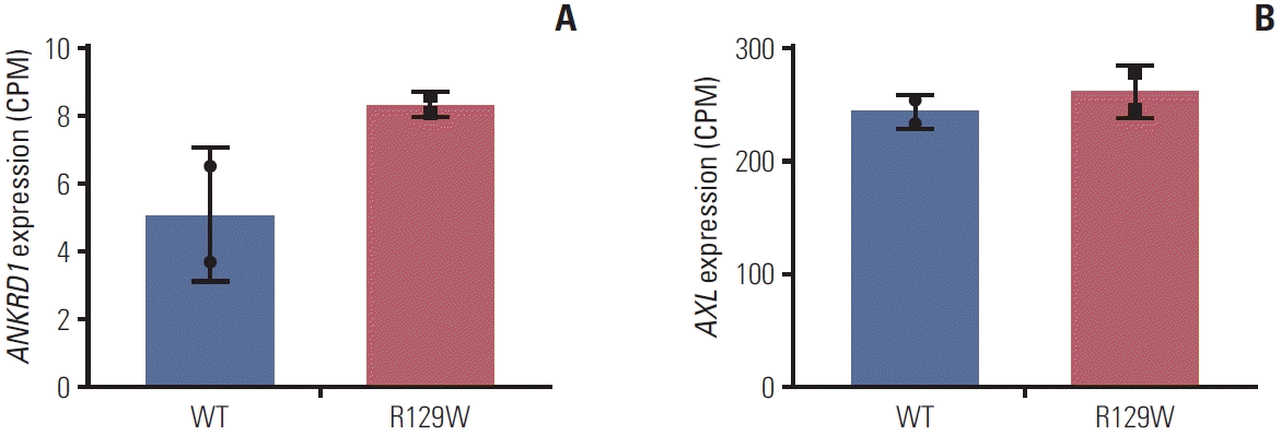
The Rho-ROCK signaling pathway is a well-known axis that regulates the reorganization of the cytoskeleton involved in cell contraction, motility, and polarity [17,21,22]. As myc-RhoAR129W–overexpressing cells showed similar cell growth to myc-RhoA–overexpressing cells, we examined ROCK1 and myosin light chain 2 (MLC2) which lies downstream of ROCK1. ROCK1-dependent phosphorylation of myosin light chain 2 (p-MLC2) is associated with stress fiber disassembly and cell detachment during migration [23]. As shown in Fig. 4D, no significant difference was observed in the expression level of ROCK1 between myc-RhoA– and myc-RhoAR129W–overexpressing cells along with no quantitative change in p-MLC2. However, the stability and activity of Yes-associated protein 1 (YAP1), which is negatively regulated by phosphorylation of Ser127/397 (p-S127/137) and regulates the extracellular matrix shape, are also known to be regulated in a Rho-ROCK–dependent manner [24]. Interestingly, p-S127/397 of YAP1 was decreased in myc-RhoAR192W–overexpressing cells (ratio, 0.34 and 0.37) compared with that in myc-RhoA–overexpressing cells (ratio, 0.52 and 0.79) compared to Mock group (ratio, 1.00 and 1.00) (Fig. 4E). We then analyzed the differential gene expression profiles of RhoAR129W versus wild-type RhoA overexpression. After confirming that the expression level of each protein was similar (Fig. 4F, left), we observed that the mRNA level (counts per million) of RhoA, which represents endogenous and ectopic mRNA, was also not significantly different after performing QuantSeq (Fig. 4F, middle). Genes that were upor down-regulated at a statistically significant (p < 0.05) level were identified. As shown in the volcano plot (Fig. 4F, right), 158 genes were upregulated in myc-RhoAR129W–overexpressing cells; conversely, 122 genes were upregulated in mycRhoA–overexpressing cells. To understand the biological significance of the158 genes upregulated by RhoAR129W, gene ontology (GO) enrichment and Kyoto Encyclopedia of Genes and Genomes (KEGG) pathway analyses were performed using Database for Annotation, Visualization and Integrated Discovery (DAVID, https://david.ncifcrf.gov/). A comparison of the transcriptome of myc-RhoAR129W–overexpressing cells with that of myc-RhoA–overexpressing cells revealed that cell migration-related gene sets (e.g., ‘GO_cell motility’, ‘GO_bone remodeling’, and ‘GO_JNK cascade’), and cytoskeleton reorganization-related gene sets (e.g., ‘KEGG_MAPK signaling pathway’, ‘KEGG_Ras signaling pathway’, and ‘KEGG_Gap junction’) were significantly enriched in myc-RhoAR129W–overexpressing cells (Fig. 4G). These results suggest that RhoAR129W overexpression is related to dynamic changes in the migration/adhesion potential of MKN1 cells. In fact, ANKRD1, which encodes the transcriptional cofactor ANKRD1 protein and is upregulated in RhoAR129W-overexpressing cells (Fig. 5A), is associated with wound healing and is regulated by YAP1 [25-27]. Since both p-S127 and p-S397 of YAP1 were reduced by RhoAR129W-overexpression (Fig. 4E), it is possible that active (non-phosphorylated Ser127/397) YAP1 was involved in the increased ANKRD1 transcript levels in a RhoAR129W-dependent manner. Its involvement in these pathways highlights the possible role of RhoAR129W in epithelial remodeling during the cellular migration of cancer cells. Considering that the RhoAR129W mutation has been identified in patients with familial gastric cancer, it is crucial to gain a comprehensive understanding of its impact on gastric cancer development and the underlying mechanisms. Therefore, further studies are required to investigate whether RhoAR129W knock-in mice develop gastric cancer and to assess the therapeutic potential of targeting RhoAR129W. The tyrosine kinase membrane receptor protein AXL plays a crucial role in YAP-mediated transcriptional activity through positive feedback mechanisms [28] and acts as a mediator of YAP-dependent oncogenic functions associated with receptor tyrosine kinase inhibitor resistance, invasion, and proliferation [28-30]. However, difference between wild-type RhoA-and RhoAR129W-overexpressing cells was not significant (Fig. 5B). Taken together, these results suggest that RhoA and AXL activate YAP through separate pathways and differentially regulate YAP target genes, which requires further investigation.
We subsequently performed WES on index patients from all other families, but none of the mutations in CDH1, CTNNA1, or RHOA were detected.

Discussion

We applied next-generation sequencing technology to identify new pathogenic variants that cause HDGC and found that germline mutations in RHOA potentially contribute to the development of cancer by affecting the YAP/ TAZ pathway, one of the downstream signaling pathways of the RhoA protein. The mutation confirmed in this study, the RHOA mutation (R129W), does not belong to a hotspot, but codes for the insert domain of the RhoA protein, which can affect effector binding [17]. This suggests that RHOA may be one of the causal genes for > 60% of patients with HDGC who do not have CHD1 or CTNNA1 mutations, and may provide evidence for prophylactic total gastrectomy for family members of the patient. To our knowledge, hereditary gastric cancer caused by RHOA germline mutations has not been reported until now, and this study is the first to reveal such possibility.
According to previous reports regarding RHOA functions in gastric cancer, among the 10 cancer hallmarks published [31], mutations in the RHOA gene are known to be related to the activation of invasion and metastasis, resistance to cell death, and sustenance of proliferative signaling [32,33]. This is because the main functions of RhoA, encoded by RHOA, include cytoskeletal organization, cell adhesion, cell migration, and cell division. The RhoA protein belongs to the Rasrelated GTPase superfamily. It is characterized by switching between activation and inactivation depending on whether it binds to GDP or GTP. When bound to GTP, RhoA is activated and interacts with effector proteins to initiate downstream signaling pathways, such as the Rho/MLC and Rho/ROCK pathways, whereas when bound to GDP, RhoA is inactivated [17,34,35]. RhoA dysregulation due to RHOA mutation results in compromised tissue integrity, disrupted intercellular adhesion, increased cell motility and invasiveness, and dysregulated cell proliferation, ultimately facilitating cancer cell development, growth, invasion, and metastasis [17,32,33].
Although there was no report on the relationship between gastric cancer and germline mutations in the RHOA gene, somatic mutations in the RHOA gene in gastric cancer have been reported in several studies [32,33,35,36]. Among various subtypes of gastric cancer, RHOA mutations are primarily observed in patients with diffuse-type gastric cancer. Wang et al. [35] reported that RHOA mutation seems to be an early event because it was detected even in intramucosal diffuse-type gastric cancer. According to a study by Kakiuchi et al. [32], somatic mutations in the RHOA gene were identified in approximately 25% of diffuse-type gastric cancers, but not in a single case in the intestinal gastric cancer cohort. In general, RHOA gene mutations exist in approximately 14%-25% of diffuse gastric cancers, and the presence of RHOA mutations increases the invasiveness of the cancer [32].
Somatic mutations in RHOA are not as frequently identified as other well-known oncogenes; however, their association with many other cancers has been reported. In colorectal cancer, it is mainly noted in microsatellite-stable colorectal cancers and has been reported to be associated with mucinous adenocarcinoma. Clinically, colorectal cancers with RHOA mutations tend to be diagnosed in younger female and are mainly located in the proximal colon. An association between mutations in the RHOA gene and aggressive metastasis has been reported in breast and lung cancers, and it is also known as a susceptibility variant of angioimmunoblastic T-cell lymphoma [37]. Although research results have been reported on the effects of somatic mutations of the RHOA gene in various cancers, the association between germline mutation of the RHOA gene and hereditary cancers, such as gastric cancer, has not been sufficiently reported to date.
Recently, as many new gene analysis techniques have been introduced, opportunities for gene analysis have increased, patients have become more accustomed to genetic testing, and the cost of testing is more realistic. Concurrent with these developments, small molecule inhibitors for targeting oncogenic genes, including those for RHOA mutations, are being explored [38,39]. If a targeted therapy for RHOA germline mutations is developed, validated for safety, and widely adopted, it could provide a significantly less invasive alternative to prophylactic total gastrectomy for family members of patients with HDGC who have not developed cancer. Such an advancement could substantially improve the management of HDGC, making a significant breakthrough in the field.
Although our study has made remarkable findings, it has several limitations. First, our results suggest that the prevalence of the RHOA mutation (R129W) as a cause of HDGC may be much lower than that of the previously known CDH1 or CTNNA1 mutations. Consequently, despite the relatively high cost, genetic screening may fail to identify the causative gene, thereby providing limited guidance for future management strategies. Second, the identification of a specific mutation as the cause of HDGC does not guarantee the development of gastric cancer in all carriers because of the variability in genetic expression and penetrance among individuals. This variability can make risk assessment challenging for both individuals and their family members despite the potential benefits knowing the genetic risk factor.
In conclusion, we identified that the RHOA mutation (R129W) may serve as a potential causative gene for HDGC. However, this mutation was only identified in a single family, leaving numerous other families for which the causative gene has yet to be determined. To comprehensively understand the role of the RHOA mutation in the pathogenesis of HDGC, further is needed.

Notes

Ethical Statement

The study protocol was approved by the Institutional Review Board of Seoul National University Hospital (IRB number: 2006-124-1133). All procedures were conducted in accordance with the Declaration of Helsinki of 1964 and later versions and relevant ethical guidelines. Informed consent for genetic testing was obtained from all participants.

Author Contributions

Conceived and designed the analysis: Oh SY, Jang G, Jeong KY, Kim HM, Kwak YJ, Kong SH, Park DJ, Lee HJ, Kim JI, Yang HK.

Collected the data: Oh SY, Jeong KY, Kim HM, Kong SH, Park DJ, Lee HJ, Yang HK.

Contributed data or analysis tools: Oh SY, Jang G, Kim J, Kong SH, Park DJ, Lee HJ, Kim JI, Yang HK.

Performed the analysis: Oh SY, Jang G, Kim J, Kwak YJ, Kim JI, Yang HK.

Wrote the paper: Oh SY, Jang G.

Review the paper: Yang HK.

Conflicts of Interest

Conflict of interest relevant to this article was not reported.

Acknowledgments

We thank all the patients and family members who participated in this study for their contributions to enrich scientific knowledge about hereditary diffuse gastric cancer.
This work was supported by the Seoul National University Hospital Research Grant (No. 05-2017-0010), the National Research Foundation of Korea (NRF) grant funded by the Korea government (MSIT) (No. 2021R1A2C3008021), and the Basic Science Research Program through the NRF funded by the Korea government (MOE) (No. RS-2023-00240042).

References

1. Ferlay J, Soerjomataram I, Dikshit R, Eser S, Mathers C, Rebelo M, et al. Cancer incidence and mortality worldwide: sources, methods and major patterns in GLOBOCAN 2012. Int J Cancer. 2015; 136:E359–86.
crossref
2. Moller H, Heseltine E, Vainio H. Working group report on schistosomes, liver flukes and Helicobacter pylori. Int J Cancer. 1995; 60:587–9.
3. Neugut AI, Hayek M, Howe G. Epidemiology of gastric cancer. Semin Oncol. 1996; 23:281–91.
4. Genta RM. Acid suppression and gastric atrophy: sifting fact from fiction. Gut. 1998; 43 Suppl 1:S35–8.
crossref
5. World Cancer Research Fund, American Institute for Cancer Research. Food, nutrition, physical activity, and the prevention of cancer: a global perspective. Washington, DC: World Cancer Research Fund, American Institute for Cancer Research;2007.
6. Ladeiras-Lopes R, Pereira AK, Nogueira A, Pinheiro-Torres T, Pinto I, Santos-Pereira R, et al. Smoking and gastric cancer: systematic review and meta-analysis of cohort studies. Cancer Causes Control. 2008; 19:689–701.
crossref
7. Matsukura N, Onda M, Tokunaga A, Yoshiyuki T, Shimizu Y, Nishi K, et al. Simultaneous gastric cancer in monozygotic twins. Cancer. 1988; 62:2430–5.
crossref
8. Kinugasa S, Matsuura H, Abe S, Yoshimura H, Tachibana M, Nakamura T, et al. Simultaneous early gastric cancer in identical twins: report of a case. Surg Today. 1996; 26:189–91.
crossref
9. Shah SC, McKinley M, Gupta S, Peek RM Jr, Martinez ME, Gomez SL. Population-based analysis of differences in gastric cancer incidence among races and ethnicities in individuals age 50 years and older. Gastroenterology. 2020; 159:1705–14.
crossref
10. van der Post RS, Vogelaar IP, Carneiro F, Guilford P, Huntsman D, Hoogerbrugge N, et al. Hereditary diffuse gastric cancer: updated clinical guidelines with an emphasis on germline CDH1 mutation carriers. J Med Genet. 2015; 52:361–74.
crossref
11. Guilford P, Hopkins J, Harraway J, McLeod M, McLeod N, Harawira P, et al. E-cadherin germline mutations in familial gastric cancer. Nature. 1998; 392:402–5.
crossref
12. Grunwald GB. The structural and functional analysis of cadherin calcium-dependent cell adhesion molecules. Curr Opin Cell Biol. 1993; 5:797–805.
crossref
13. Shiozaki H, Oka H, Inoue M, Tamura S, Monden M. E-cadherin mediated adhesion system in cancer cells. Cancer. 1996; 77:1605–13.
crossref
14. Majewski IJ, Kluijt I, Cats A, Scerri TS, de Jong D, Kluin RJ, et al. An alpha-E-catenin (CTNNA1) mutation in hereditary diffuse gastric cancer. J Pathol. 2013; 229:621–9.
15. Rimm DL, Koslov ER, Kebriaei P, Cianci CD, Morrow JS. Alpha 1(E)-catenin is an actin-binding and -bundling protein mediating the attachment of F-actin to the membrane adhesion complex. Proc Natl Acad Sci U S A. 1995; 92:8813–7.
crossref
16. Lien WH, Klezovitch O, Fernandez TE, Delrow J, Vasioukhin V. alphaE-catenin controls cerebral cortical size by regulating the hedgehog signaling pathway. Science. 2006; 311:1609–12.
crossref
17. Jose L. Orgaz VS-M: RhoA. In: Choi S, editor. Encyclopedia of signaling molecules. Cham: Springer; 2018. p. 4681-91.
18. Oliveira C, Pinheiro H, Figueiredo J, Seruca R, Carneiro F. Familial gastric cancer: genetic susceptibility, pathology, and implications for management. Lancet Oncol. 2015; 16:e60–70.
crossref
19. Luo X, Maciaszek JL, Thompson BA, Leong HS, Dixon K, Sousa S, et al. Optimising clinical care through CDH1-specific germline variant curation: improvement of clinical assertions and updated curation guidelines. J Med Genet. 2023; 60:568–75.
crossref
20. de Assumpcao PB, de Assumpcao PP, Moreira FC, Ribeiro-Dos-Santos A, Vidal AF, Magalhaes L, et al. Incidence of hereditary gastric cancer may be much higher than reported. Cancers (Basel). 2022; 14:6125.
crossref
21. Amano M, Nakayama M, Kaibuchi K. Rho-kinase/ROCK: a key regulator of the cytoskeleton and cell polarity. Cytoskeleton (Hoboken). 2010; 67:545–54.
crossref
22. Sadok A, Marshall CJ. Rho GTPases: masters of cell migration. Small GTPases. 2014; 5:e29710.
23. Shi J, Wu X, Surma M, Vemula S, Zhang L, Yang Y, et al. Distinct roles for ROCK1 and ROCK2 in the regulation of cell detachment. Cell Death Dis. 2013; 4:e483.
crossref
24. Li Y, Wang J, Zhong W. Regulation and mechanism of YAP/TAZ in the mechanical microenvironment of stem cells (Review). Mol Med Rep. 2021; 24:506.
25. Almodovar-Garcia K, Kwon M, Samaras SE, Davidson JM. ANKRD1 acts as a transcriptional repressor of MMP13 via the AP-1 site. Mol Cell Biol. 2014; 34:1500–11.
26. Samaras SE, Almodovar-Garcia K, Wu N, Yu F, Davidson JM. Global deletion of Ankrd1 results in a wound-healing phenotype associated with dermal fibroblast dysfunction. Am J Pathol. 2015; 185:96–109.
crossref
27. Jimenez AP, Traum A, Boettger T, Hackstein H, Richter AM, Dammann RH. The tumor suppressor RASSF1A induces the YAP1 target gene ANKRD1 that is epigenetically inactivated in human cancers and inhibits tumor growth. Oncotarget. 2017; 8:88437–52.
28. Saab S, Chang OS, Nagaoka K, Hung MC, Yamaguchi H. The potential role of YAP in Axl-mediated resistance to EGFR tyrosine kinase inhibitors. Am J Cancer Res. 2019; 9:2719–29.
29. Xu MZ, Chan SW, Liu AM, Wong KF, Fan ST, Chen J, et al. AXL receptor kinase is a mediator of YAP-dependent oncogenic functions in hepatocellular carcinoma. Oncogene. 2011; 30:1229–40.
crossref
30. He L, Lei Y, Hou J, Wu J, Lv G. Implications of the receptor tyrosine kinase Axl in gastric cancer progression. Onco Targets Ther. 2020; 13:5901–11.
31. Hanahan D, Weinberg RA. Hallmarks of cancer: the next generation. Cell. 2011; 144:646–74.
crossref
32. Kakiuchi M, Nishizawa T, Ueda H, Gotoh K, Tanaka A, Hayashi A, et al. Recurrent gain-of-function mutations of RHOA in diffuse-type gastric carcinoma. Nat Genet. 2014; 46:583–7.
crossref
33. Nam S, Kim JH, Lee DH. RHOA in gastric cancer: functional roles and therapeutic potential. Front Genet. 2019; 10:438.
crossref
34. Dvorsky R, Blumenstein L, Vetter IR, Ahmadian MR. Structural insights into the interaction of ROCKI with the switch regions of RhoA. J Biol Chem. 2004; 279:7098–104.
crossref
35. Wang K, Yuen ST, Xu J, Lee SP, Yan HH, Shi ST, et al. Wholegenome sequencing and comprehensive molecular profiling identify new driver mutations in gastric cancer. Nat Genet. 2014; 46:573–82.
crossref
36. Cho SY, Park JW, Liu Y, Park YS, Kim JH, Yang H, et al. Sporadic early-onset diffuse gastric cancers have high frequency of somatic CDH1 alterations, but low frequency of somatic RHOA mutations compared with late-onset cancers. Gastroenterology. 2017; 153:536–49.
crossref
37. Manso R, Sanchez-Beato M, Monsalvo S, Gomez S, Cereceda L, Llamas P, et al. The RHOA G17V gene mutation occurs frequently in peripheral T-cell lymphoma and is associated with a characteristic molecular signature. Blood. 2014; 123:2893–4.
crossref
38. Chang HR, Nam S, Lee J, Kim JH, Jung HR, Park HS, et al. Systematic approach identifies RHOA as a potential biomarker therapeutic target for Asian gastric cancer. Oncotarget. 2016; 7:81435–51.
crossref
39. Yoon C, Cho SJ, Aksoy BA, Park DJ, Schultz N, Ryeom SW, et al. Chemotherapy resistance in diffuse-type gastric adenocarcinoma is mediated by RhoA activation in cancer stem-like cells. Clin Cancer Res. 2016; 22:971–83.
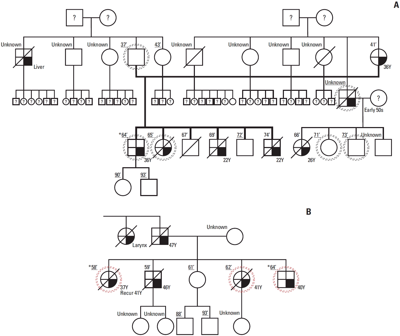
Fig. 1.
Flowchart showing the family selection for whole genome sequencing in this study.
crt-2024-328f1.tif
Fig. 2.
(A, B) Pedigrees of two families with hereditary diffuse gastric cancer who underwent whole genome sequencing (WGS) to identify new candidate variants of hereditary diffuse gastric cancer. Where known, the individual’s age is presented to the right of the symbols. The age is underlined if DNA from blood sample was available. Not all relatives consented to undergo genetic screening. A dotted circle is drawn around the symbol of the individual for whom WGS was performed and red dotted circle means individuals with RHOA R129W germline mutation (square, male; circle, female; oblique line, deceased; black, disease; *, index patient; number’, year of birth; unknown, year of birth unknown; numberY, age at diagnose; ?, all unknown).
crt-2024-328f2.tif
Fig. 3.
RhoA expression level in each gastric cancer cell line. RhoA expression level was the lowest in the MKN1 gastric cancer cell line.
crt-2024-328f3.tif
Fig. 4.
Functional validation of RHOA mutation (R129W). (A) Levels of exogenous Myc-tagged wild-type RhoA (myc-RhoA) and Myc-tagged mutant RhoA (myc-RhoAR129W). (B) Overexpression of myc-RhoA and myc-RhoAR129W exhibited a similar inhibitory effect on cell growth. (C) RhoAR129W has a higher potential to exist in a GTP-bound state than the wild-type RhoA. (D) Expression levels of ROCK1 and p-MLC2 showed no difference between cells overexpressing myc-RhoA- and myc-RhoAR129W. (E) Phosphorylation of Ser127/397 was lower in the myc-RhoAR129W–overexpressing cells than in the myc-RhoA–overexpressing cells. (F) Although the expression levels of RhoA and the mRNA were similar in wild-type RhoA and RhoAR129W overexpressing cells, up- or down-regulated genes were differed. (G) Genes related cell migration and cytoskeleton reorganization were enriched in RhoAR129W overexpressing cells. CPM, counts per million; DAVID, Database for Annotation, Visualization and Integrated Discovery; GO, Gene Ontology; KEGG, Kyoto Encyclopedia of Genes and Genomes; WT, wild type.
crt-2024-328f4.tif
Fig. 5.
ANKRD1 (A) and AXL (B) level in wild-type RhoA and RhoAR129W overexpressing cells. Among ANKRD1 and AXL, only ANKRD1 is upregulated in RhoAR129W-overexpressing cells. CPM, counts per million.
crt-2024-328f5.tif
Table 1.
Characteristics of index patients of familial gastric cancer
Variable No. (%) (n=13)
Age at gastric cancer diagnosis (yr), mean±SD 36.8±10.7
WHO classification
 Moderate differentiated adenocarcinoma 1 (7.6)
 Poorly differentiated adenocarcinoma 6 (46.2)
 Signet ring cell carcinoma 6 (46.2)
Lauren classification
 Diffuse type 10 (76.9)
 Intestinal type 1 (7.7)
 Mixed type 1 (7.7)

SD, standard deviation; WHO, World Health Organization.

Table 2.
Pathogenicity predictions for the RHOA mutation (R129W)
Tool Result
Sorting intolerant from tolerant (SIFT) Affect protein function
Polymorphism phenotyping v2 (PolyPhen2) Benign
Mutation taster Disease-causing
Combined annotation dependent depletion (CADD) score 33
Protein variation effect analyzer Deleterious
I-mutant suite Disease
TOOLS
Similar articles