Journal List > Cancer Res Treat > v.56(4) > 1516088696

Chen, Zeng, Wang, Sun, Huang, Zhu, Lu, Liao, Zhang, Chen, Yuan, Yang, Guo, Yang, Wen, Zhang, Li, Luo, Wu, Yang, Liu, Zheng, He, Sun, and Zhen: Stratified Treatment in Pediatric Anaplastic Large Cell Lymphoma: Result of a Prospective Open-Label Multiple-Institution Study

Abstract

Purpose

The risk stratification of pediatric anaplastic large cell lymphoma (ALCL) has not been standardized. In this study, new risk factors were included to establish a new risk stratification system for ALCL, and its feasibility in clinical practice was explored.

Materials and Methods

On the basis of the non-Hodgkin’s lymphoma Berlin–Frankfurt–Munster 95 (NHL-BFM-95) protocol, patients with minimal disseminated disease (MDD), high-risk tumor site (multiple bone, skin, liver, and lung involvement), and small cell/lymphohistiocytic (SC/LH) pathological subtype were enrolled in risk stratification. Patients were treated with a modified NHL-BFM-95 protocol combined with an anaplastic lymphoma kinase inhibitor or vinblastine (VBL).

Results

A total of 136 patients were enrolled in this study. The median age was 8.8 years. The 3-year event-free survival (EFS) and overall survival of the entire cohort were 77.7% (95% confidence interval [CI], 69.0% to 83.9%) and 92.3% (95% CI, 86.1% to 95.8%), respectively. The 3-year EFS rates of low-risk group (R1), intermediate-risk group (R2), and high-risk group (R3) patients were 100%, 89.5% (95% CI, 76.5% to 95.5%), and 67.9% (95% CI, 55.4% to 77.6%), respectively. The prognosis of patients with MDD (+), stage IV cancer, SC/LH lymphoma, and high-risk sites was poor, and the 3-year EFS rates were 45.3% (95% CI, 68.6% to 19.0%), 65.7% (95% CI, 47.6% to 78.9%), 55.7% (95% CI, 26.2% to 77.5%), and 70.7% (95% CI, 48.6% to 84.6%), respectively. At the end of follow-up, one of the five patients who received maintenance therapy with VBL relapsed, and seven patients receiving anaplastic lymphoma kinase inhibitor maintenance therapy did not experience relapse.

Conclusion

This study has confirmed the poor prognostic of MDD (+), high-risk site and SC/LH, but patients with SC/LH lymphoma and MDD (+) at diagnosis still need to receive better treatment (ClinicalTrials.gov number, NCT03971305).

Introduction

Anaplastic large cell lymphoma (ALCL) is a unique type of non-Hodgkin’s lymphomas (NHLs) and accounts for 10%-15% of all pediatric lymphomas [1-3]. ALCL is a mature T cell lymphoma that is characterized by t(2;5)(p23;q35) translocation in most cases, thus resulting in the abnormal expression of the anaplastic lymphoma kinase (ALK) gene, which is related to disease prognosis, tumorigenesis, and other biological characteristics. According to ALK expression, ALCL can be divided into two types: ALK– and ALK+ [4]. More than 95% of pediatric ALCL cases are ALK+, and most of them have advanced diseases, a high incidence of extranodal involvement, and frequent presence of B-symptoms.
The treatment of pediatric ALCL varies greatly among study groups, and the intensity and duration of chemotherapy have not been unified. The protocols of Berlin–Frankfurt–Munster (BFM) and ALCL 99 are widely used internationally [5,6].
Studies showed that the BFM study group used short pulse chemotherapy to treat children with ALCL, and the 5-year survival rate could reach 70%-85% [3]. Nevertheless, 30% of pediatric ALCL patients will relapse, and about 50% of relapse patients can survive and be rescued by aggressive salvage therapy [6-8]. Therefore, the early identification and intervention of the poor prognostic factors of pediatric ALCL will help reduce recurrence and death and improve the quality of life and prognosis of patients.
Owing to the low incidence of pediatric ALCL, few large-scale clinical studies have been conducted on this topic; thus, unfavorable prognostic factors have not been unified. The stratified criteria for risk groups in the BFM Cooperative Group for pediatric ALCL included the stage, and multiple bone involvement. Recent studies have found that the pathological type of ALCL, minimal disseminated disease (MDD), in the peripheral blood or bone marrow at diagnosis are significantly associated with poor prognosis [5,9-12]. ALCL with multiple skin or solid organ involvement also has a poor prognosis [7,9,13,14]. However, no prospective study has incorporated these prognostic factors into the risk stratification of pediatric ALCL to guide treatment.
To improve the individualized treatment of pediatric ALCL, the Pediatric Lymphoma Collaboration Group in Southern China conducted a prospective multicenter clinical study to establish a new risk stratification of pediatric ALCL by incorporating more prognostic factors on the basis of the non-Hodgkin’s lymphoma Berlin–Frankfurt–Munster 95 (NHL-BFM-95) protocol. A modified NHL-BFM-95 protocol was used to improve the prognosis of pediatric ALCL and validate the feasibility of the new risk stratification system to guide clinical practice.

Materials and Methods

1. Patients

Between February 2015 to January 2022, the Pediatric Lymphoma Collaboration Group in Southern China was initiated and led by the Sun Yat-sen University Cancer Center, and 22 hospitals in south China participated in this study. Children and adolescents with ALCL who were younger than 18 years of age at diagnosis were included. Patients with secondary immunodeficiency disorders, secondary cancers, post-transplantation lymphoma, or human immunodeficiency virus infections were excluded.

2. Diagnosis

The diagnosis of ALCL was based on pathological and immunohistochemical studies of the tumor tissue. According to morphology, the tumor can be divided into common, lymphohistiocytic (LH), small cell (SC), Hodgkin-like, and mixed subtypes. In addition, immunohistochemistry was performed to detect CD30, ALK, CD2, CD3, CD5, CD7 TIA-1, granzyme B, perforin, and other T cell–related antigens. Histology was centrally reviewed. MDD was measured by reverse transcription polymerase chain reaction for the NPM-ALK fusion gene in the bone marrow or peripheral blood, and an MDD greater than 0.001% was considered positive.

3. Stage

The staging was based on positron emission tomography (PET)/computed tomography (CT) and bone marrow. Efficacy was evaluated using ultrasound, X-ray, CT, magnetic resonance imaging, and PET/CT according to the disease condition. The staging system was based on the St Jude staging system [15].

4. Risk stratification

In this study, more prognostic factors were introduced on the basis of the NHL-BFM-95 risk stratification to establish a new stratification system. The low-risk group (R1) was defined as patients with disease stage I/II. The intermediate-risk group (R2) was defined as patients with disease stage III. The high-risk group (R3) was defined as patients with disease stage IV or multiple involvements of the bone or liver or lung or skin and soft tissue (Table 1). Patients who were not tested with MDD were stratified by clinical prognostic factors and histologic subtype assigned to the corresponding risk category.
All patients were treated with chemotherapy of different intensities according to the risk stratification (Table 1). Details of the chemotherapy regimens are shown in S1 and S2 Tables. Low-risk (R1) patients did not need the prephase of chemotherapy at diagnosis but directly received four courses of chemotherapy with course A alternating with course B without intrathecal chemotherapy. Patients with intermediate risk (R2) received the prephase of chemotherapy at diagnosis, followed by alternating courses of AA and BB for a total of six courses. High-risk (R3) patients received the prephase of chemotherapy at diagnosis, followed by six courses of alternating courses of AA, BB, and CC. Efficacy was evaluated every two courses.

5. Maintenance treatment

In patients with suspected viable residual lesions detected by PET/CT at the end of chemotherapy, biopsy was strongly recommended to determine the presence of viable tumors. If the patient had viable residual tumors proved by pathology, we nonrandomly assigned patients to use crizotinib or vinblastine (VBL) on the basis of factors such as patient tolerance to previous chemotherapy and age. Patients with poor bone marrow function should take crizotinib at 280 mg/m2 twice a day for 1 year as much as possible, and patients who are too young and inconvenient to take medicine should use intravenous VBL 6 mg/m2 once a week as much as possible.

6. Response evaluation

The tumor response was evaluated after every two courses of therapy. Complete remission (CR) was defined as the clinical disappearance of the disease, as confirmed by a CT scan and bone marrow (BM) and cerebrospinal fluid (CSF) screening. Partial remission (PR) was defined as 50% tumor regression. Steady disease (SD) was defined as tumor lesions that were reduced by < 50% and the absence of new lesion. Progressive disease (PD) was defined as the appearance of new disease during treatment or the incomplete regression of the local tumor, followed by progression during chemotherapy. Relapse was defined as evidence of disease 1 month after CR without treatment.

7. Statistical analysis

The main endpoints of this study were event-free survival (EFS) and overall survival (OS). OS refers to the length of time from diagnosis to death caused by any reason or last follow-up. EFS refers to the length of time from diagnosis to tumor recurrence or progression, occurrence of a secondary tumor, death caused by any reason, or last follow-up. The secondary endpoint was the association between the prognostic factors and survival. Kaplan-Meier survival analysis was performed using the log-rank test. Multivariate analyses were performed using Cox regression. All comparisons were two-sided, and p-values < 0.05 were considered statistically significant. Statistical analyses were performed using SPSS ver. 25.0 (IBM Corp., Armonk, NY).

Results

1. Patient characteristics

A total of 136 patients were included in this study, with a male to female ratio of 1.3:1 and median age of 8.8 years (1.0-15.8 years). There were 128 patients aged 14 years old or younger and 8 aged more than 14 years old. There were 0, 10, 91, and 35 patients with stages I, II, III, and IV cancer, respectively. There were 9, 49, and 78 patients in R1, R2, and R3, respectively. Lymph node involvement was the most common manifestation (81.6%), and extranodal organ involvement accounted for 77.7% of the cases. The ALK fusion gene status was evaluated by immunohistochemistry in 130 patients, of whom 119 (87.5%) were ALK (+). The detailed patient characteristics are shown in Table 2.

2. Chemotherapy efficacy

Treatment response was evaluated by a CT scan and BM and CSF screening 3 weeks after the end of first-line chemotherapy. Among the 136 patients, 112 (82.4%) achieved CR, 17 (12.5%) achieved PR, one (0.7%) achieved SD, and six (4.4%) achieved PD; the overall response rate (CR+PR) was 94.9%. Seventeen patients showed suspected viable residual small lesions detected by PET/CT at the end of first-line treatment. Among these patients, two patients continued chemotherapy for two more courses, and three patients underwent biopsy of the residual lesions; however, no viable tumor was detected by pathology. Biopsies were not available for the remaining 12 patients for specific reasons, and the patients were treated with maintenance therapy after the end of first-line treatment (S3 Table). Among them, seven patients (five at R2 and two at R3) received crizotinib, and five patients (all at R3) received VBL. All patients received maintenance therapy for 1 year of each type. At the end of maintenance treatment, PR was found in one case, and CR was found in the other cases.

3. Survival

The median follow-up time of the whole cohort was 39.1 months (0.3-87.3 months), the 3-year EFS and OS were 77.7% (95% confidence interval [CI], 69.0 to 83.9), and 92.3% (95% CI, 86.1 to 95.8), respectively (Fig. 1A and B). The 3-year EFS of patients at stages II, III, and IV were 100%, 79.8% (95% CI, 68.8 to 87.3), and 65.7% (95% CI, 47.6 to 78.9), respectively (p=0.05), and the 3-year OS rates were 100%, 94.2% (95% CI, 86.6 to 97.6), and 85.7% (95% CI, 79.0 to 93.8), respectively (p=0.19) (Fig. 1C and D). The 3-year EFS rates of patients with R1, R2, and R3 were 100%, 89.5% (95% CI, 76.5 to 95.5), and 67.9% (95% CI, 55.4 to 77.6), respectively (p=0.01), and the 3-year OS rates were 100%, 95.9% (95% CI, 84.7 to 99.0), and 89.2% (95% CI, 79.5 to 94.5) (p=0.31), respectively (Fig. 1E and F).
Among the 136 patients, 79 could be classified into definite pathological subtypes, and the rest could not be classified because of pathological difficulties or limitations of the research conditions. We stratified 79 patients with definite pathological types into two groups: SC/LH group and non-SC/LH group. The 3-year EFS was 55.7% (95% CI, 26.2 to 77.4) and 87.4% (95% CI, 75.2 to 93.4), respectively (p=0.01) (Fig. 2A and B), and the 3-year OS rates were 93.8% (95% CI, 63.2 to 99.1) and 96.5% (95% CI, 86.7 to 99.1), respectively (p=0.71).
Among the 136 patients, 31 (22.8%) had tumor involvement at high-risk sites, including multiple bone, liver, lung, and/or skin involvement. The 3-year EFS rates of patients with or without high-risk tumor sites were 70.7% and 79.5%, respectively (p=0.48) (Fig. 2C and D). A total of 76 patients were tested for MDD in the bone marrow or peripheral blood at diagnosis, and 22 patients (28.9%) were positive. The 3-year EFS rates of patients with negative and positive MDD were 84.8% (95% CI, 71.8 to 92.1) and 45.3% (95% CI, 18.9 to 68.5), respectively (p=0.001). The 3-year OS rates were 96.1% (95% CI, 85.3 to 99.0) and 81.8% (95% CI, 58.5 to 92.8), respectively (p=0.02) (Fig. 2E and sF).
At the end of follow-up, one of the five patients who received maintenance therapy with VBL relapsed within 3 years but survived after salvaged treatment. At the end of the follow-up, seven patients receiving ALK inhibitor maintenance therapy did not experience relapse, and the 3-year EFS and OS rates were 100%.
Univariate analysis results showed that MDD (+), high-risk site, stage, and SC/LH ratio were not independent prognostic factors for EFS and OS (Table 3). Multivariate analysis revealed similar findings.

4. Relapsed or refractory patients

At the end of follow-up, there were 20 patients with relapsed or refractory diseases, with a median age of 12 years (1.4-18 years). Among them, there were 13 and 7 patients at stages III and IV, respectively, and there were 4 and 16 patients at R2 and R3, respectively. All patients received 1 to 3 lines of salvage chemotherapy regimens, including single drug or combination with gemcitabine, vinblastine, crizotinib, and CC course of the BFM regimen, e.g., etoposide, cytarabine, vindesine, dexamethasone, and intrathecal dexamethasone, cytarabine and methotrexate). One patient received high-dose chemotherapy with rescued autologous stem cell transplantation after CR.

5. Adverse reaction

Among the entire cohort, 99 patients (61.2%) had grades 3 to 4 hematologic toxicity, 37 patients (27.2%) had infection, 29 patients (21.3%) had grades 3 to 4 mucositis, and 18 patients (13.2%) had mild liver function damage. One treatment-related death occurred in a 5-year-old boy with stage III, high-risk, and unclear pathological subtype cancer. The tumor achieved CR after six courses, but the patient died of multiple organ dysfunction caused by sepsis.

Discussion

In this study, several prognostic factors, including tumor site, pathological subtype, and PET/CT and MDD findings, were added to risk stratification for pediatric ALCL to guide clinical treatment. At the same time, chemotherapy was modified on the basis of the NHL-BFM-95 protocol and achieved a good treatment outcome. The 3-year EFS and OS rates were 77.7% and 92.3%, respectively, which is consistent with the survival data reported by most studies [16,17]. To the best of our knowledge, this is the largest prospective clinical trial of pediatric ALCL in Asia that has adopted a new risk stratification system for the precise diagnosis and treatment of pediatric ALCL.
Our study showed that pediatric ALCL with R1-R2 had good survival, with a 3-year EFS of 89.5%-100%. For these patients, future research is mainly aimed at reducing the intensity of treatment and improving their quality of life. We recommend that patients at R1 should be treated with a reduced intensity of treatment, and the prephase of chemotherapy should be omitted because of their small tumor burden. Patients at R1 also have a low risk of tumor involvement in the central nervous system and do not require lumbar puncture or intrathecal chemotherapy [16]. These changes may alleviate both short- and long-term side effects. In addition, the introduction of PET/CT for risk stratification and efficacy evaluation has higher sensitivity and specificity than CT [18-20].
However, the prognosis of pediatric ALCL at R3 and stage IV remains poor, with a 3-year EFS of only 65.7% to 67.9%, thus suggesting that these patients still need improved treatment. Fortunately, most patients still have the potential to be cured after salvage treatment following relapse, and the 3-year OS of each subgroup in this study could reach 81.8%-100%. This benefits from the development and application of new therapies, such as targeted therapy for ALK and CD30 [21-24]. Therefore, the early identification of patients with poor prognosis and an increase in treatment intensity can help reduce recurrence and avoid additional treatment and subsequent side effects.
This study attempted to incorporate a novel first-line therapy. At the end of first-line treatment, 12 patients with suspected viable residual tumors detected by PET/CT did not undergo biopsy, and all of them received ALK inhibitor or VBL maintenance therapy to reduce the incidence of tumor progression. This study confirmed that, at the end of maintenance therapy, the CR rate of 12 patients was 92.3%, which was higher than that of most previous first-line regimens. Some studies have shown that the application of VBL in the first-line treatment of pediatric ALCL can increase the one-year EFS, but the 2-year EFS remains unchanged [25]. In the current study, one of the five patients who received VBL maintenance therapy had disease progression within 3 years, whereas 13 patients who treated with ALK inhibitors had no disease progression. The preliminary results of these novel treatments are promising.
At present, few prognostic factors can be used for risk stratification in pediatric ALCL. A European study involving 235 children/adolescents with ALCL showed that mediastinal, visceral (lung, liver, or spleen), and skin involvement were identified as adverse prognostic factors, and patients with one of these factors had a significantly lower 5-year EFS rate (61% vs. 89%) [13]. Another study showed that liver and lung involvement increased the risk of disease recurrence or progression, and the 5-year EFS and OS were only 47.6% and 64.8%, respectively [26]. In the current study, patients with multiple skin, bone, liver, and/or lung involvement were enrolled in R3 and enhanced intensity treatment, and the 3-year EFS and OS were 70.7% and 93.2%, respectively. These values were significantly higher than those in previous studies. We included these high-risk sites as R3 and received more intensive treatment, and patients’ survival were improved, so there was no difference in EFS and OS between high-risk sites and non-high-risk sites patients. Therefore, it is appropriate to enroll these patients in R3 and for these patients to receive more intensive chemotherapy.
There are several pathological subtypes of ALCL. Recently, a study by the European Inter-Group for Childhood Non-Hodgkin Lymphoma involving 420 pediatric ALCL cases reported that among the pathological subtypes of pediatric ALCL, only SC/LH subtypes were independently associated with treatment failure, and the 10-year progression-free survival was only 50% compared with 79% for the other pathological subtypes [5,9]. Therefore, patients with SC/LH were included in R3. However, although the treatment intensity of these patients has improved, the results show that the 3-year EFS rate is only 55.7%, which is much lower than that of other pathological types. There was no significant difference between the OS rates of the two groups possibly because of the small number of SC/LH cases and the short follow-up time. Patients with SC/LH may have unique biological characteristics, and simply increasing the intensity of chemotherapy does not improve prognosis; therefore, it is necessary to develop new treatment methods.
In recent years, several studies have suggested that MDD (+) is a poor prognostic factor, and this finding was proved by the detection of the NPM-ALK fusion gene in the bone marrow or peripheral blood of pediatric ALCL patients at diagnosis. The cumulative recurrence rate of MDD (+) is as high as 61%, whereas that of MDD (–) is only approximately 20% [27-30]. Therefore, we enrolled patients with MDD (+) in R3. However, despite receiving intensified treatment, the 3-year EFS rate (45.3% vs. 84.8%, p=0.001) and OS rate (81.8% vs. 96.1%, p=0.02) in patients with MDD (+) were much lower than those in patients with MDD (–), and 46.3% of the patients relapsed within 3 years. Therefore, it is reasonable to enroll these patients in R3 for management, but our results suggest that it is not appropriate to treat patients with MDD (+) with regimens that are similar to the NHL-BFM-95 protocol. Even intensive therapies, such as hematopoietic stem cell transplantation [6,31], or targeted therapies [21,22] are needed as first-line therapies. In addition, studies have shown that MDD detection in the peripheral blood and bone marrow is highly correlated (r=0.74) [30]. Therefore, using peripheral blood instead of bone marrow for quantitative MDD evaluation can reduce the discomfort caused by bone marrow aspiration.
Since some patients do not have MDD testing, and some patients do not subdivide into pathological subtypes, it is possible that MDD positivity and LH/SC were classified as R1 or R2, leading to insufficient treatment, which potentially affecting the prognosis of these patients. Therefore, we hypothesized that R3 may have more patients and R1 and R2 may have a better response. Patients with confirmed poor prognostic factors were classified into R3, and with the strongest treatment, the survival rate did not exceed efficacy of R3 in previous studies, which indirectly indicates that the prognosis of these patients is really poor, and this stratification method is appropriate. It also shows that we did not overtreat, and that the effect might have been worse with less severe treatment.
Multivariate analysis showed that MDD (+), high-risk site, stage, and SC/LH ratio were not independent prognostic factors for EFS and OS, probably because some patients do not have MDD testing, and some patients do not subdivide into pathological subtypes. We should design more rigorous clinical studies to demonstrate the prognostic value of these factors in the future.
Thus far, the risk stratification method of ALCL in children has not been standardized [1], and there may be many reasons. Most studies had a small sample size or retrospective design, with some degree of study bias. The enrollment criteria of the different studies varied greatly. There is also a lack of uniform definitions for some clinical features, such as mediastinal involvement and liver/spleen involvement. Furthermore, the tumor control and supportive treatment strategies differ. Overall, the establishment of an appropriate risk stratification system will help develop accurate treatment strategies to avoid undertreatment or overtreatment.
This study has some limitations, with rather short follow-up time and small case number. Especially, the case number receiving maintenance treatment was extremely small, and the result was not convincing. However, as a prospective multicenter study with the largest sample size in China, it still provides important information for future study. In conclusion, this study incorporated new prognostic predictors and established a new risk stratification system for the precise treatment of pediatric patients with ALCL, which is convenient and feasible in clinical practice. In high-risk patients, the treatment of SC/LH pathological types and MDD (+) in peripheral blood/bone marrow at diagnosis still needs to be improved.

Electronic Supplementary Material

Supplementary materials are available at Cancer Research and Treatment website (https://www.e-crt.org).

Notes

Ethical Statement

This study was approved by the ethics Committee of Sun Yat-sen University of Cancer Center (approval number: B2019-006-01), and the study was conducted according to the principles of the Declaration of Helsinki and the Guidelines for Good Clinical Practice (ClinicalTrials.gov number, NCT03971305). All children’s guardians signed an informed consent.

Author Contributions

Conceived and designed the analysis: Sun X, Zhen Z.

Collected the data: Wang J, Sun F, Huang J, Zhu J, Lu S, Liao N, Zhang X, Chen Z, Yuan X, Yang Z, Guo H, Yang L (Yang Liangchun), Wen C, Zhang W, Li Y, Luo X, Wu Z, Yang L (Yang Lihua), Liu R, Zheng M, He X.

Contributed data or analysis tools: Wang J, Sun F, Huang J, Zhu J, Lu S, Liao N, Zhang X, Chen Z, Yuan X, Yang Z, Guo H, Yang L (Yang Liangchun), Wen C, Zhang W, Li Y, Luo X, Wu Z, Yang L (Yang Lihua), Liu R, Zheng M, He X.

Performed the analysis: Chen T, Zeng C.

Wrote the paper: Chen T, Zeng C.

Reivew: Sun X, Zhen Z.

Conflict of Interest

Conflict of interest relevant to this article was not reported.

ACKNOWLEDGMENTS

The authors would like to thank all members of the Pediatric Lymphoma Collaboration Group in Southern China, the medical teams in each investigator center for their contributions and all the patients for their participation.

References

1. Lowe EJ, Gross TG. Anaplastic large cell lymphoma in children and adolescents. Pediatr Hematol Oncol. 2013; 30:509–19.
crossref
2. Sandlund JT, Downing JR, Crist WM. Non-Hodgkin’s lymphoma in childhood. N Engl J Med. 1996; 334:1238–48.
crossref
3. Seidemann K, Tiemann M, Schrappe M, Yakisan E, Simonitsch I, Janka-Schaub G, et al. Short-pulse B-non-Hodgkin lymphoma-type chemotherapy is efficacious treatment for pediatric anaplastic large cell lymphoma: a report of the Berlin-Frankfurt-Munster Group Trial NHL-BFM 90. Blood. 2001; 97:3699–706.
crossref
4. Yi SH, Zou DH, Ken LG. Decipher the 2016 revision of the World Health Organization classification of lymphoid neoplasms. Zhonghua Yi Xue Za Zhi. 2016; 96:3365–9.
5. Lamant L, McCarthy K, d’Amore E, Klapper W, Nakagawa A, Fraga M, et al. Prognostic impact of morphologic and phenotypic features of childhood ALK-positive anaplastic large-cell lymphoma: results of the ALCL99 study. J Clin Oncol. 2011; 29:4669–76.
crossref
6. Furqan F, Ahn KW, Chen Y, Kaur M, Abutalib SA, Ahmed N, et al. Allogeneic haematopoietic cell transplant in patients with relapsed/refractory anaplastic large cell lymphoma. Br J Haematol. 2023; 200:54–63.
crossref
7. Knorr F, Brugieres L, Pillon M, Zimmermann M, Ruf S, Attarbaschi A, et al. Stem cell transplantation and vinblastine monotherapy for relapsed pediatric anaplastic large cell lymphoma: results of the international, prospective ALCL-relapse trial. J Clin Oncol. 2020; 38:3999–4009.
crossref
8. Woessmann W, Zimmermann M, Lenhard M, Burkhardt B, Rossig C, Kremens B, et al. Relapsed or refractory anaplastic large-cell lymphoma in children and adolescents after BerlinFrankfurt-Muenster (BFM)-type first-line therapy: a BFMgroup study. J Clin Oncol. 2011; 29:3065–71.
crossref
9. Mussolin L, Le Deley MC, Carraro E, Damm-Welk C, Attarbaschi A, Williams D, et al. Prognostic factors in childhood anaplastic large cell lymphoma: long term results of the international ALCL99 trial. Cancers (Basel). 2020; 12:2747.
crossref
10. Damm-Welk C, Mussolin L, Zimmermann M, Pillon M, Klapper W, Oschlies I, et al. Early assessment of minimal residual disease identifies patients at very high relapse risk in NPM-ALK-positive anaplastic large-cell lymphoma. Blood. 2014; 123:334–7.
crossref
11. Mussolin L, Pillon M, d’Amore ES, Santoro N, Lombardi A, Fagioli F, et al. Prevalence and clinical implications of bone marrow involvement in pediatric anaplastic large cell lymphoma. Leukemia. 2005; 19:1643–7.
crossref
12. Damm-Welk C, Busch K, Burkhardt B, Schieferstein J, Viehmann S, Oschlies I, et al. Prognostic significance of circulating tumor cells in bone marrow or peripheral blood as detected by qualitative and quantitative PCR in pediatric NPM-ALK-positive anaplastic large-cell lymphoma. Blood. 2007; 110:670–7.
crossref
13. Le Deley MC, Reiter A, Williams D, Delsol G, Oschlies I, McCarthy K, et al. Prognostic factors in childhood anaplastic large cell lymphoma: results of a large European intergroup study. Blood. 2008; 111:1560–6.
crossref
14. Yang HB, Li J, Shen T. Primary anaplastic large cell lymphoma of the lung: report of two cases and literature review. Acta Haematol. 2007; 118:188–91.
15. Murphy SB. Classification, staging and end results of treatment of childhood non-Hodgkin’s lymphomas: dissimilarities from lymphomas in adults. Semin Oncol. 1980; 7:332–9.
16. Brugieres L, Le Deley MC, Rosolen A, Williams D, Horibe K, Wrobel G, et al. Impact of the methotrexate administration dose on the need for intrathecal treatment in children and adolescents with anaplastic large-cell lymphoma: results of a randomized trial of the EICNHL Group. J Clin Oncol. 2009; 27:897–903.
crossref
17. Lowe EJ, Sposto R, Perkins SL, Gross TG, Finlay J, Zwick D, et al. Intensive chemotherapy for systemic anaplastic large cell lymphoma in children and adolescents: final results of Children’s Cancer Group Study 5941. Pediatr Blood Cancer. 2009; 52:335–9.
crossref
18. Ram-Wolff C, Vercellino L, Brice P, La Selva R, Bagot M. (18) F-fluorodeoxyglucose-positron emission tomography is more sensitive than computed tomography in initial staging of patients with an anaplastic T-cell lymphoma first presenting in the skin. Eur J Dermatol. 2017; 27:496–504.
crossref
19. Cheson BD, Fisher RI, Barrington SF, Cavalli F, Schwartz LH, Zucca E, et al. Recommendations for initial evaluation, staging, and response assessment of Hodgkin and non-Hodgkin lymphoma: the Lugano classification. J Clin Oncol. 2014; 32:3059–68.
crossref
20. Johnson SA, Kumar A, Matasar MJ, Schoder H, Rademaker J. Imaging for staging and response assessment in lymphoma. Radiology. 2015; 276:323–38.
crossref
21. Mosse YP, Voss SD, Lim MS, Rolland D, Minard CG, Fox E, et al. Targeting ALK with crizotinib in pediatric anaplastic large cell lymphoma and inflammatory myofibroblastic tumor: a Children’s Oncology Group Study. J Clin Oncol. 2017; 35:3215–21.
crossref
22. Mediwake H, Morris K, Curley C, Butler J, Kennedy G. Use of brentuximab vedotin as salvage therapy pre-allogeneic stem cell transplantation in relapsed/refractory CD30 positive lympho-proliferative disorders: a single centre experience. Intern Med J. 2017; 47:574–8.
23. Lowe EJ, Reilly AF, Lim MS, Gross TG, Saguilig L, Barkauskas DA, et al. Brentuximab vedotin in combination with chemotherapy for pediatric patients with ALK+ ALCL: results of COG trial ANHL12P1. Blood. 2021; 137:3595–603.
crossref
24. Lowe EJ, Reilly AF, Lim MS, Gross TG, Saguilig L, Barkauskas DA, et al. Crizotinib in combination with chemotherapy for pediatric patients with ALK+ anaplastic large-cell lymphoma: the results of Children’s Oncology Group Trial ANHL12P1. J Clin Oncol. 2023; 41:2043–53.
crossref
25. Le Deley MC, Rosolen A, Williams DM, Horibe K, Wrobel G, Attarbaschi A, et al. Vinblastine in children and adolescents with high-risk anaplastic large-cell lymphoma: results of the randomized ALCL99-vinblastine trial. J Clin Oncol. 2010; 28:3987–93.
crossref
26. Chen SH, Chen JS, Jou ST, Wu KH, Hung IJ, Sheen JM, et al. Outcome and prognosis of anaplastic large cell lymphoma in children: a report from the Taiwan Pediatric Oncology Group. Leuk Lymphoma. 2019; 60:1942–9.
crossref
27. Mussolin L, Damm-Welk C, Pillon M, Zimmermann M, Franceschetto G, Pulford K, et al. Use of minimal disseminated disease and immunity to NPM-ALK antigen to stratify ALK-positive ALCL patients with different prognosis. Leukemia. 2013; 27:416–22.
crossref
28. Iijima-Yamashita Y, Mori T, Nakazawa A, Fukano R, Takimoto T, Tsurusawa M, et al. Prognostic impact of minimal disseminated disease and immune response to NPM-ALK in Japanese children with ALK-positive anaplastic large cell lymphoma. Int J Hematol. 2018; 107:244–50.
crossref
29. Damm-Welk C, Lovisa F, Contarini G, Ludersen J, Carraro E, Knorr F, et al. Quantification of minimal disease by digital PCR in ALK-positive anaplastic large cell lymphoma: a step towards risk stratification in international trials? Cancers (Basel). 2022; 14:1703.
crossref
30. Damm-Welk C, Kutscher N, Zimmermann M, Attarbaschi A, Schieferstein J, Knorr F, et al. Quantification of minimal disseminated disease by quantitative polymerase chain reaction and digital polymerase chain reaction for NPM-ALK as a prognostic factor in children with anaplastic large cell lymphoma. Haematologica. 2020; 105:2141–9.
crossref
31. Brink M, Meeuwes FO, van der Poel MW, Kersten MJ, Wondergem M, Mutsaers P, et al. Impact of etoposide and ASCT on survival among patients aged <65 years with stage II to IV PTCL: a population-based cohort study. Blood. 2022; 140:1009–19.
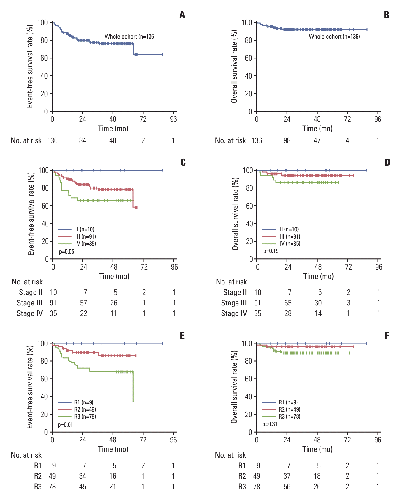
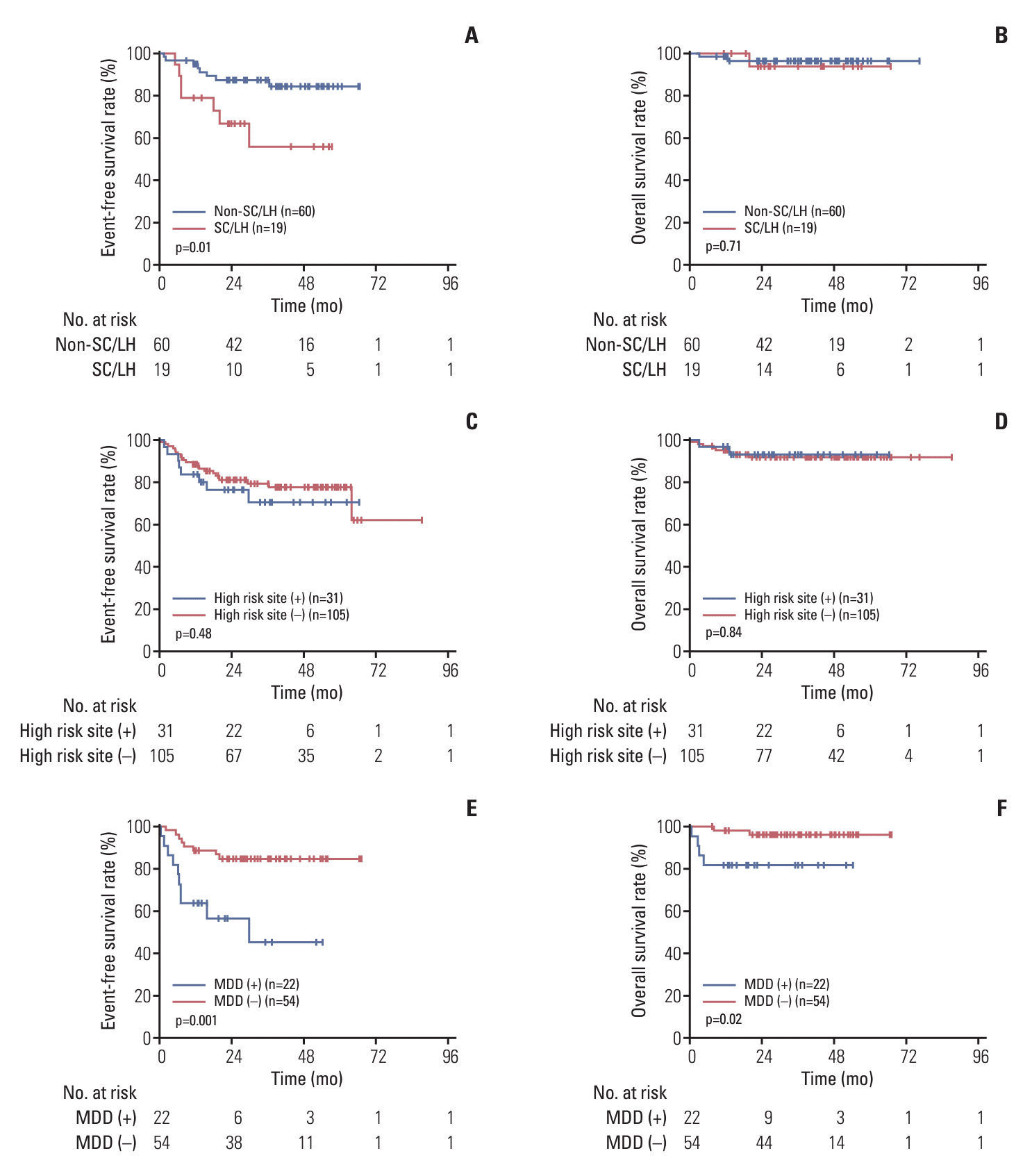
Fig. 1.
Survival rates of 136 patients with anaplastic large cell lymphoma. (A) Kaplan-Meier estimate of event-free survival for the whole cohort. (B) Kaplan-Meier estimate of overall survival for the whole cohort. (C) Kaplan-Meier estimate of event-free survival stratified by stage. (D) Kaplan-Meier estimate of overall survival stratified by stage. (E) Kaplan-Meier estimate of event-free survival stratified by risk stratification. (F) Kaplan-Meier estimate of overall survival stratified by risk stratification.
crt-2024-104f1.tif
Fig. 2.
Survival rates of 136 patients with anaplastic large cell lymphoma stratified by new risk factors. (A) Kaplan-Meier estimate of event-free survival stratified by pathological type. (B) Kaplan-Meier estimate of overall survival according to pathological type. (C) Kaplan-Meier estimate of event-free survival stratified by high-risk site of multiple bone/lung/liver/skin involvement. (D) Kaplan-Meier estimate of overall survival stratified by high-risk site. (E) Kaplan-Meier estimate of event-free survival stratified by minimal disseminated disease (MDD). (F) Kaplan-Meier estimate of overall survival stratified by MDD. LH, lymphohistiocytic; SC, small cell.
crt-2024-104f2.tif
Table 1.
Risk stratification and therapeutic schedule
Group Definition Schedulea)
Low risk (R1) Stages I and II A-B-A-Bb)
Intermediate risk (R2) Stage III v-AA-BB-AA-BB-AA-BB
High risk (R3) Stage IV; Involvements of multiple bone, lung, liver, skin; pathological type of SC/LH; positive MDD in peripheral blood and/or bone marrow at diagnosis v-AA-BB-CC-AA-BB-CC

LH, lymphohistiocytic; MDD, minimal disseminated disease; SC, small cell.

a) Chemotherapy regimens are shown in detail in S1 and S2 Tables,

b) Intrathecal therapy is omitted.

Table 2.
Characteristics of 136 children with anaplastic large cell lymphoma
Characteristic No. (%)
Sex
 Male 77 (56.6)
 Female 59 (43.4)
Age (yr)
 ≤ 14 128 (94.1)
 > 14 8 (5.9)
Stage
 I 0
 II 10 (7.4)
 III 91 (66.9)
 IV 35 (25.7)
Risk stratification
 R1 9 (6.6)
 R2 49 (36.0)
 R3 78 (57.4)
Tumor site
 Lymph node 111 (81.6)
 Bone 36 (26.4)
 Skin 24 (17.6)
 Lung 15 (11.0)
 Bone marrow 11 (8.0)
 Soft tissue 8 (5.9)
 Spleen 6 (4.4)
 CNS 5 (3.7)
 Liver 1 (0.7)
ALK status (IHC)
 Positive 119 (87.5)
 Negative 11 (8.1)
 Unmeasured 6 (4.4)
Histological subtype
 Common 58 (42.6)
 Small cell 11 (8.1)
 Lymphohistiocytic 8 (5.9)
 Hodgkin 2 (1.5)
 Unsure 57 (41.9)
Bone marrow/Pepripheral blood MDDa)
 + 22 (16.2)
 – 54 (39.7)
 Unmeasured 60 (44.1)

ALK, anaplastic lymphoma kinase; CNS, central nervous system; IHC, immunohistochemistry.

a) Minimal disseminated disease (MDD) analyzed by real time-PCR for NPM-ALK fusion gene in bone marrow or peripheral blood.

Table 3.
Multivariate analyses of potential prognostic factors
Characteristic Event-free survival
Overall survival
HR (95% CI) p-value HR (95% CI) p-value
Stage
 II NR 0.613 NR 0.917
 III 0.128-1.966 0.128-1.966
 IV
MDD
 (+) - 0.055 - 0.765
 (–) 0.049-1.031 0.013-24.772
High risk site
 (+) - 0.859 - 0.906
 (–) 0.187-4.043 0.025-26.127
SC/LH
 (+) - 0.120 - 0.518
 (–) 0.082-1.332 0.014-8.513

CI, confidence interval; HR, hazard ratio; LH, lymphohistiocytic; MDD, minimal disseminated disease; NR, not reached; SC, small cell.

TOOLS
Similar articles