Journal List > Cancer Res Treat > v.56(4) > 1516088695

Kim, Cho, Park, Lee, Lee, Kim, Lee, Yoo, Kim, Kim, Kim, Shin, Maeng, Kim, Kwon, Kim, Jun, Kwon, Choi, Lee, Choi, An, Choi, Kim, Kim, Kim, Shim, Gong, and Bae: A Nationwide Study on HER2-Low Breast Cancer in South Korea: Its Incidence of 2022 Real World Data and the Importance of Immunohistochemical Staining Protocols

Abstract

Purpose

Notable effectiveness of trastuzumab deruxtecan in patients with human epidermal growth factor receptor 2 (HER2)–low advanced breast cancer (BC) has focused pathologists’ attention. We studied the incidence and clinicopathologic characteristics of HER2-low BC, and the effects of immunohistochemistry (IHC) associated factors on HER2 IHC results.

Materials and Methods

The Breast Pathology Study Group of the Korean Society of Pathologists conducted a nationwide study using real-world data on HER2 status generated between January 2022 and December 2022. Information on HER2 IHC protocols at each participating institution was also collected.

Results

Total 11,416 patients from 25 institutions included in this study. Of these patients, 40.7% (range, 6.0% to 76.3%) were classified as HER2-zero, 41.7% (range, 10.5% to 69.1%) as HER2-low, and 17.5% (range, 6.7% to 34.0%) as HER2-positive. HER2-low tumors were associated with positive estrogen receptor and progesterone receptor statuses (p < 0.001 and p < 0.001, respectively). Antigen retrieval times (≥ 36 minutes vs. < 36 minutes) and antibody incubation times (≥ 12 minutes vs. < 12 minutes) affected on the frequency of HER2 IHC 1+ BC at institutions using the PATHWAY HER2 (4B5) IHC assay and BenchMark XT or Ultra staining instruments. Furthermore, discordant results between core needle biopsy and subsequent resection specimen HER2 statuses were observed in 24.1% (787/3,259) of the patients.

Conclusion

The overall incidence of HER2-low BC in South Korea concurs with those reported in previously published studies. Significant inter-institutional differences in HER2 IHC protocols were observed, and it may have impact on HER2-low status. Thus, we recommend standardizing HER2 IHC conditions to ensure precise patient selection for targeted therapy.

Introduction

Breast cancer (BC) is the most commonly occurring cancer in women worldwide, with an estimated 2.3 million new patients annually [1]. The treatment of invasive BC has advanced significantly, particularly with the discovery and characterization of human epidermal growth factor receptor 2 (HER2). Patients with HER2 gene amplification and/or protein overexpression consistently showed a robust response to current anti-HER2 treatments with improved survival rates [2]. Therefore, accurately determining HER2 status in BC become crucial. The American Society of Clinical Oncology and College of American Pathologists (ASCO/CAP) have provided HER2 testing guidelines that include pre-analytic, analytic, and post-analytic considerations since 2007 [3-5]. In a parallel effort, the Korean Society of Pathologists (KSP) and the Korean Institute of Genetic Testing Evaluation (KIGTE) have conducted periodic quality assessments of the laboratories’ HER2 testing performances.
The evolution of HER2-targeted therapies highlights the vital importance of precise HER2 evaluation. Since the introduction of trastuzumab, the first targeted drug for HER2-positive BC, pertuzumab and lapatinib have been granted approval by the U.S. Food and Drug Administration (FDA) for treating HER2-positive BC [6]. Recent progress has focused on HER2-low BC, defined as HER2 immunohistochemical (IHC) expression of 1+ or 2+ without amplification by in-situ hybridization (ISH). The DESTINY-Breast04 trial demonstrated that the newly introduced antibody-drug conjugate, trastuzumab deruxtecan (T-DXd), significantly prolongs progression-free and overall survival [7]. Based on these data, the FDA has approved T-DXd for patients with metastatic or unresectable HER2-low BC [6]. These advancements in HER2-targeted therapies have increased the burden on pathologists to accurately diagnose both HER2-positive and HER2-low BC.
Many studies have been undertaken to understand the characteristics of HER2-low BC [8-15]. Although studies have reported that approximately one-third to a half of all BCs belong to the HER2-low category, the reported prevalences of HER2-low BCs vary considerably. Furthermore, determining the incidence of HER2-low BC accurately is challenging due to variations in the IHC staining methods used and interobserver variability for interpreting HER2-low expression [9,10,16].
This study had two objectives. The first was to investigate the nationwide incidence of HER2-low BC in South Korea on real-world cases from 2022. The rationale for concentrating on data from 2022 lies in the objective to collect information from clinical practice following the introduction of the HER2-low BC concept. Within this objective, we analyzed the clinicopathologic characteristics of HER2-low BC by comparing the features of HER2-zero and HER2-positive BCs. Additionally, we compared HER2 test results obtained by preoperative core needle biopsy (CNB) and subsequent resection. The second objective was to examine the HER2 IHC staining methods used at across various institutions and identify contributing factors to inter-institutional variations in HER2 status.

Materials and Methods

The Breast Pathology Study Group of the Korean Society of Pathologists (BPKSP) is an academic organization within the KSP framework, and aims to facilitate scholarly exchange among pathologists specializing in breast pathology to ensure the quality of breast pathology diagnoses. Members of BPKSP were invited to this study if their pathology laboratories were accredited by the KSP Quality Assessment (QA) program for IHC staining and had successfully passed the KIGTE QA program for HER2 ISH. All 25 institutions participating in this study obtained prior approval from their local ethics committees.
Each institution collected consecutive cases of invasive BC diagnosed between January 2022 and December 2022. Patients with microinvasive carcinoma were excluded. Pre-treatment results were included for analysis when the patient had received neoadjuvant systemic therapy. In instances of ipsilateral multiple tumors, the largest mass was considered representative for the results. Bilateral cases were counted as separate instances. Baseline clinicopathological data, including sex, age at diagnosis, type of surgery, estrogen receptor (ER) status, progesterone receptor (PR) status, HER2 IHC result, and HER2 fluorescence ISH (FISH) or silver ISH (SISH) results (if conducted), and Ki-67 labeling indices, were collected from pathology reports or medical records. ER, PR, and HER2 results were interpreted according to the most recently issued ASCO/CAP guidelines [3,17].
HER2 statuses are classified as HER2-zero, HER2-low, and HER2-positive. Tumors were defined as HER2-positive when they showed IHC 3+ or IHC 2+ with HER2 gene amplification by FISH or SISH. HER2-zero was defined by an IHC result of 0. HER2-low status was defined as an IHC 1+ or 2+ in the absence of HER2 gene amplification. HER2 testing was conducted on preoperative CNB and subsequent resection specimens for certain patients, and the results were included for comparison analysis. In the assessment of HER2 low incidence and clinicopathologic factors, HER2 status was defined based on the results from the surgical specimen, in cases where both CNB and surgical specimen results were available.
Each institution was mandated to provide comprehensive details of their HER2 IHC method, including the name of staining device, the source and clone of the primary antibody, the antigen retrieval method, the incubation temperature and duration of the primary antibody, as well as the detection kit used.
Statistical analysis was performed using SPSS ver. 27.0 (IBM Corp., Armonk, NY). The chi-square test was used to compare clinicopathological characteristics based on HER2 status and to determine the significance of correlation between HER2 IHC analytic factors and the incidence of HER2-low BC. p-values of < 0.05 were considered statistically significant.

Results

1. Patients characteristics

Twenty-five institutions participated in this study, and 11,416 patients were included. Patient median age at diagnosis was 53 years (range of 22-100 years). The clinicopathologic characteristics of the patients are summarized in Table 1.

2. HER2 testing results

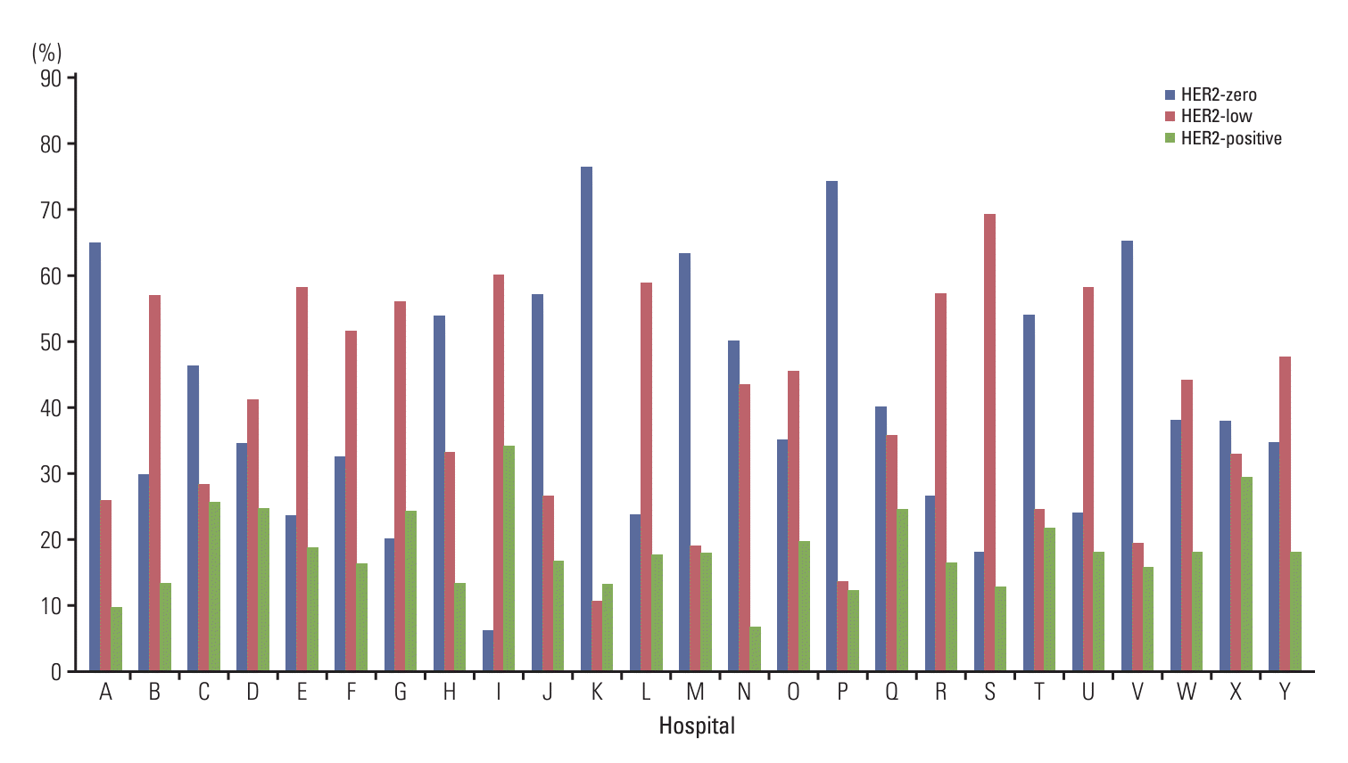
Four thousand six hundred seventy-four (40.9%) patients had only CNB HER2 test results, 3,483 (30.5%) only resection specimen, and 3,259 (28.6%) had both. Based on HER2 IHC results, 4,650 patients (40.7%) were scored as 0, 2,763 (24.2%) as 1+, 2,321 (20.3%) as 2+, and 1,682 (14.7%) as 3+. For all IHC 2+ patients, SISH or FISH tests were performed as a reflex test, revealing gene amplification in 321 patients (13.9%), including one case with amplification while IHC score was 0. Consequently, 4,649 patients (40.7%) were classified as HER2-zero, 4,764 (41.7%) as HER2-low, and 2,003 (17.5%) as HER2-positive. Variations in HER2 statuses were observed across institutions (S1 Table), with frequency ranges for HER2-zero, HER2-low, and HER2-positive patients from 6.0% to 76.3%, 10.5% to 69.1%, and 6.7% to 34.0%, respectively (Fig. 1).

3. Relationships between clinicopathologic characteristics and HER2 statuses

On comparing the HER2-low group to the HER2-zero group or the HER2-positive group, HER2-low tumors were more frequently subtyped as no special type than as special type (p=0.011). In addition, HER2-low status was more frequently exhibited ER and PR positivity (p < 0.001 for all). The HER2-low group was also significantly associated with low Ki-67 levels when the cutoff value was set at 20% (p < 0.001) (Table 1). The clinicopathological differences between HER2-zero and HER2-low status, following stratification by ER status or triple-negative status were described in S2 Table.

4. HER2 statuses of CNB and resection specimens

Three thousand two hundred fifty-nine patients had preoperative CNB and resection specimen HER2 test results. Out of the 1,292 patients initially classified as HER2-zero by CNB, 967 (74.8%) were also HER2-zero by resection specimen results, but 316 (24.4%) were reclassified as HER2-low and 9 (0.7%) as HER2-positive (Table 2, S3 Fig.). Of 1,687 patients with HER2-low CNB result, 420 (24.9%) were reclassified as HER2-zero, 1,243 (73.7%) remained HER2-low, and 24 (1.4%) were reclassified as HER2-positive. Finally, of 280 patients with a HER2-positive CNB, 3 (1.1%) were downgraded to HER2-zero, 15 (5.4%) to HER2-low, and 262 (93.6%) remained HER2-positive. HER2 IHC score changes are provided in S4 Table. HER2 statuses were discrepant for 787 (24.1%) patients, with the most common changes from HER2-low to HER2-zero in 420 patients and being from HER2-zero to HER2-low in 316 patients.

5. Relations between analytic factors of IHC and HER2 statuses

Most institutions (88.0%) favored the Ventana 4B5 clone for HER2 IHC. Sixteen (64.0%) institutions used the Ventana BenchMark Ultra instrument, six (24.0%) used the BenchMark XT unit, and three used other instruments. Details on IHC methods, including staining devices, antigen retrieval solutions, primary antibody sources, antibody incubation conditions, and detection kits used in each laboratory, are provided in Table 3. No two laboratories utilized identical HER2 IHC protocols, as evidenced by the varied settings for the parameters listed in Table 3.
Among the numerous parameters affecting IHC results, this study focused on analytic factors such as antibody incubation time and antigen retrieval time because they are easily adjusted using an automated staining device. To determine whether the inter-institutional variability in HER2-low frequency was influenced by these analytic factors, the data of 21 institutions that used the BenchMark XT or Ultra and the antibody clone 4B5 were subjected to analysis. In resected surgical specimens, when a 36-minute cutoff was applied to antigen retrieval time, the HER2 1+ rate was significantly higher in institutions using a ≥ 36-minute protocol than those using a < 36-minute protocol (p < 0.001) (Table 4). Similarly, when a 12-minute cutoff was applied to antibody incubation time, the longer time group showed higher HER2 1+ rate (p < 0.001). In the CNB group, incubation time did not statistically significant in 12-minute cutoff (p=0.224), and longer antigen retrieval group showed lower HER2 1+ rate (p=0.028). Despite the limited number of cases using the DAKO antibody, they revealed a higher incidence of HER2 1+ rates compared to those using the Ventana clone 4B5 antibody across all specimen types (Table 5).

Discussion

The importance of assessing HER2 status in BC is now universally acknowledged, as HER2 status serves not only as a prognostic marker but also in determining treatment regimens. Before the HER2-low era, the primary concern of pathologists was the identification of HER2-positive BC, with quality assessments centered on this detection [18]. After the development of T-DXd and successful clinical trial for the HER2-low BC, pathologists now pay attention to detecting HER2-low from real world practice. Recently, the ASCO/CAP released updated guidelines for HER2 interpretation, which included a recommendation that pathologists should add a footnote to HER2 test reports about the possible eligibility for T-DXd [19].
Several studies have reported HER2-low BC incidences ranging from 35% to 65%. However, many of these studies were retrospective and conducted before the HER2-low issue gained prominence or used previous HER2 interpretation guidelines [8-15]. However, the data used in the present study was collected in 2022 according to the latest HER2 testing guidelines and was interpreted carefully with an understanding of the importance of HER2-low status. Thus, our study provides an up-to-date overview of real-world conditions. Additionally, we focused on the overall incidence of HER2-low within our extensive cohort, rather than placing primary emphasis on differentiation of HER2-low and HER2-zero BC. Nevertheless, our findings that HER2-low BC was significantly associated with a hormone-positive status and a low Ki-67 proliferation index are consistent with previous research.
Comparing preoperative CNB and surgical specimen results showed that significant cases were reclassified between HER2-zero and HER2-low, reinforcing previously reported studies [12,20]. The underlying reason for these inconsistencies is probably multifactorial, which raises concerns about the specimen type for determining the HER2-low status. The current ASCO/CAP HER2 guidelines recommend repeat testing on the excision specimen if the initial HER2 result is negative, though this is advised only under specific circumstances [19]. Since this strategy is suitable for distinguishing between HER2-positive and false-negative cases as determined by biopsy, it may not be effective in HER2-low cases. Additionally, there is evidence of dynamic changes in HER2-low expression over the course of clinical treatment. Miglietta et al. [20] reported an overall discordance rate of 38% between primary and secondary tumors, with most cases transitioning from HER2-zero to HER2-low status, or vice versa. Consensus on this matter is necessary to optimize the selection of patient treatment options. Until then, re-testing of the excision specimens would be helpful for uncovering more HER2-low cases.
Interobserver variability is one of the many factors that influence the interpretation of HER2 IHC and could account for the broad range of HER2-low BC incidence reported in earlier studies. As the concordance rate for identifying HER2 3+ cases is high, non-3+ cases present a challenge and can cause the problem in HER2-low era [21,22]. One of our authors (E.Y.C.), who has over 20 years of experience in breast pathology, re-evaluated 1,063 HER2 IHC 1+ cases (unpublished data not included in this study) initially diagnosed by seven pathologists with varying levels of experience. Among these cases, 143 were reclassified as HER2 0, 653 remained HER2 1+, and 267 were classified as HER2-ultralow (weak, incomplete membranous staining in ≤ 10% of tumor cells). These results underscore the difficulty and potential variability among pathologists interpreting faint HER2 staining.
Antibody selection can contribute to variability in HER2-low interpretations. Our investigation revealed that the majority of institutions (21 out of 25) utilize the Ventana 4B5 clone. Standardizing the use of antibodies from a single company could promote more consistent results across laboratories. Notably, the clone 4B5 antibody was employed in the DESTINY-04 trial, suggesting its potential for precise detection of HER2-low in the application of T-DXd. However, some laboratories use the Dako polyclonal antibody for HER2 IHC. Karakas et al. [23] reported significant interantibody variation in a cohort of low-HER2-expressing BC patients. They compared two antibodies, HercepTest (Dako Agilent) and PATHWAY anti-HER2 (4B5) (Ventana) and found an overall agreement rate of 57.8%. Layfield et al. [24] also observed differences in staining properties between the two antibodies, highlighting the need for a more comprehensive evaluation of their use in diagnosing HER2-low.
This study also reveals that longer antigen retrieval times or antibody incubation periods may alter the incidence of 1+ staining results in the resected surgical specimens. Inconsistent analysis in CNB needs further elucidation, but it may correlate with numerous factors that can affect HER2 IHC results from pre-analytic to post-analytic stages. Limited cellularity or artifacts, such as edge effect, may also contributed to the complexity of the analysis [25]. When focusing on analytic factors, antigen retrieval time, antibody incubation time, and antibody incubation temperature are easily adjustable parameters when suboptimal staining is observed. Increasing antigen retrieval time may intensify IHC staining, but there comes a point that the tissue degeneration begins [26]. Similarly, higher incubation temperature and longer incubation time may enhance quality, but could weaken antigenantibody binding, resulting false-negative stains [27,28]. Thus, appropriately adjusting these factors is important for IHC staining protocols. Recently, Chen et al. [29] examined the impact of modifying staining methods on the incidence of HER2-low expression in IHC and observed that omitting antigen retrieval reduced the intensity of HER2 staining, and affected the IHC scores of 30.1% of tissue cores. In addition, prolonging the duration of antibody incubation enhanced HER2 staining intensity and increased the number of HER2-low cases, but it did not alter the final categorization of HER2-positivity. Garrido et al. [30] also published a study confirming that changes in the staining protocol can alter HER2 IHC results, especially for IHC 1+ cases.
One limitation of our study is the lack of accurate documentation regarding the pathologists responsible for reviewing the HER2 IHC. Interobserver variability in interpreting HER2-low status presents a significant challenge. In smaller institutions, interpretations were predominantly made by a single pathologist, while larger institutions often relied on multiple pathologists. Recognizing the critical relevance of HER2-low, the BPKSP has continuously worked to educate its members on HER2-low interpretation and enhance quality assessment practices. We believe these efforts can reduce interobserver variability. Additionally, we plan to explore the potential of artificial intelligence to further reduce variability and enhance the accuracy of HER2-low interpretations.
In summary, we evaluated the overall incidence of HER2-low BC in South Korea, in accordance with the most recent interpretation guidelines. Our study revealed that 41.7% (10.5%-69.1%) of the BC cases were categorized as HER2-low. Considering that laboratories utilize a range of settings for HER2 IHC, inter-institutional variabilities can have a significant impact on the accuracy of HER2-low identification. To address this issue, we recommend expanding the scope of quality assessment programs for HER2 IHC to include traditional quality control for HER2-positive cases, and the accurate identification of HER2-low cases. In addition, we propose continuous training programs be implemented to reduce interobserver variability.

Electronic Supplementary Material

Supplementary materials are available at Cancer Research and Treatment website (https://www.e-crt.org).

Notes

Ethical Statement

This study was reviewed and approved by the Institutional Review Board and Ethics Committee of Asan Medical Center (No. 2023-0386) and Yeungnam University College of Medicine (YUMC 2023-03-004), as well as the committees of all other participating centers. The informed consent was waived.

Author Contributions

Conceived and designed the analysis: Gong G, Bae YK.

Collected the data: Cho EY, Park SY, Lee HJ, Lee JS, Kim JY, Lee HC, Yoo JY, Kim HS (Hee Sung Kim), Kim B, Kim WS, Shin N, Maeng YH, Kim HS (Hun Soo Kim), Kwon SY, Kim C, Jun SY, Kwon GY, Choi HJ, Lee SM, Choi JE, An AR, Choi HJ, Kim E, Kim A, Kim JY, Shim JY, Gong G.

Contributed data or analysis tools: Kim MC, Bae YK.

Performed the analysis: Kim MC, Cho EY, Bae YK.

Wrote the paper: Kim MC, Cho EY, Gong G, Bae YK.

Conflicts of Interest

Conflict of interest relevant to this article was not reported.

ACKNOWLEDGMENTS

This study was supported by Korean Society of Pathologists Grant.

References

1. Sung H, Ferlay J, Siegel RL, Laversanne M, Soerjomataram I, Jemal A, et al. Global cancer statistics 2020: GLOBOCAN estimates of incidence and mortality worldwide for 36 cancers in 185 countries. CA Cancer J Clin. 2021; 71:209–49.
crossref
2. Kim H, Lee SB, Kim J, Chung IY, Kim HJ, Ko BS, et al. Improvement of survival in Korean breast cancer patients over a 14-year period: a large-scale single-center study. PLoS One. 2022; 17:e0265533.
crossref
3. Wolff AC, Hammond ME, Allison KH, Harvey BE, Mangu PB, Bartlett JM, et al. Human epidermal growth factor receptor 2 testing in breast cancer: American Society of Clinical Oncology/College of American Pathologists clinical practice guideline focused update. J Clin Oncol. 2018; 36:2105–22.
crossref
4. Wolff AC, Hammond ME, Schwartz JN, Hagerty KL, Allred DC, Cote RJ, et al. American Society of Clinical Oncology/College of American Pathologists guideline recommendations for human epidermal growth factor receptor 2 testing in breast cancer. Arch Pathol Lab Med. 2007; 131:18–43.
crossref
5. Wolff AC, Hammond ME, Hicks DG, Dowsett M, McShane LM, Allison KH, et al. Recommendations for human epidermal growth factor receptor 2 testing in breast cancer: American Society of Clinical Oncology/College of American Pathologists clinical practice guideline update. J Clin Oncol. 2013; 31:3997–4013.
crossref
6. Tozbikian G, Krishnamurthy S, Bui MM, Feldman M, Hicks DG, Jaffer S, et al. Emerging landscape of targeted therapy of breast cancers with low human epidermal growth factor receptor 2 protein expression. Arch Pathol Lab Med. 2024; 148:242–55.
crossref
7. Modi S, Jacot W, Yamashita T, Sohn J, Vidal M, Tokunaga E, et al. Trastuzumab deruxtecan in previously treated HER2-low advanced breast cancer. N Engl J Med. 2022; 387:9–20.
crossref
8. Tarantino P, Hamilton E, Tolaney SM, Cortes J, Morganti S, Ferraro E, et al. HER2-low breast cancer: pathological and clinical landscape. J Clin Oncol. 2020; 38:1951–62.
crossref
9. Gampenrieder SP, Rinnerthaler G, Tinchon C, Petzer A, Balic M, Heibl S, et al. Landscape of HER2-low metastatic breast cancer (MBC): results from the Austrian AGMT_MBC-Registry. Breast Cancer Res. 2021; 23:112.
crossref
10. Horisawa N, Adachi Y, Takatsuka D, Nozawa K, Endo Y, Ozaki Y, et al. The frequency of low HER2 expression in breast cancer and a comparison of prognosis between patients with HER2-low and HER2-negative breast cancer by HR status. Breast Cancer. 2022; 29:234–41.
crossref
11. Zhang G, Ren C, Li C, Wang Y, Chen B, Wen L, et al. Distinct clinical and somatic mutational features of breast tumors with high-, low-, or non-expressing human epidermal growth factor receptor 2 status. BMC Med. 2022; 20:142.
crossref
12. Baez-Navarro X, van Bockstal MR, Andrinopoulou ER, van Deurzen CH. HER2-low breast cancer: incidence, clinicopathologic features, and survival outcomes from real-world data of a large nationwide cohort. Mod Pathol. 2023; 36:100087.
crossref
13. Bergeron A, Bertaut A, Beltjens F, Charon-Barra C, Amet A, Jankowski C, et al. Anticipating changes in the HER2 status of breast tumours with disease progression-towards better treatment decisions in the new era of HER2-low breast cancers. Br J Cancer. 2023; 129:122–34.
crossref
14. Schettini F, Chic N, Braso-Maristany F, Pare L, Pascual T, Conte B, et al. Clinical, pathological, and PAM50 gene expression features of HER2-low breast cancer. NPJ Breast Cancer. 2021; 7:1.
crossref
15. Yang L, Liu Y, Han D, Fu S, Guo S, Bao L, et al. Clinical genetic features and neoadjuvant chemotherapy response in HER2-low breast cancers: a retrospective, multicenter cohort study. Ann Surg Oncol. 2023; 30:5653–62.
crossref
16. Tarantino P, Gandini S, Nicolo E, Trillo P, Giugliano F, Zagami P, et al. Evolution of low HER2 expression between early and advanced-stage breast cancer. Eur J Cancer. 2022; 163:35–43.
crossref
17. Allison KH, Hammond ME, Dowsett M, McKernin SE, Carey LA, Fitzgibbons PL, et al. Estrogen and progesterone receptor testing in breast cancer: American Society of Clinical Oncology/College of American Pathologists guideline update. Arch Pathol Lab Med. 2020; 144:545–63.
crossref
18. Ruschoff J, Lebeau A, Kreipe H, Sinn P, Gerharz CD, Koch W, et al. Assessing HER2 testing quality in breast cancer: variables that influence HER2 positivity rate from a large, multicenter, observational study in Germany. Mod Pathol. 2017; 30:217–26.
crossref
19. Wolff AC, Somerfield MR, Dowsett M, Hammond ME, Hayes DF, McShane LM, et al. Human epidermal growth factor receptor 2 testing in breast cancer. Arch Pathol Lab Med. 2023; 147:993–1000.
crossref
20. Miglietta F, Griguolo G, Bottosso M, Giarratano T, Lo Mele M, Fassan M, et al. Evolution of HER2-low expression from primary to recurrent breast cancer. NPJ Breast Cancer. 2021; 7:137.
crossref
21. Al Haddabi I, Qureshi A, Saparamadu A, Al Hamdani A, Al Riyami M, Ganguly S. Inter-observer agreement in reporting HER 2 Neu protein over expression by immunohistochemistry. Indian J Pathol Microbiol. 2014; 57:201–4.
crossref
22. Md Pauzi SH, Masir N, Yahaya A, Mohammed F, Tizen Laim NM, Mustangin M, et al. HER2 testing by immunohistochemistry in breast cancer: a multicenter proficiency ring study. Indian J Pathol Microbiol. 2021; 64:677–82.
23. Karakas C, Tyburski H, Turner BM, Wang X, Schiffhauer LM, Katerji H, et al. Interobserver and interantibody reproducibility of HER2 immunohistochemical scoring in an enriched HER2-low-expressing breast cancer cohort. Am J Clin Pathol. 2023; 159:484–91.
crossref
24. Layfield LJ, Frazier S, Esebua M, Schmidt RL. Interobserver reproducibility for HER2/neu immunohistochemistry: a comparison of reproducibility for the HercepTest and the 4B5 antibody clone. Pathol Res Pract. 2016; 212:190–5.
crossref
25. Hicks DG, Schiffhauer L. Standardized assessment of the HER2 status in breast cancer by immunohistochemistry. Lab Med. 2011; 42:459–67.
crossref
26. Krenacs L, Krenacs T, Stelkovics E, Raffeld M. Heat-induced antigen retrieval for immunohistochemical reactions in routinely processed paraffin sections. Methods Mol Biol. 2010; 588:103–19.
crossref
27. Boenisch T. Heat-induced antigen retrieval restores electrostatic forces: prolonging the antibody incubation as an alternative. Appl Immunohistochem Mol Morphol. 2002; 10:363–7.
crossref
28. Ramos-Vara JA, Miller MA. When tissue antigens and antibodies get along: revisiting the technical aspects of immunohistochemistry--the red, brown, and blue technique. Vet Pathol. 2014; 51:42–87.
29. Chen YY, Yang CF, Hsu CY. The impact of modified staining method on HER2 immunohistochemical staining for HER2-low breast cancer. Pathology. 2024; 56:122–4.
crossref
30. Garrido C, Manoogian M, Ghambire D, Lucas S, Karnoub M, Olson MT, et al. Analytical and clinical validation of PATHWAY Anti-HER-2/neu (4B5) antibody to assess HER2-low status for trastuzumab deruxtecan treatment in breast cancer. Virchows Arch. 2024; 484:1005–14.
crossref

Fig. 1.
Proportions of human growth factor receptor 2 (HER2) statuses at the participating institutions.
crt-2024-092f1.tif
Table 1.
Clinicopathologic characteristics of the three HER2 statuses
Clinicopathologic factor Total HER2 category
p-value
HER2-zero (n=4,649) HER2-low (n=4,764) HER2-positive (n=2,003) Zero vs. low Low vs. High
Age (yr)
 < 60 7,624 3,133 (67.4) 3,174 (66.6) 1,317 (65.8) 0.429 0.488
 ≥ 60 3,792 1,516 (32.6) 1,590 (33.4) 686 (34.2)
Specimen type
 Needle biopsy 4,674 1,894 (40.7) 1,627 (34.2) 1,153 (57.6) < 0.001 < 0.001
 Resection 3,483 1,365 (29.4) 1,563 (32.8) 555 (27.7)
 Needle biopsy and resection 3,259 1,390 (29.9) 1,574 (33.0) 295 (14.7)
Histologic type
 No special type 10,319 4,093 (88.0) 4,273 (89.7) 1,953 (97.5) 0.011 < 0.001
 Special types 1,097 556 (12.0) 491 (10.3) 50 (2.5)
ER status
 Negative 2,727 1,133 (24.4) 611 (12.8) 983 (49.1) < 0.001 < 0.001
 Positive 8,689 3,516 (75.6) 4,153 (87.2) 1,020 (50.9)
PR status
 Negative 4,100 1,591 (34.2) 1,203 (25.3) 1,306 (65.1) < 0.001 < 0.001
 Positive 7,316 3,058 (65.8) 3,561 (74.7) 697 (34.9)
Ki-67 index (%)
 < 20 5,207 2,306 (49.6) 2,586 (54.3) 315 (15.7) < 0.001 < 0.001
 ≥ 20 6,209 2,343 (50.4) 2,178 (45.7) 1,688 (84.3)

Values are presented as number (%). ER, estrogen receptor; HER2, human epidermal growth factor receptor 2; PR, progesterone receptor.

Table 2.
Comparison of HER2 statuses as determined by preoperative core needle biopsy and surgical specimen
HER2 results in core needle biopsies
Total
HER2-zero HER2-low HER2-positive
HER2 results in surgical specimen
 HER2-zero 967 420 3 1,390
 HER2-low 316 1,243 15 1,574
 HER2-positive 9 24 262 295
Total 1,292 1,687 280 3,259

HER2, human epidermal growth factor receptor 2.

Table 3.
HER2 immunohistochemistry staining conditions used at the participating institutions
Institution Automated staining device Primary antibody
Antigen retrieval
Antibody incubation
Detection kit
Source Clone Dilution Buffer Time (min) Temperature (℃) Time (min)
A BenchMarkXT Ventana 4B5 Prediluted CC1 36 36 16 UltraView DAB
B BenchMarkUltra Ventana 4B5 Prediluted CC1 16 37 16 OptiView DAB
C BenchMarkUltra Ventana 4B5 Prediluted CC1 36 42 32 UltraView DAB
D BenchMarkUltra Ventana 4B5 Prediluted CC1 16 36 8 OptiView DAB
E BenchMarkUltra Ventana 4B5 Prediluted CC1 36 36 32 OptiView DAB
F BenchMarkUltra Ventana 4B5 Prediluted CC1 36 42 16 UltraView DAB
G BenchMarkXT Dako Polyclonal 1:500 CC1 32 37 16 OptiView DAB
H BenchMarkUltra Ventana 4B5 Prediluted CC1 36 37 20 OptiView DAB
I DAKO-Omnis GI100 Dako Polyclonal 1:600 TRS-Hi pH9 30 37 20 FLEX
J BenchMarkUltra Ventana 4B5 Prediluted CC1 36 37 16 UltraView DAB
K BenchMarkXT Ventana 4B5 Prediluted CC1 16 37 28 OptiView DAB
L BenchMarkUltra Ventana 4B5 Prediluted CC1 24 37 16 OptiView DAB
M BenchMarkUltra Ventana 4B5 Prediluted CC1 52 42 32 UltraView DAB
N BenchMarkXT Ventana Polyclonal 1:250 CC1 8 37 16 OptiView DAB
O BenchMarkUltra Ventana 4B5 Prediluted CC1 16 37 56 OptiView DAB
P BenchMarkUltra Ventana 4B5 Prediluted CC1 16 37 4 OptiView DAB
Q BenchMarkUltra Ventana 4B5 Prediluted CC1 24 37 12 OptiView DAB
R BondMax Dako A0485 1:600 Bond ER2 20 25 15 DAB
S BenchMarkXT Ventana 4B5 Prediluted CC1 30 37 20 UltraView DAB
T BenchMarkUltra Ventana 4B5 Prediluted CC1 24 37 12 OptiView DAB
U BenchMarkUltra Ventana 4B5 Prediluted CC1 36 37 28 UltraView DAB
V BenchMarkUltra Ventana 4B5 Prediluted CC1 16 36 16 OptiView DAB
W BenchMarkXT Ventana 4B5 Prediluted CC1 32 37 24 OptiView DAB
X BenchMarkUltra Ventana 4B5 Prediluted CC1 24 36 8 OptiView DAB
Y BenchMarkUltra Ventana 4B5 Prediluted CC1 36 36 32 UltraView DAB

HER2, human epidermal growth factor receptor 2.

Table 4.
Comparison of antigen retrieval times and antibody incubation times used for HER2 immunohistochemistry between IHC 0 and 1+ in surgical and biopsy specimen
Total (n=4,366) Surgical specimen
p-value Total (n=2,757) Biopsy specimen
p-value
IHC 0 (%) IHC 1+ (%) IHC 0 (%) IHC 1+ (%)
Antigen retrieval time (min)
 < 36 1,653 1,199 (72.5) 454 (27.5) < 0.001 1,999 1,337 (66.9) 662 (33.1) 0.028
 ≥ 36 2,713 1,477 (54.4) 1,236 (45.6) 758 540 (71.2) 218 (28.8)
Antibody incubation time (min)
 < 12 202 168 (83.2) 34 (16.8) < 0.001 246 159 (64.6) 87 (35.4) 0.224
 ≥ 12 4,164 2,508 (60.2) 1,656 (39.8) 2,511 1,718 (68.4) 793 (31.6)

Values are presented as number (%). HER2, human growth factor receptor 2; IHC, immunohistochemistry.

Table 5.
Comparison of antibody clones used for HER2 immunohistochemistry between IHC 0 and 1+ in surgical and biopsy specimen
Antibody clone Total (n=4,576) Surgical specimen
p-value Total (n=2,810) Biopsy specimen
p-value
IHC 0 (%) IHC 1+ (%) IHC 0 (%) IHC 1+ (%)
Ventana 4,366 2,676 (61.3) 1,690 (38.7) < 0.001 2,757 1,877 (68.1) 880 (31.9) < 0.001
Dako 210 67 (31.9) 143 (68.1) 53 15 (28.3) 38 (71.7)

Values are presented as number (%). HER2, human growth factor receptor 2; IHC, immunohistochemistry.

TOOLS
Similar articles