Journal List > Cancer Res Treat > v.56(4) > 1516088687

Kim, Keam, Kang, Kim, Kim, Lee, Kwon, Lee, Yang, Choi, Kim, Ji, Yun, Choi, Lee, Kim, and Ahn: Analysis of Response and Progression Patterns of Tyrosine Kinase Inhibitors in Recurrent or Metastatic Adenoid Cystic Carcinoma: A Post Hoc Analysis of Two KCSG Phase II Trials

Abstract

Purpose

In this study, we evaluated 66 patients diagnosed with adenoid cystic carcinoma (ACC) enrolled in two Korean Cancer Study Group trials to investigate the response and progression patterns in recurrent and/or metastatic ACC treated with vascular endothelial growth factor receptor tyrosine kinase inhibitors (VEGFR-TKIs).

Materials and Methods

We evaluated 66 patients diagnosed with ACC who were enrolled in the Korean Cancer Study Group trials. The tumor measurements, clinical data, treatment outcomes, and progression patterns of therapy were analyzed.

Results

In the 66 patients (53 receiving axitinib and 13 receiving nintedanib), the disease control rate was 61%, and three patients achieved partial response. The median follow-up, median progression-free survival (PFS), overall survival, and 6-month PFS rate were 27.6%, 12.4%, and 18.1% months and 62.1%, respectively. Among 42 patients who experienced progression, 27 (64.3%) showed target lesion progression. Bone metastasis was an independent poor prognostic factor.

Conclusion

Overall, most patients demonstrated stable disease with prolonged PFS; however, prominent target lesion progression occurred in some patients. Thus, PFS may capture VEGFR-TKI efficacy better than the objective response rate.

Introduction

Adenoid cystic carcinoma (ACC) is a rare malignancy that typically originates in the salivary glands, comprising only 1% of malignant tumors in the head and neck region, and 10% of all salivary gland neoplasms [1]. ACC is characterized by unpredictable recurrence, slow tumor growth, and a prolonged clinical course, often with delayed development of distant metastasis [1,2]. Despite surgical resection of the primary tumor, recurrence is common both locally and distantly. Unfortunately, effective systemic treatment options for recurrent and/or metastatic (R/M) ACC are limited.
The vascular endothelial growth factor receptor (VEGFR) has emerged as a potential therapeutic target for ACC [3] because of its role in tumor angiogenesis. VEGFR presents a potential avenue for addressing this unfulfilled medical requirement. Several clinical trials have evaluated the efficacy of anti-angiogenic multi-target tyrosine kinase inhibitors (TKIs), including lenvatinib [4], sunitinib [5], sorafenib [6], nintedanib [7], dovitinib [8], axitinib [9], regorafenib [10], and rivoceranib [11]. The primary endpoint of these trials was the overall response rate (RR). However, the results of these trials were mostly negative, with RRs ranging from 0% to 16%. VEGFR-TKIs have shown modest activity in R/M ACC, primarily achieving disease stabilization rather than significant tumor shrinkage. Limited research has been conducted on the response and progression patterns after VEGFR-TKI treatment in ACC. As such, it is important to identify the best endpoint for capturing the true clinical benefits of VEGFR-TKI in patients with R/M ACC.
We analyzed the data from two trials conducted by the Korean Cancer Study Group (KCSG) that investigated the use of VEGFR-TKIs in patients with R/M ACC. The purpose of this study was to examine the response and progression patterns in patients with R/M ACC who underwent VEGFR-TKI therapy, and further evaluate its clinical implications.

Materials and Methods

1. Study design and patient eligibility

The data for this analysis were obtained from two prospective phase II clinical trials that investigated the efficacy of axitinib or nintedanib as palliative treatments for R/M ACC. Detailed descriptions of the study designs for these trials have been previously documented [7,12]. In brief, the axitinib study (KCSG HN16-08 trial, NCT02859012) [12], conducted between December 2016 and October 2017, was a multicenter, randomized phase II trial, with 30 patients each assigned to the axitinib and observation arms. After excluding three patients who did not receive axitinib for more than two cycles and one patient who did not undergo evaluation for tumor response, the per-protocol analysis included 27 patients from the axitinib arm and 26 patients from the observation arm who crossed over to the axitinib arm.
The nintedanib study (KCSG HN14-01, NCT02558387) [7], conducted between November 2014 and November 2015, was a multicenter single-arm study that enrolled 20 patients. Thirteen patients diagnosed with ACC were included in the analysis. The final dataset comprised 66 patients.
Treatment response and disease progression were assessed using the Response Evaluation Criteria in Solid Tumors (RECIST) ver. 1.1. Both trials adhered to the Declaration of Helsinki and International Conference on Harmonization Guidelines for Good Clinical Practice. The study protocol received approval from the ethics committees of all participating centers, and written informed consent was obtained from all patients.

2. Statistical analysis

Descriptive statistics were applied to summarize continuous or categorical variables. The statistical significance of clinical and biochemical parameters was assessed using the chi-square test or Fisher’s exact test. The best overall response was determined based on the most favorable responses observed across all response categories. Disease progression was defined at the date of the scan with documented radiological progression. Progression-free survival (PFS) was calculated from the first dose of VEGFR-TKIs to the first documentation of progressive disease, or death from any cause, or loss to follow-up. Overall survival (OS) was calculated from the first dose of VEGFR-TKIs to the last visit, or death from any cause. Survival curves were estimated using the Kaplan-Meier method. Multivariate Cox proportional hazards regression analysis was performed to identify independent prognostic factors. All statistical tests were two-sided, and a significance level of p < 0.05 was considered statistically significant. Data analysis was conducted using IBM SPSS Statistics for Windows, ver. 22 (IBM Corp., Armonk, NY).

Results

1. Patient characteristics

A total of 66 patients diagnosed with R/M ACC were included in this analysis. Among them, 53 received axitinib and 13 received nintedanib. The detailed clinical characteristics of the patients are summarized in Table 1; the median age was 56 years (range, 49 to 61 years), and 52% were female. Most patients (n=63, 95.5%) had distant metastases. All the patients had an Eastern Cooperative Oncology Group performance status of 0-1. Lung metastases were observed in 91% of patients (n=60), whereas liver and bone metastases were present in 28.8% and 21.2% of patients, respectively. Overall, 30% of the patients (n=22) had not received any prior systemic chemotherapy for R/M ACC, whereas 41% (n=30) had undergone two or more cycles of chemotherapy. The median follow-up period for this population was 27.6 months (95% confidence interval [CI], 11.5 to 43.9 months).

2. Response patterns of VEGFR-TKI treatment

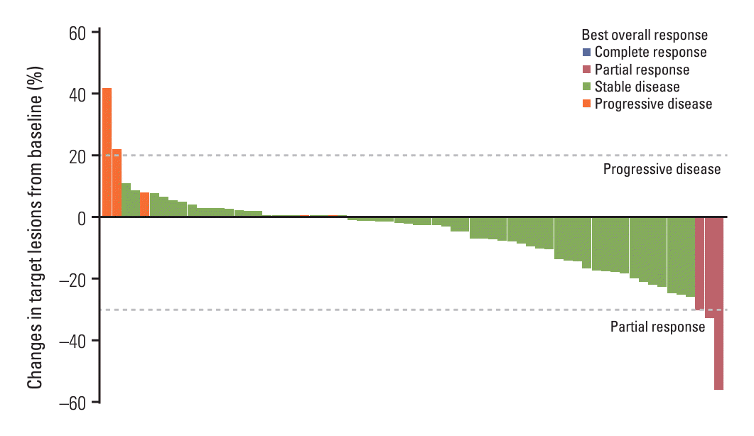
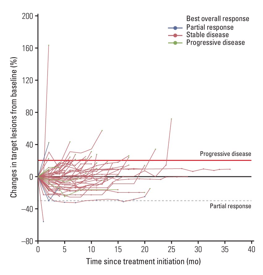
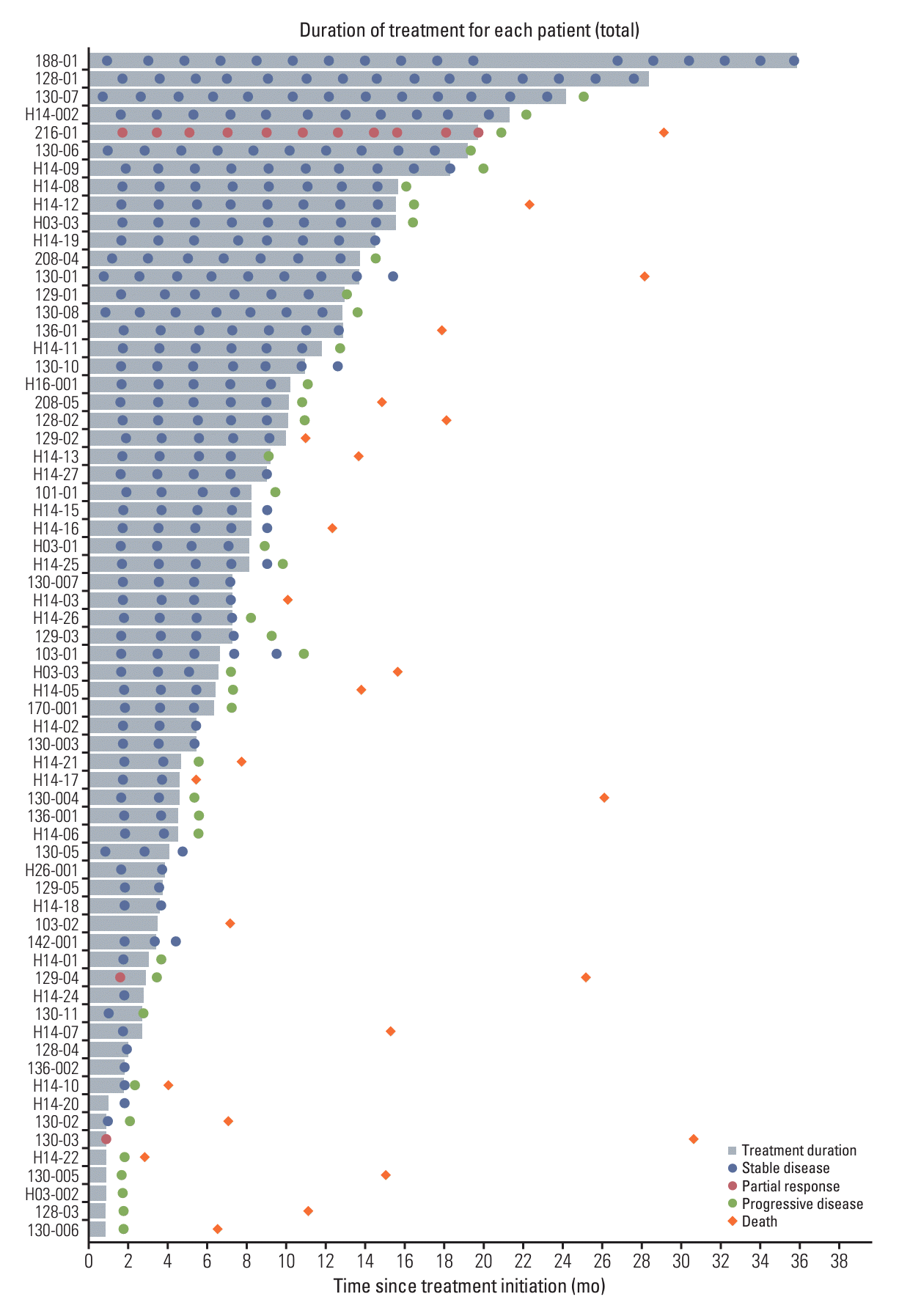
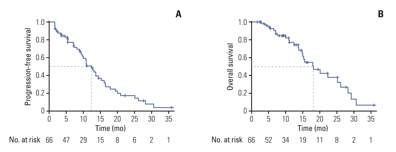
The response pattern was evaluated in the entire cohort. The efficacy results are summarized in Table 2. A waterfall plot (Fig. 1) illustrates the change in tumor size from baseline for each patient, showcasing a range of tumor burden changes from –56% to +42% at the best overall response. According to the RECIST 1.1 criteria, three patients achieved a partial response (PR), 58 patients had stable disease (SD), and five patients had progressive disease. The disease control rate (DCR) was thus 92.4% (61/66 patients), with a DCR of 96.2% (51/53) in the axitinib group and 76.9% (10/13) in the nintedanib group. Spider plots (Fig. 2) further illustrate the dynamics of tumor burden during VEGFR-TKI therapy in the cohort, reflecting the range of tumor burden changes observed at the best overall response. In 31.8% of patients (n=21), the tumor burden remained below a 20% increase from baseline throughout therapy. A swimmer plot depicting the duration of treatment and PFS with VEGFR-TKI treatment for each patient is shown in Fig. 3. PFS events followed a consistent pattern, with the PFS curve declining steadily at nearly a 45° angle, resulting in a median PFS of 12.4 months (95% CI, 10.0 to 14.8 months), and a median OS of 18.1 months (95% CI, 12.7 to 23.5 months) (Fig. 4). In the axitinib group, the median PFS was 11.0 months (95% CI, 7.7 to 12. 5 months) and the median OS was 17.9 months (95% CI, 13.7 to 19.7 months) (S1 Fig.). In the nintedanib group, the median PFS was 20.5 months (95% CI, 18.0 to 22.4 months) and the median OS was 26.1 months (95% CI, 22.5 to 29.0 months) (S2 Fig.). The 6-month PFS rate was 62.1% (95% CI, 51.5 to 75.0).

3. Pattern of treatment failure according to RECIST

All measurable lesions were monitored using regular computed tomography scans until RECIST 1.1–defined progression. Of the 66 patients included in the analysis, 42 exhibited disease progression. The progression patterns are summarized in Table 3. Of these, 36 patients (85.8%) experienced progression of existing lesions, target lesions, non-target lesions, or both. Among the 42 patients who experienced disease progression, 27 (64.3%) exhibited progression in the target lesions. Moreover, three patients (7.1%) showed progression only in new lesions, while another three patients (7.1%) had combined progression involving both existing and novel lesions.

4. Independent predictors of survival in the study population

We further investigated the clinical factors that could serve as significant risk factors for PFS and OS. Initially, univariate Cox regression analysis was performed to identify significant risk factors in the training set; the detailed results are presented in Table 4. Subsequently, multivariate analysis was conducted using potential predictors of survival outcomes, including sex, presence of liver metastasis, presence of bone metastasis, and number of metastatic organs (≥ 2). Univariate Cox regression analysis indicated that male sex, presence of liver metastasis, and presence of bone metastasis were associated with a shorter OS. The multivariate logistic regression model further confirmed that bone metastasis (hazard ratio, 3.35; 95% CI, 1.54 to 7.27; p=0.02) remained an independent risk factor for OS, suggesting that patients with bone metastasis had a higher risk of mortality (Table 4).

Discussion

This study provides insights into the response and progression patterns observed in patients with R/M ACC undergoing VEGFR-TKI treatment. Firstly, a noteworthy portion of the patients demonstrated SD, with extended PFS during VEGFR-TKI treatment. The median PFS of 12.4 months signifies a substantial postponement in progression or death. Furthermore, the considerable DCR of 92% observed within the analyzed cohort highlights the effectiveness of VEGFR-TKIs in suppressing tumor progression rather than tumor shrinkage in R/M ACC. Secondly, our analysis unveiled a substantial progression of target lesions in patients with R/M ACC. Specifically, 64.3% of patients experienced progression of the target lesions. Finally, bone metastasis emerged as an independent adverse prognostic factor in individuals with R/M ACC.
Several VEGFR-TKIs, including dovitinib, axitinib, sunitinib, sorafenib, and regorafenib, have previously been studied in clinical trials. These drugs demonstrated a limited number of PRs, no complete responses, and varying degrees of disease stabilization. No objective response was observed with sunitinib [5], regorafenib [10], and nintedanib [7], whereas a 9%-16% objective RR was observed with sorafenib [6], axitinib [9], and lenvatinib [4]. Therefore, they often failed to meet the primary endpoint of RRs.
Given the rarity of this disease and its varying natural history, it is crucial to consider novel phase II clinical trial design strategies to efficiently use patient resources. Further, it is essential to carefully select the appropriate endpoints to capture the unique characteristics of this rare and slow-growing tumor in a small patient population. Given these findings, alternative primary endpoints, such as a median PFS, may be more appropriate for ACC trials using VEGFR-TKIs. Implementing such an endpoint in future clinical trials can provide valuable insights into the efficacy of cytostatic molecular targeted agents in ACC and improve the overall efficiency of drug investigations for this rare and challenging disease.
We further observed a consistent decline in PFS in patients treated with VEGFR-TKIs, as evidenced by the intriguing pattern revealed by the Kaplan-Meier curves. However, it appears that this trend is mainly driven by the axitinib cohort, acknowledging the limitations associated with this observation. PFS declined at a rate of 45°, suggesting an evenly distributed progression of the disease over time. The absence of a specific time-point for disease progression further emphasizes the unpredictable nature of its occurrence. These findings provide valuable details regarding the effectiveness of VEGFR-TKIs and elucidate the time-dependent nature of disease progression. By understanding these dynamics, we can gain important information that contributes to a better understanding of treatment outcomes, enabling the optimization of therapeutic strategies for patients.
In this study, the most common cause of radiologic progression in R/M ACC was related to changes in target lesions rather than to changes in non-target diseases or new lesions. Understanding the type of progression can provide valuable information when considering subsequent treatment strategies after the failure of previous chemotherapy. Although lung metastasis is the most common site, the presence of bone metastasis predicts a worse prognosis. Previous studies have reported that the median survival time after metastasis to the vertebrae (20.0-20.6 months) is worse than that after lung metastasis (32.3-54.0 months) [2,13,14]. Bone metastasis, which is not measurable, can contribute to non-target lesion progression. Metastatic lesions with higher progression potential are often found in the liver, bone, and brain/central nervous system, which are either immune-privileged or tolerogenic organs [2]. This underscores the importance of the findings of the present study, as patients with highly progressive lesions in the bones experience worse survival outcomes, likely necessitating more effective and targeted therapeutics.
While our study contributes valuable insights, it is essential to recognize several inherent limitations. Firstly, the discrepancy in sample sizes between the axitinib and nintedanib studies is a notable concern, potentially affecting the robustness and generalizability of our findings. Secondly, the small sample size may have constrained the statistical power and generalizability of our results. Thirdly, the relatively short follow-up duration might not fully capture the long-term disease course and late recurrence commonly observed in adrenocortical carcinoma (ACC). Fourth, the heterogeneity of treatments received by enrolled patients, including variations in surgical extent, postoperative radiotherapy, and chemotherapy regimens, could have introduced confounding factors influencing outcomes. Moreover, we acknowledge the limitations of post hoc analyses and observational studies, such as the potential for reporting bias and methodological constraints. Nevertheless, it is noteworthy that our study contributes to the limited body of research investigating the response and progression patterns in this patient population, while the relatively large sample size strengthens the significance of our findings.
In conclusion, our study revealed that most patients with R/M ACC treated with VEGFR-TKIs showed SD and prolonged PFS. These results suggest that PFS should be considered a more suitable primary endpoint in clinical trials assessing the effectiveness of VEGFR-TKIs for R/M ACC, rather than relying solely on the commonly used objective RR. Additionally, the significant occurrence of progression in target lesions highlights the importance of close monitoring and the evaluation of target lesion progression in patients undergoing VEGFR-TKI treatment. Overall, these findings hold important implications for optimizing future trial designs and for assessing the clinical efficacy of VEGFR-TKIs in R/M ACC.

Electronic Supplementary Material

Supplementary materials are available at Cancer Research and Treatment website (https://www.e-crt.org).

Notes

Ethical Statement

This study was performed in line with the principles of the Declaration of Helsinki and International Conference on Harmonization Guidelines for Good Clinical Practice. The study protocol received approval from the ethics committees of all participating centers, and written informed consent was obtained from all patients.

Author Contributions

Conceived and designed the analysis: Keam B, Ahn MJ.

Collected the data: Keam B, Kang EJ, Kim HR, Kwon JH, Yang Y, Kim MK, Yun T, Lee KH, Ahn MJ.

Contributed data or analysis tools: Kim Y, Keam B, Kim JS, Lee KW, Lee KE, Choi YH, Ji JH, Choi MY, Kim SB.

Performed the analysis: Kim Y, Keam B, Ahn MJ.

Wrote the paper: Kim Y, Keam B.

Conflicts of Interest

Bhumsuk Keam received research funding from MSD, AstraZeneca, and Ono Pharmaceutical Co., Ltd., and has served as an advisor for Handok, NeoImmuneTec, Trialinformatics, and ImmuneOncia, outside of the current work. This study was supported by BI. BI had no role in the design, analysis, or interpretation of the results in this study. BI was given the opportunity to review the manuscript for medical and scientific accuracy as it relates to BI substances, as well as intellectual property considerations. Otherwise, the authors declare that they have no relevant conflicts of interest regarding the publication of this manuscript.

ACKNOWLEDGMENTS

We would like to acknowledge all the patients and their families for their contributions to this study. We thank Young Chul Cho of Trialinformatics for supporting the data management. This research was performed using the KCSG Trial Data Hub and supported by the KCSG Data Center.
This study was supported by the National R&D Program for Cancer Control through the National Cancer Center funded by the Ministry of Health and Welfare, Republic of Korea (HA22C0011) and (HA22C0012). The funder had no role in the study design, data collection, analyses, and interpretation of data, in the writing of the report, or in the decision to submit the article for publication.

References

1. Laurie SA, Ho AL, Fury MG, Sherman E, Pfister DG. Systemic therapy in the management of metastatic or locally recurrent adenoid cystic carcinoma of the salivary glands: a systematic review. Lancet Oncol. 2011; 12:815–24.
crossref
2. Sung MW, Kim KH, Kim JW, Min YG, Seong WJ, Roh JL, et al. Clinicopathologic predictors and impact of distant metastasis from adenoid cystic carcinoma of the head and neck. Arch Otolaryngol Head Neck Surg. 2003; 129:1193–7.
crossref
3. Park S, Nam SJ, Keam B, Kim TM, Jeon YK, Lee SH, et al. VEGF and Ki-67 overexpression in predicting poor overall survival in adenoid cystic carcinoma. Cancer Res Treat. 2016; 48:518–26.
crossref
4. Tchekmedyian V, Sherman EJ, Dunn L, Tran C, Baxi S, Katabi N, et al. Phase II study of lenvatinib in patients with progressive, recurrent or metastatic adenoid cystic carcinoma. J Clin Oncol. 2019; 37:1529–37.
crossref
5. Chau NG, Hotte SJ, Chen EX, Chin SF, Turner S, Wang L, et al. A phase II study of sunitinib in recurrent and/or metastatic adenoid cystic carcinoma (ACC) of the salivary glands: current progress and challenges in evaluating molecularly targeted agents in ACC. Ann Oncol. 2012; 23:1562–70.
crossref
6. Thomson DJ, Silva P, Denton K, Bonington S, Mak SK, Swindell R, et al. Phase II trial of sorafenib in advanced salivary adenoid cystic carcinoma of the head and neck. Head Neck. 2015; 37:182–7.
crossref
7. Kim Y, Lee SJ, Lee JY, Lee SH, Sun JM, Park K, et al. Clinical trial of nintedanib in patients with recurrent or metastatic salivary gland cancer of the head and neck: a multicenter phase 2 study (Korean Cancer Study Group HN14-01). Cancer. 2017; 123:1958–64.
crossref
8. Keam B, Kim SB, Shin SH, Cho BC, Lee KW, Kim MK, et al. Phase 2 study of dovitinib in patients with metastatic or unresectable adenoid cystic carcinoma. Cancer. 2015; 121:2612–7.
crossref
9. Ho AL, Dunn L, Sherman EJ, Fury MG, Baxi SS, Chandramohan R, et al. A phase II study of axitinib (AG-013736) in patients with incurable adenoid cystic carcinoma. Ann Oncol. 2016; 27:1902–8.
crossref
10. Ho AL, Sherman EJ, Baxi SS, Haque S, Ni A, Antonescu CR, et al. Phase II study of regorafenib in progressive, recurrent/metastatic adenoid cystic carcinoma. J Clin Oncol. 2016; 34(15 Suppl):6096.
crossref
11. Hanna GJ, Ahn MJ, Muzaffar J, Keam B, Bowles DW, Wong DJ, et al. A phase II trial of rivoceranib, an oral vascular endothelial growth factor receptor 2 inhibitor, for recurrent or metastatic adenoid cystic carcinoma. Clin Cancer Res. 2023; 29:4555–63.
crossref
12. Kang EJ, Ahn MJ, Ock CY, Lee KW, Kwon JH, Yang Y, et al. Randomized phase II study of axitinib versus observation in patients with recurred or metastatic adenoid cystic carcinoma. Clin Cancer Res. 2021; 27:5272–9.
crossref
13. Jang S, Patel PN, Kimple RJ, McCulloch TM. Clinical outcomes and prognostic factors of adenoid cystic carcinoma of the head and neck. Anticancer Res. 2017; 37:3045–52.
14. van der Wal JE, Becking AG, Snow GB, van der Waal I. Distant metastases of adenoid cystic carcinoma of the salivary glands and the value of diagnostic examinations during follow-up. Head Neck. 2002; 24:779–83.
crossref

Fig. 1.
Waterfall plot of the best overall response of the 66 patients with evaluable disease, with overall best responses broken down by the administration of either vascular endothelial growth factor receptor tyrosine kinase inhibitor (axitinib [n=53] or nintedanib [n=13]).
crt-2024-008f1.tif
Fig. 2.
Spider plot of the changes in tumor size over time for each patient, starting from the initiation of vascular endothelial growth factor receptor tyrosine kinase inhibitor treatment.
crt-2024-008f2.tif
Fig. 3.
Swimmer plot illustrating the duration of vascular endothelial growth factor receptor tyrosine kinase inhibitor (VEGFR-TKI) treatment and progression-free survival (PFS) for each patient. Each horizontal line represents an individual patient, with the length of the line indicating the duration of VEGFR-TKI treatment. PFS events are denoted by vertical lines, providing insight into the timing of disease progression or treatment discontinuation for each patient.
crt-2024-008f3.tif
Fig. 4.
Kaplan-Meier curves depicting progression-free survival (A) and overall survival (B) for the entire patient population. Progression-free survival and overall survival were analyzed across the entire cohort, resulting in a median progression-free survival of 12.4 months (95% confidence interval [CI], 10.0 to 14.8 months) and a median overall survival of 18.1 months (95% CI, 12.7 to 23.5 months).
crt-2024-008f4.tif
Table 1.
Baseline demographic and clinical characteristics of the patients
Variable Value (n=66)
Age (yr), median (range) 56 (49-61)
Sex
 Male 32 (48.5)
 Female 34 (51.5)
ECOG PS
 0 17 (25.8)
 1 49 (74.2)
VEGFR-TKIs
 Axitinib 53 (80.3)
 Nintedanib 13 (19.7)
Disease distribution at enrollment
 Locoregional disease only 3 (4.5)
 Distant metastasis 63 (95.5)
Site of distant metastasis
 Lung 60 (90.9)
 Liver 19 (28.8)
 Bone 14 (21.2)
 Brain 1 (1.5)
No. of distant metastatic organs
 0-1 51 (77.3)
 ≥ 2 15 (22.7)
Prior lines of systemic therapy
 0 19 (28.8)
 1 19 (28.8)
 ≥ 2 28 (42.4)

Values are number (%) unless otherwise indicated. ECOG PS, Eastern Cooperative Oncology Group performance status; VEGFR-TKI, vascular endothelial growth factor receptor tyrosine kinase inhibitor.

Table 2.
Summary of the drug efficacy results
Best overall response (n=66) Value
Partial response 3 (4.5)
Stable disease 58 (87.9)
Progressive disease 5 (7.6)
Disease control rate 61 (92.4)
Progression-free survival (mo), median (95% CI) 12.4 (10.0-14.8)
Overall survival, median (95% CI) 18.1 (12.7-23.5)
6-Month progression-free survival rate (%) (95% CI) 62.1 (51.5-75.0)

Values are number (%) unless otherwise indicated. CI, confidence interval.

Table 3.
Patterns of disease progression by RECIST ver. 1.1
Patterns of disease progression (n=66) No. (%)
Progression of TL only 15 (22.7)
Progression of TL and NTL 11 (16.7)
Progression of NTL only 10 (15.2)
Progression of TL and new lesion 1 (1.5)
Progression of NTL and new lesion 2 (3.0)
New lesion only 3 (4.5)
Ongoing 24 (36.4)

NTL, non-target lesions; RECIST, Response Evaluation Criteria in Solid Tumors; TL, target lesions.

Table 4.
Uni- and multivariate analyses of the clinical parameters affecting PFS of VEGFR-TKI
Variable Univariate analysis
Multivariate analysis
HR (95% CI) p-value HR (95% CI) p-value
Age (yr)
 < 60 1
 ≥ 60 1.05 (0.58-1.90) 0.88 - -
Sex
 Female 1
 Male 1.92 (1.02-3.61) 0.04 1.23 (0.39-1.57) 0.16
Prior line of systemic therapy
 0-1 1
 ≥ 2 1.48 (0.79-2.79) 0.22 - -
VEGFR-TKIs
 Axitinib 1
 Nintedanib 1.6 (0.57-4.50) 0.37 - -
Dose reduction
 No 1
 Yes 0.91 (0.49-1.66) 0.75 - -
Liver metastasis
 Absence 1
 Presence 2.17 (1.08-4.36) 0.04 2.34 (0.86-6.35) 0.09
Bone metastasis
 Absence 1
 Presence 4.08 (2.01-8.30) < 0.01 3.35 (1.54-7.27) 0.02
No. of metastatic organs
 0-1 1
 ≥ 2 2.1 (0.73-6.02) 0.17 1.65 (0.84-2.64) 0.18

CI, confidence interval; HR, hazard ratio; PFS, progression-free survival; TKI, tyrosine kinase inhibitor; VEGFR, vascular endothelial growth factor receptor.

TOOLS
Similar articles