Journal List > Cancer Res Treat > v.56(4) > 1516088686

Yang, Yu, Li, Peng, Zhou, Yao, Lv, Fang, Shi, Wei, Wang, Cai, Zhang, Zhang, and Zhou: Evaluation of Molecular Residual Disease by a Fixed Panel in Resectable Colorectal Cancer

Abstract

Purpose

Molecular residual disease (MRD) is a promising biomarker in colorectal cancer (CRC) for prognosis and guiding treatment, while the whole-exome sequencing (WES) based tumor-informed assay is standard for evaluating MRD based on circulating tumor DNA (ctDNA). In this study, we assessed the feasibility of a fixed-panel for evaluating MRD in CRC.

Materials and Methods

Seventy-five patients with resectable stage I-III CRC were enrolled. Tumor tissues obtained by surgery, and preoperative and postoperative day 7 blood samples were collected. The ctDNA was evaluated using the tumor-agnostic and tumor-informed fixed assays, as well as the WES-based and panel-based personalized assays in randomly selected patients.

Results

The tumor-informed fixed assay had a higher preoperative positive rate than the tumor-agnostic assay (73.3% vs. 57.3%). The preoperative ctDNA status failed to predict disease-free survival (DFS) in either of the fixed assays, while the tumor-informed fixed assay-determined postoperative ctDNA positivity was significantly associated with worse DFS (hazard ratio [HR], 20.74; 95% confidence interval [CI], 7.19 to 59.83; p < 0.001), which was an independent predictor by multivariable analysis (HR, 28.57; 95% CI, 7.10 to 114.9; p < 0.001). Sub-cohort analysis indicated the WES-based personalized assay had the highest preoperative positive rate (95.1%). The two personalized assays and the tumor-informed fixed assay demonstrated same results in postoperative landmark (HR, 26.34; 95% CI, 6.01 to 115.57; p < 0.001), outperforming the tumor-agnostic fixed panel (HR, 3.04; 95% CI, 0.94 to 9.89; p=0.052).

Conclusion

Our study confirmed the prognostic value of the ctDNA positivity at postoperative day 7 by the tumor-informed fixed panel. The tumor-informed fixed panel may be a cost-effective method to evaluate MRD, which warrants further studies in future.

Introduction

Colorectal cancer (CRC), ranked as the third most prevalent cancer, remains one of the leading causes of cancer-related mortality worldwide [1,2]. Significant advancements in early detection have facilitated the diagnosis of CRC at early tumor stages, enabling more patients to undergo curative resection and experience improved outcomes. In early-stage CRCs, curative resection is often performed, resulting in a remarkable 5-year survival rate surpassing 80% [1,3]. Standard treatment for stage II-III CRC involves postoperative adjuvant chemotherapy (ACT) or follow-up, considering clinical risk factors and mismatch repair status after radical surgery. However, over 80% of stage II and around 50% of stage III patients achieve clinical cure through surgery alone, while 15%-20% of stage II/III patients experience disease recurrence despite ACT administration [4-6]. These findings highlight the existing dilemma of over- and under-treatment for early-stage CRC. Therefore, there is an imminent necessity for a reliable biomarker to distinguish high-risk patients from those with a favorable prognosis and minimal risk of recurrence. This will enable the implementation of personalized and effective treatment strategies.
Circulating tumor DNA (ctDNA) detection to assess the presence of molecular residual disease (MRD) in patients after curative-intent surgery has emerged as a promising tool for predicting cancer recurrence [7,8]. Numerous studies have consistently reported the association between ctDNA positivity and poor prognosis, whether after curative surgery or ACT [8]. Moreover, the dynamic changes in ctDNA during ACT have been shown to reflect treatment efficacy, as patients who achieve ctDNA clearance exhibit significantly longer disease-free survival (DFS) compared to those with persistent ctDNA positivity [9]. The groundbreaking DYNAMIC II study revealed the potential of ctDNA-guided approach in clinical practice. Researchers showed that the ctDNA-guided adjuvant therapy was non-inferior to standard management in terms of 2-year recurrence-free survival. Additionally, the ctDNA-guided approach led to a reduction in the utilization of ACT compared to the standard-management group (15% vs. 28%). These findings demonstrate the clinical significance of ctDNA-based MRD in predicting cancer recurrence and guiding treatment decisions for patients with CRC after curative treatment [10].
Next-generation sequencing (NGS) has been widely utilized to evaluate ctDNA-MRD in various cancer types. Tumor-agnostic assays targeting specific genes have been used for ctDNA analysis, with a limited detection (LOD) ranging from 0.2% to 0.5% [11]. Meta-analysis has shown that ctDNA fraction is usually lower than 0.01% after curative treatment in different cancers [8]. This suggests that the sensitivity of tumor-agnostic assays may not be accurate enough for MRD detection. To improve sensitivity, genomic mutations from tumor tissues are called prior to detect minute amounts of ctDNA after curative treatment. Zhou et al. [12] reported a study in 106 patients with locally advanced rectal cancer who underwent neoadjuvant chemoradiotherapy followed by surgery. Using a 1021-gene liquid biopsy panel with special bioinformatic methods for peak calling and positivity determination, they achieved an improved LOD of 0.03% and found that 40% of stage II and 76.8% of stage III patients were ctDNA-positive before the operation. Notably, all patients who achieved a pathologic complete response were tested negative for preoperative ctDNA [12]. Moreover, personalized panels designed based on patients’ specific tumor tissue mutations can further enhance sensitivity by increasing the number of effective sites and sequencing depth. One representative assay utilizing this tumor-informed strategy is the Signatera platform by Natera, which customizes a panel of 16 single nucleotide variants based on whole-exome sequencing of tumors. The LOD of Signatera is around 0.01%. Reinert et al. [13] has published a multicenter study involving 130 stage I-III colon cancer patients assessed by the Signatera platform. They reported a preoperative ctDNA-positive rate of 90.6% in stage II-III patients and found that ctDNA-positive patients had a significantly higher risk of recurrence at 30 days postoperation (hazard ratio [HR], 7.2; 95% confidence interval [CI], 2.7 to 19.0; p < 0.001) [13]. Similarly, in the GALAXY study of the CIRCULATE-Japan project that enrolled resectable CRC patients, researchers observed 95% and 18% ctDNA positivity rates before and after the operation, respectively. Additionally, ctDNA status at 4 weeks postoperation was found to be an independent factor to predict recurrence (HR, 15.3; p < 0.001) [9]. Growing evidences support the important role of MRD in guiding precise and personalized treatment. Although the tumor-informed strategy has been widely accepted and favored for evaluating ctDNA-MRD due to its superior sensitivity, several questions remain to be answered. For instance, the appropriate time for postoperative landmark in clinical application is not yet well-established, and the requirements of MRD testing platforms in different clinical scenarios are yet to be distinguished. Furthermore, regarding detection technologies, a unified consensus on assessing different MRD detection strategies and assays (including sensitivity, specificity, positive predictive value, negative predictive value, LOD, etc) has not been reached.
Additionally, for tumor-informed assay, whole-exome sequencing (WES) and whole genome sequencing are mostly applied to obtain tumor mutations and are usually followed by personalized panels. However, it remains unclear whether patient-specific mutations from a relatively large CRC fixed panel is sufficient and cost-effective for MRD detection, considering the relatively high release of ctDNA in CRCs. Here we report the results from a prospective study conducted in patients with stage I-III CRC, in which we aimed to evaluate the feasibility of a fixed panel in assessing MRD in CRC by comparing WES and panel-based personalized assays.

Materials and Methods

1. Study design

Seventy-six patients with resectable stage I-III CRC who provided written informed consent between May 2019 and July 2020 were enrolled in this study. To be eligible for enrollment, patients needed to have a pathologically confirmed diagnosis of CRC and were able to provide enough tumor tissues for sequencing. Exclusion criteria included a history of another primary cancer within the previous 3 years or treatment with neoadjuvant chemoradiotherapy.
The histopathological features, clinical characteristics and treatment of the patients were collected. Tumor tissue samples were obtained from each patient during the surgical procedure, and blood samples were collected preoperation and approximately 7 days postoperation for ctDNA analysis. The genomic profiles of both tumor tissues and blood samples were initially assessed using a 41-gene panel through capture-based DNA sequencing. The ctDNA in the blood samples was analyzed using two distinct strategies: a tumor-agnostic and a tumor-informed approach. Additionally, two personalized panels were used in randomly selected 41 patients to detect MRD in the blood samples. Detailed information of the MRD assays are described below, with technical parameters depicted in S1 Table.

2. DNA isolation

Genomic DNA of tissue specimens was extracted using QIAamp DNA FFPE tissue kits (Qiagen, Hilden, Germany). The extracted DNA was then quantified using the Qubit 2.0 fluorimeter (Life Technologies, Carlsbad, CA). Approximately 10 mL of blood were collected in streck tubes (DNA preserving tubes) and processed within 72 hours of collection. Whole blood was centrifuged at 2,200 ×g for 10 minutes at 4°C. After discarding the red blood cells and buffy coat, we centrifuged the plasma at 16,000 ×g for another 10 minutes at 4°C. Supernatants were subsequently stored at −80°C until further analysis. Cell-free DNA (cfDNA) from plasma was purified using a QIAamp Circulating Nucleic Acid kit according to the manufacturer’s instructions.

3. MRD calling using fixed panel

In our previous report, we introduced the fixed panel ColonCore developed by Burning Rock Biotech in Guangzhou, China [14]. This panel has undergone enhancements, now encompassing the complete exons of 41 genes associated with colon cancer, spanning 213 kb of the human genome (S2 Table). This upgraded version of the ColonCore panel was utilized in the current study. The raw sequencing data analysis and tumor-agnostic somatic mutation calling were conducted using the in-house bioinformatics pipeline, as previously reported. A ctDNA-positive status is defined as the detection of any somatic mutation. In the tumor-informed caller, variants were treated as false-positive and were first filtered out if they were not detected in paired tumor tissues. ctDNA analysis was conducted using a previously published algorithm called PROPHET (Burning Rock Biotech, Guangzhou, China) [15]. A single-stranded consensus sequence approach followed by duplex consensus sequence was used to create read families. This algorithm utilizes the maximum likelihood (ML) method by monitoring multiple loci to estimate the ctDNA fraction in plasma samples. In PROPHET, the probability (p) of the read count for each informed mutation, following a Poisson distribution, is calculated. If the p-value is less than 0.05, the mutation is considered significant and true-positive. Subsequently, a likelihood ratio test is performed to determine the sample-level p-value. In this context, the tumor-informed fixed panel strategy defines a sample as ctDNA-positive if it possesses at least one significant site and a sample-level p < 0.0005.

4. ctDNA analysis through personalized assays

Tissue samples were subjected to sequencing using a panel that included 520 cancer-related genes (OncoScreen plus, Burning Rock Biotech). Gene list of the OncoScreen plus panel is reported in S3 Table. Alternatively, WES was performed using the Twist Human Core Exome kit (Twist Bioscience, South San Francisco, CA), following the manufacturer’s recommendations. Based on the previously reported screening criteria, two sets of individualized probes were designed according to the mutations detected in the tissue samples, and ctDNA was analyzed using the PROPHET algorithm. For the panel-based personalized assay, up to 25 mutation loci were included. Hotspot variants with a variant allele frequency (VAF) ≥ 0.3% or non-hotspot mutations with a VAF ≥ 2% were selected. Priority was given to hotspot variants, followed by those with high VAF. ctDNA positivity was defined as having one significant site and a sample-level p < 0.0005 or having at least two significant sites and a sample-level p < 0.005. On the other hand, for the WES-based personalized assay, up to 50 highly ranked variants with a VAF above 3.0% were selected. Mutations with high VAF and high-impact were prioritized. Essentially, variants situated in the repetitive regions, regions with a high GC content (> 75%), and homologous regions were filtered out. The biotinylated capture probe pool was produced in-house based on each personalized panel design. ctDNA positivity was defined as having two or more significant sites and a sample-level p < 0.005. Detailed information of the NGS experiments is reported in our previous study [14,15].

5. Statistical analysis

Continuous variables were described with mean (standard deviation) or median (interquartile range [IQR]) and categorical variables were described with number (%). Statistical analyses were conducted using the R software (ver. 4.0.3, https://www.r-project.org/). Categorical variables were compared among groups using the chi-square test or Fisher’s exact test, while the Kruskal-Wallis test and Wilcoxon test were employed for comparing differences of continuous variables between multiple or two groups, respectively. To account for confounding factors, multivariable Cox regression analysis was performed. Survival estimates were obtained using Kaplan-Meier analysis, and differences in survival metrics between groups were assessed using the log-rank test. A significance level of p < 0.05 was considered statistically significant.

Results

1. Patient characteristics and genomic profiling of tumor tissues

After excluding one patient with no trackable mutation detected using the target panel in tumor tissues, a total of 75 patients were included in this study for MRD analysis using four different ctDNA-MRD detection assays (Fig. 1). The baseline characteristics of the 75 patients are summarized in Table 1 and the characteristics for each patient are provided in S4 Table. In brief, the median age at diagnosis was 67 years old (IQR, 57 to 71 years). Of all the patients, 56.0% (42/75) were stage II, and 41.3% (31/75) were stage III. A total of 46 (61.3%) patients received adjuvant therapies after surgery while 29 (38.7%) patients did not. The follow-up information was available for 71 patients (4 patients were missing) with a median follow-up time of 1,173 days (range, 1,015 to 1,239 days). Till their last follow-up, 21 patients (28.0%) experienced disease recurrence, and 12 patients (16.0%) died (Table 1).
Genomic profiling of tumor tissue samples identified a total of 349 somatic mutations in 75 patients, with a median of 4 mutations per patient. Missense mutations were the most common mutation type, followed by stop-gained mutations (Fig. 2A and B). TP53, APC, KRAS, PIK3A, and SMAD4 were the most frequently mutated genes (Fig. 2B), consistent with a previous report in the Chinese CRC population [16] and The Cancer Genome Atlas Program-Colon Adenocarcinoma/Rectum adenocarcinoma (TCGA-COAD/READ) cohort (S5A Fig.). KRAS G12D, KRAS G13D, and KRAS G12V were identified as the most frequent variations (S6 Table).

2. Preoperative ctDNA positivity detected by fixed assays and its prognostic value

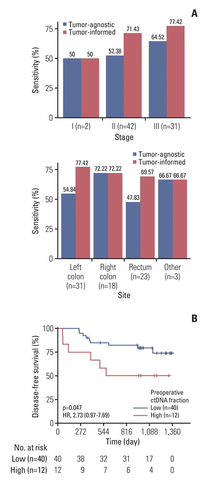
Preoperative blood samples were collected from 75 patients, and ctDNA status was assessed by a fixed panel targeting 41 CRC-related genes. We evaluated the performance of the tumor-agnostic and tumor-informed fixed assays in detecting preoperative ctDNA positivity and predicting recurrence. Quality control data of the blood samples including DNA concentrations are reported in S7 Table. The tumoragnostic and tumor-informed fixed assays identified 43 (57.3%) and 55 (73.3%) preoperative ctDNA-positive patients, respectively (S8 Table). Among the 55 baseline ctDNA-positive patients defined by the tumor-informed fixed assay, a total of 178 somatic mutations were detected, with a median of 3 mutations per patient. The most frequently altered genes and sites detected in ctDNA were similar to those observed in tumor tissues (Fig. 2C and D). More than half of the mutations (51%) identified from tissue samples were detected in blood samples (S5B Fig.). The tumor-informed fixed assay showed higher ctDNA-positive rates than the tumor-agnostic fixed assay stratified by different stages and tumor sites (Fig. 3A). By contrast, the standard carcinoembryonic antigen (CEA) blood test for CRC screening and recurrence monitoring only yielded positive results in 29.3% (22/75) of the patients (S8 Table). These results strongly suggest that the tumor-informed assay exhibits higher sensitivity in ctDNA detection compared to the tumor-agnostic panel and CEAbased testing in CRC patients.
The prognostic value of ctDNA before curative treatment remains controversial in various cancer types [17-20]. We then examined whether the preoperative ctDNA status can identify patients at a higher risk of recurrence. We observed no association between DFS/overall survival (OS) and preoperative ctDNA status, assessed by either the tumoragnostic (DFS: HR, 1.68; 95% CI, 0.68 to 4.18; p=0.255 [S5C Fig.]; OS: HR, 0.95; 95% CI, 0.30 to 3.01; p=0.934 [S5D Fig.]) or tumor-informed fixed assays (DFS: HR, 0.98; 95% CI, 0.38 to 2.53; p=0.967 [S5E Fig.]; OS: HR, 0.69; 95% CI, 0.21 to 2.30; p=0.548 [S5F Fig.]). However, based on the fraction of ctDNA in plasma samples estimated by the ML method, the ctDNA-positive patients were stratified into two subgroups (top 25%: high ctDNA group; bottom 75%: low ctDNA group, cutoff=0.3484%), and the high ctDNA group exhibited poorer DFS (HR, 2.73; 95% CI, 0.97 to 7.69; p=0.047) (Fig. 3B), indicating that preoperative ctDNA levels may be a more sensitive prognostic factor than preoperative ctDNA positivity.

3. Postoperative landmark ctDNA predicts recurrence

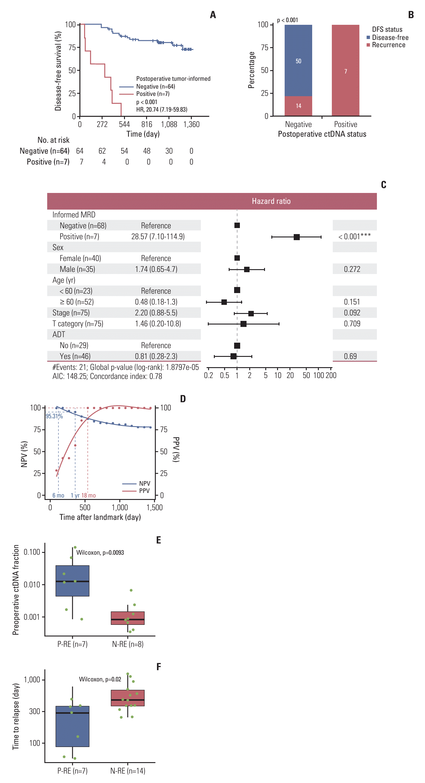
To further explore the postoperative ctDNA status and its prognostic value, plasma samples were collected 7 days post-surgery. The landmark MRD status determined by the tumor-informed fixed assay at postoperative day 7 was significantly associated with DFS (HR, 20.74; 95% CI, 7.19 to 59.83; p < 0.001) (Fig. 4A). Among these patients, seven out of seven with postoperative ctDNA positivity (100%) and 14 out of 64 with postoperative ctDNA negativity (21.9%) experienced relapse during the follow-up (Fig. 4B), yielding a positive predictive value (PPV) of 100% and a negative predictive value (NPV) of 78.1%, respectively. Conversely, no significant association was observed for the tumor-agnostic fixed assay with DFS (HR, 1.71; 95% CI, 0.69 to 4.20; p=0.239) (S9A Fig.).
Multivariable Cox regression analysis showed that tumor-informed ctDNA positivity at postoperative day 7 was an independent risk factor for predicting recurrence after adjusting sex, age, TNM stage and adjuvant therapy (HR, 28.57; 95% CI, 7.10 to 114.9) (Fig. 4C). Furthermore, MRD-positive patients in stage II have a worse prognosis compared to MRD-negative patients in stage III (HR, 6.22; 95% CI, 1.46 to 26.53; p=0.005) (S9B Fig.). These results suggest that the postoperative landmark ctDNA status can outperform traditional TNM staging in providing a more effective prognostic stratification.
In the application of ctDNA-MRD tests, interpreting the predictive power for prognosis based on MRD status at the landmark and its time-dependent changes is crucial but often overlooked. To address this gap, we plotted the NPV and PPV of the landmark MRD test over time (Fig. 4D). Our findings demonstrated that the postoperative ctDNA negativity determined by the tumor-informed assay initially maintained 100% NPV within 6 months, which gradually decreased thereafter. However, it still remained above 95% within 1 year. In contrast, the PPV increased to 100% at the 18th month.
Additionally, we examined the characteristics of the ctDNA-negative patients who relapsed during the follow-up, referred to as “Negative-relapsed” (N-RE) patients. Using the tumor-informed fixed assay, we identified 14 out of 71 patients as N-RE. Moreover, we also determined 50 patients who were postoperative ctDNA-negative and non-recurrent (N-NR), as well as seven patients who were postoperative ctDNA-positive and recurrent (P-RE). Further analysis of the clinical features revealed that these N-RE patients had a higher ratio of CEA positivity (S10 Table). Besides, compared to P-RE patients, N-RE patients exhibited a lower preoperative ctDNA fraction (p=0.0093) (Fig. 4E), as well as a lower prevalence of preoperative ctDNA positivity (57.1% vs. 100.0%) (S9C Fig.), though no significant difference was observed, suggesting a less amount of ctDNA was released from the tumor cells in N-RE patients. Consistently, the N-RE patients exhibited longer time from surgery to disease recurrence than P-RE patients (p=0.02) (Fig. 4F), suggesting the necessity of longitudinal MRD monitoring in these patients.
We further examined whether genomic alterations might contribute to the ability of ctDNA to be released into plasma. Molecular screening of tumor tissues from N-RE patients revealed a trend of fewer genomic alterations with lower maximum allelic fraction (MaxAF) (S9D-F Fig.). Previous studies have indicated that tumor cells harboring mutations in cell cycle-related genes were less likely to release DNA [19]. We did observe a higher percentage of patients harboring mutant genes related to cell cycling (including TP53, SMAD4, CHEK2, and ATM) in N-RE than in N-NR and P-RE patients (92.9% vs. 80.0% vs. 71.4%) (S9G Fig.). This trend became more pronounced when considering mutations in SMAD4 alone (42.9% vs. 18% vs. 14.3%) (S9H Fig.). Collectively, our results indicated that the genomic background of CRC patients may play a role in ctDNA release, potentially leading to false-negative results.

4. Postoperative landmark ctDNA in guiding adjuvant therapy

In this cohort, 61.3% of patients (46/75) received ACT following curative surgery, including nine patients with FOLFOX (fluorouracil, leucovorin, and oxaliplatin), 22 patients with CAPEOX (capecitabine and oxaliplatin), three patients with RALOX (raltitrexed plus oxaliplatin), and eight patients with a single-agent capecitabine. The information regarding the chemotherapy regimen was missing for four patients. Consistent with previous studies [21,22], the landmark ctDNA-negative patients did not experience a DFS benefit from adjuvant therapy (HR, 0.83; 95% CI, 0.28 to 2.40; p=0.725) (S11A Fig.). In subgroup analysis, stage IIA patients with negative postoperative ctDNA did not derive any significant advantage from intensified chemotherapy regimens (p=0.183) (S11B Fig.). Similarly, in stage III patients with landmark negative ctDNA, no significant difference in DFS was observed between the patients receiving FOLFOX and CAPEOX regimens (HR, 0.35; 95% CI, 0.04 to 3.17; p=0.332) (S11C Fig.). Due to the limited sample size, no significant difference in DFS was observed between the postoperative ctDNA-positive patients with and without ACT (HR, 1.41; 95% CI, 0.28 to 7.12; p=0.673) (S11D Fig.).

5. Subgroup ctDNA analysis using two different personalized assays

To facilitate a direct comparison, we assessed the detection sensitivity and prognostic value of two personalized assays using a sub-cohort of 41 randomly selected patients. Tumor tissue samples were analyzed for variants calling using WES or a targeted panel-based approach with 520 genes. We identified a total of 2,022 variants with WES, of which 85.7% were unique to individual patients. The targeted panel assay identified 524 mutations, with 57.3% being individual unique (S12A Fig.). Notably, the WES-based assay covered a higher median number of variants compared to the panel-based assay, regardless of all mutations (51 vs. 11) or individual unique mutations (46 vs. 5).
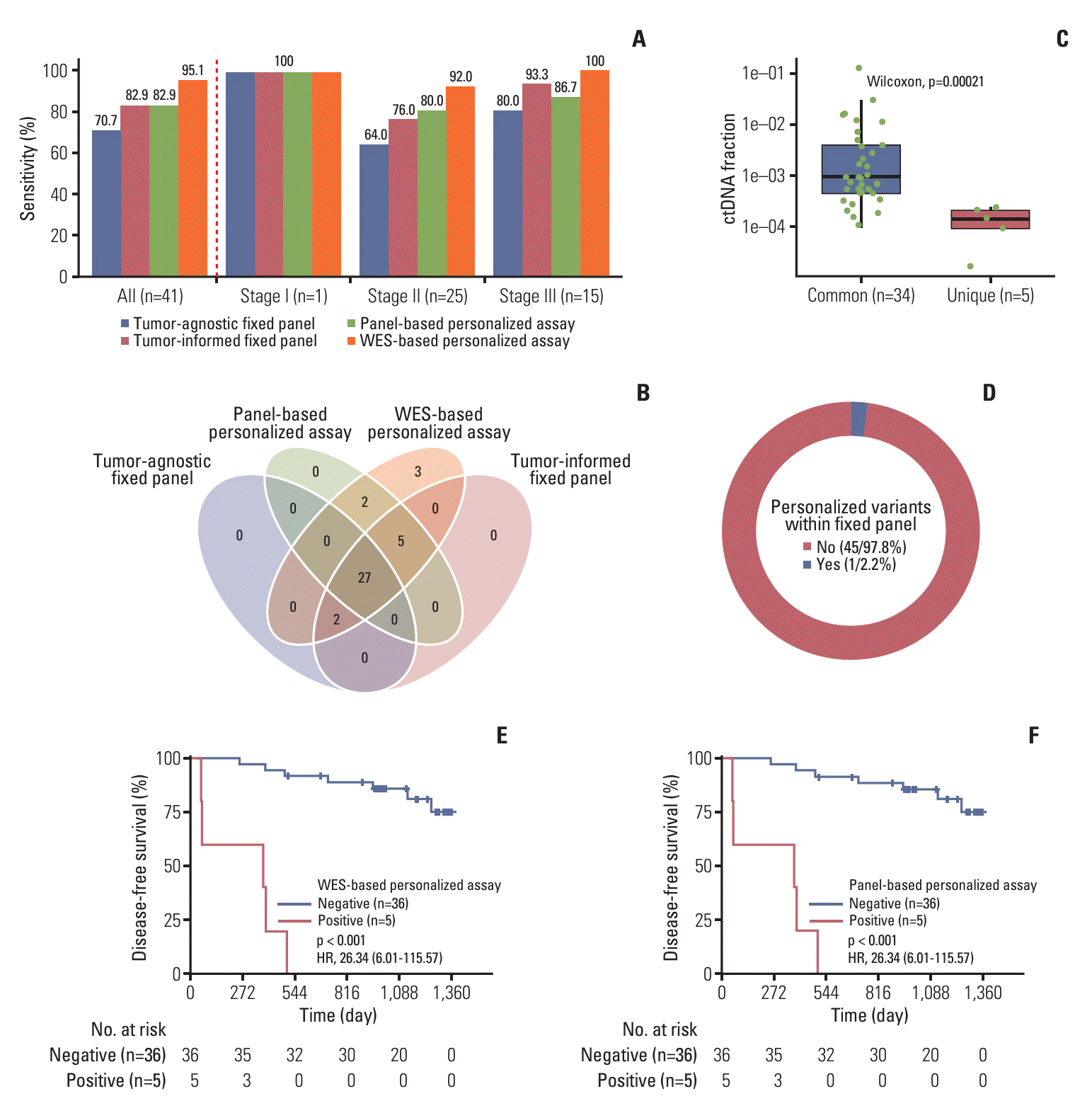
The WES-based personalized assay exhibited superior sensitivity in the preoperative test across different stages (total, 95.1%; stage I, 100.0%; stage II, 92.0%; stage III, 100.0%), while the tumor-agnostic fixed assay identified the fewest cases of preoperative ctDNA positivity (total sensitivity, 70.7%; stage I, 100.0%; stage II, 64.0%; stage III, 80.0%) (Fig. 5A). The tumor-informed fixed panel and the panel-based personalized assay displayed similar performance with total preoperative ctDNA positivity of 82.9% and 100% for stage I cancers, while the panel-based personalized assay performed better in stage II (80.0% vs. 76.0%) but less sensitive in stage III diseases (86.7% vs. 93.3%) (Fig. 5A). In addition to the 34 patients detected by the tumor-informed fixed assay, the WES-based personalized assay identified additional five patients with relatively lower ctDNA fractions due to its lower LOD (Fig. 5B and C). A total of 46 variants were determined in these five patients, and only one variant was also covered by tumor-informed fixed assay (Fig. 5D). Thirty-two patients were positive for preoperative ctDNA test according to both the tumor-informed fixed assay and personalized assays, and the WES-based assay detected the most unique variants in these cases (S12B Fig.). These findings suggest that the fixed panel, even with a tumor-informed strategy, does not adequately cover the positive mutant sites, highlighting the advantages of the WES-based personalized strategy for capturing individual mutations.
Consistent with the above results for fixed assays, no association between preoperative ctDNA positivity and DFS was observed by using the personalized assays (panel-based personalized assay: HR, 0.60; 95% CI, 0.16 to 2.22; p=0.440 [S12C Fig.]; WES-based personalized assay: HR, 0.81; 95% CI, 0.10 to 6.34; p=0.837 [S12D Fig.]). For postoperative landmark analysis, the WES-based personalized and panel-based personalized assays had the same results with the tumor-informed fixed panel (HR, 26.34; 95% CI, 6.01 to 115.57; p < 0.001) (Fig. 5E and F, S12E Fig.), all better than the tumor-agnostic fixed panel (HR, 3.04; 95% CI, 0.94 to 9.89; p=0.052) (S12F Fig.). Comparing the performance of predicting recurrence, the WES-based and panel-based assays exhibited same NPV and PPV, as did the tumor-informed fixed assay, indicating comparable performances of the ctDNA assays with the tumor-informed strategies. The tumor-agnostic fixed assay exhibited the worst performance (S13 Table). The seven N-RE patients were tested negative in all four assays, revealing the inherent limitations of these assays in predicting recurrence at the 7-day postoperative landmark. Nevertheless, these limitations could potentially be overcome by implementing longitudinal monitoring.

Discussion

ctDNA-based MRD detection, as an emerging prognostic biomarker, can accurately identify CRC patients at a higher risk of recurrence after curative therapy. While tumor-informed strategies have been recognized for their improved sensitivity in MRD detection, the clinical utility and standardized guidelines for selecting appropriate assays to predict recurrence and guide treatment decisions are still unclear.
In our study, we firstly assessed the prognostic value of the tumor-informed and tumor-agnostic fixed panels. In concordance with previously published studies, we observed that the tumor-informed strategy, even using the same fixed panel with the tumor-agnostic test, yielded improved sensitivity, as indicated by the higher baseline positivity rate (73.3% vs. 57.3%). Although the preoperative ctDNA status did not show any association with DFS, patients in the preoperative ctDNA-positive group with elevated ctDNA levels demonstrated a significantly increased risk of recurrence compared to those with lower ctDNA levels. This demonstrates that the ctDNA level, even before the operation, possesses potential predictive value for patient recurrence.
Traditionally, most studies assessing MRD are designed with the postoperative landmark around 1 month after surgery. However, the average hospital stay for CRC patients undergoing resection surgery is typically under 10 days [23]. This extended gap between patient discharge and MRD evaluation necessitates readmission post-discharge, leading to follow-up challenges and potential patient dropout. Furthermore, adhering to National Comprehensive Cancer Network guidelines, clinical practice stresses administering ACT as soon as medically feasible [24], while MRD testing entails additional time, potentially delaying the provision of timely ctDNA results prior to ACT initiation. Given these considerations, early identification and detection of potentially high-risk patients after surgery can greatly assist in decision-making and the timely initiation of intensive treatments in postsurgical management. Notably, Chen et al. [25] have highlighted the significance of early ctDNA detection, specifically at 3-7 days after surgery, which might provide novel insights into the postoperative MRD test for CRC management. Taken together, the postoperative day 7 landmark was selected in this study for further exploration. As a result, we have confirmed the significant potential of using tumor-informed fixed assay for identifying patients with poor prognosis. However, a previous study has revealed the association between surgery and elevated cfDNA levels, persisting for up to 4 weeks, which might result a dilution of ctDNA in the blood and introduce false-negative results in the early landmark MRD tests [26]. A relatively high N-RE rate was also found in our study. Interestingly, we observed that N-RE patients determined by the tumor-informed fixed assay had a higher ratio of SMAD4 mutation in tumor tissues. A previous research has revealed that there are transcriptional factors take over the regulatory role of SMAD4 when it is mutant, and consequently keep the functional of transforming growth factor β on tumor invasion and metastasis [27]. Additionally, a previous study has discovered that subclones carrying driver mutations related to cell cycling are less likely to release ctDNA [28]. Our results suggest that mutant SMAD4 may impact the ctDNA release or stronger malignancy, which may introduce false negativity. This indicates that the influence of genomic background of cohort should be also considered when evaluating the performance of ctDNA assays.
By using a sub-cohort, our study evaluated the ctDNA-MRD using personalized assays with prior WES or fixed panel sequencing on tumor tissues. To our knowledge, this is the first study that performed head-to-head comparisons of these four panels using various strategies and approaches. We observed the highest preoperative ctDNA-positive rate with WES-based personalized assays, which indicated its highest sensitivity for ctDNA detection. In the term of postoperation, we noticed a similar prognostic capacity with tumor-informed personalized assays (WES-based or panel-based) and the tumor-informed fixed assay. It could be attributed to the presence of hotspot mutations in CRC that contribute to high ctDNA release. Our results indicate the potential utility of tumor-informed fixed panels in CRC. However, clinical studies with larger sample sizes are required in the future for further validation.
There are also limitations of this study. According to the small cohort, the number of postoperative ctDNA-positive patients was not sufficient to observe a significant difference of clinical outcome in patients with or without adjuvant therapy. Additionally, the postoperative day 7 time point chosen may not be optimal, and long-term monitoring was not conducted due to a lack of multiple sampling. Therefore, the median lead-time of MRD detection leading to radioactive relapse can not be measured. Future clinical trials are highly recommended to further assess the clinical utility and benefits for patients using ctDNA-MRD assays employing different strategies and approaches.
Taken together, our study further establishes the prognostic value of ctDNA positivity at postoperative day 7 and confirms the prognostic value of the ctDNA positivity at postoperative day 7 by the tumor-informed fixed panel. The tumor-informed fixed panel may be a cost-effective method to evaluate MRD in clinic, which warrants further studies in future. These findings may contribute to the advancement of personalized medicine and emphasize the potential of ctDNA-MRD as a reliable biomarker for CRC recurrence prediction.
Our study confirmed the prognostic value of the ctDNA positivity at postoperative day 7 by the tumor-informed fixed panel. The tumor-informed fixed panel may be a cost-effective method to evaluate MRD in clinic, which warrants further studies in future.

Notes

Ethical Statement

This study was approved by the ethics committee of the First Affiliated Hospital of Soochow University (No. 2021270), and was performed in accordance with the Declaration of Helsinki. Informed consent was taken from all patients before participating in any study-related procedure.

Author Contributions

Conceived and designed the analysis: Yang J, Zhou J.

Collected the data: Yu C, Li H.

Contributed data or analysis tools: Yang J, Yao J, Wei Y.

Performed the analysis: Peng D, Zhou Q.

Wrote the paper: Yang J, Yu C, Li H, Peng D, Zhou Q, Lv J, Fang S, Shi J, Wei Y, Wang G, Cai S, Zhang Z (Zhihong Zhang), Zhang Z (Zixiang Zhang), Zhou J.

Study supervision: Zhang Z (Zhihong Zhang), Zhang Z (Zixiang Zhang), Zhou J.

Conflicts of Interest

The authors declare no potential conflicts of interest, except the employment of Di Peng, Qiaoxia Zhou, Juan Lv, Shuai Fang, Jiaochun Shi, Guoqiang Wang, Shangli Cai and Zhihong Zhang in Burning Rock Biotech.

ACKNOWLEDGMENTS

This work was supported by the Project of the Suzhou Science and Technology Bureau medical health technology innovation project (SKYD2022106), the Project of the State Key Laboratory of Radiation Medicine and Protection, Soochow University, (No. GZK12023037), the Project of Medical Applied and Basic Research Foundation of Suzhou Science & Technology Bureau (grant number: SKY2023156), the Project of the First Hospital of Soochow University Natural Science Foundation Incubation Programme for Doctoral Trainees (grant number: BXQN202218) and the Project of Extracurricular Academic Research of Soochow University (grant number: KY2022132A, KY2023104A).

References

1. Biller LH, Schrag D. Diagnosis and treatment of metastatic colorectal cancer: a review. JAMA. 2021; 325:669–85.
crossref
2. Sung H, Ferlay J, Siegel RL, Laversanne M, Soerjomataram I, Jemal A, et al. Global cancer statistics 2020: GLOBOCAN estimates of incidence and mortality worldwide for 36 cancers in 185 countries. CA Cancer J Clin. 2021; 71:209–49.
crossref
3. Figueredo A, Charette ML, Maroun J, Brouwers MC, Zuraw L. Adjuvant therapy for stage II colon cancer: a systematic review from the Cancer Care Ontario Program in evidence-based care’s gastrointestinal cancer disease site group. J Clin Oncol. 2004; 22:3395–407.
crossref
4. Andre T, de Gramont A, Vernerey D, Chibaudel B, Bonnetain F, Tijeras-Raballand A, et al. Adjuvant fluorouracil, leucovorin, and oxaliplatin in stage II to III colon cancer: updated 10-year survival and outcomes according to BRAF mutation and mismatch repair status of the MOSAIC study. J Clin Oncol. 2015; 33:4176–87.
5. Bockelman C, Engelmann BE, Kaprio T, Hansen TF, Glimelius B. Risk of recurrence in patients with colon cancer stage II and III: a systematic review and meta-analysis of recent literature. Acta Oncol. 2015; 54:5–16.
crossref
6. Quasar Collaborative Group, Gray R, Barnwell J, McConkey C, Hills RK, Williams NS, et al. Adjuvant chemotherapy versus observation in patients with colorectal cancer: a randomised study. Lancet. 2007; 370:2020–9.
crossref
7. Dasari A, Morris VK, Allegra CJ, Atreya C, Benson AB 3rd, Boland P, et al. ctDNA applications and integration in colorectal cancer: an NCI Colon and Rectal-Anal Task Forces whitepaper. Nat Rev Clin Oncol. 2020; 17:757–70.
crossref
8. Moding EJ, Nabet BY, Alizadeh AA, Diehn M. Detecting liquid remnants of solid tumors: circulating tumor DNA minimal residual disease. Cancer Discov. 2021; 11:2968–86.
crossref
9. Kotani D, Oki E, Nakamura Y, Yukami H, Mishima S, Bando H, et al. Molecular residual disease and efficacy of adjuvant chemotherapy in patients with colorectal cancer. Nat Med. 2023; 29:127–34.
crossref
10. Tie J, Cohen JD, Lahouel K, Lo SN, Wang Y, Kosmider S, et al. Circulating tumor DNA analysis guiding adjuvant therapy in stage II colon cancer. N Engl J Med. 2022; 386:2261–72.
crossref
11. Chen K, Shields MD, Chauhan PS, Ramirez RJ, Harris PK, Reimers MA, et al. Commercial ctDNA assays for minimal residual disease detection of solid tumors. Mol Diagn Ther. 2021; 25:757–74.
crossref
12. Zhou J, Wang C, Lin G, Xiao Y, Jia W, Xiao G, et al. Serial circulating tumor DNA in predicting and monitoring the effect of neoadjuvant chemoradiotherapy in patients with rectal cancer: a prospective multicenter study. Clin Cancer Res. 2021; 27:301–10.
crossref
13. Reinert T, Henriksen TV, Christensen E, Sharma S, Salari R, Sethi H, et al. Analysis of plasma cell-free DNA by ultradeep sequencing in patients with stages I to III colorectal cancer. JAMA Oncol. 2019; 5:1124–31.
crossref
14. Zhu L, Huang Y, Fang X, Liu C, Deng W, Zhong C, et al. A novel and reliable method to detect microsatellite instability in colorectal cancer by next-generation sequencing. J Mol Diagn. 2018; 20:225–31.
crossref
15. Chen K, Yang F, Shen H, Wang C, Li X, Chervova O, et al. Individualized tumor-informed circulating tumor DNA analysis for postoperative monitoring of non-small cell lung cancer. Cancer Cell. 2023; 41:1749–62.
crossref
16. Ye ZL, Qiu MZ, Tang T, Wang F, Zhou YX, Lei MJ, et al. Gene mutation profiling in Chinese colorectal cancer patients and its association with clinicopathological characteristics and prognosis. Cancer Med. 2020; 9:745–56.
crossref
17. Tie J, Wang Y, Cohen J, Li L, Hong W, Christie M, et al. Circulating tumor DNA dynamics and recurrence risk in patients undergoing curative intent resection of colorectal cancer liver metastases: a prospective cohort study. PLoS Med. 2021; 18:e1003620.
crossref
18. Schraa SJ, van Rooijen KL, Koopman M, Vink GR, Fijneman RJA. Cell-free circulating (tumor) DNA before surgery as a prognostic factor in non-metastatic colorectal cancer: a systematic review. Cancers (Basel). 2022; 14:2218.
crossref
19. Li N, Wang BX, Li J, Shao Y, Li MT, Li JJ, et al. Perioperative circulating tumor DNA as a potential prognostic marker for operable stage I to IIIA non-small cell lung cancer. Cancer. 2022; 128:708–18.
crossref
20. Gianni C, Palleschi M, Merloni F, Bleve S, Casadei C, Sirico M, et al. Potential impact of preoperative circulating biomarkers on individual escalating/de-escalating strategies in early breast cancer. Cancers (Basel). 2022; 15:96.
crossref
21. Tie J, Cohen JD, Wang Y, Christie M, Simons K, Lee M, et al. Circulating tumor DNA analyses as markers of recurrence risk and benefit of adjuvant therapy for stage III colon cancer. JAMA Oncol. 2019; 5:1710–7.
crossref
22. Taieb J, Taly V, Vernerey D, Bourreau C, Bennouna J, Faroux R, et al. Analysis of circulating tumour DNA (ctDNA) from patients enrolled in the IDEA-FRANCE phase III trial: prognostic and predictive value for adjuvant treatment duration. Ann Oncol. 2019; 30(Suppl 5):V867.
crossref
23. Geltzeiler CB, Rotramel A, Wilson C, Deng L, Whiteford MH, Frankhouse J. Prospective study of colorectal enhanced recovery after surgery in a community hospital. JAMA Surg. 2014; 149:955–61.
crossref
24. NCCN Clinical Practice Guidelines in Oncology (NCCN Guidelines) for Colon Cancer. Version 1.2024. Plymouth Meeting, PA: National Comprehensive Cancer Network;2024.
25. Chen G, Peng J, Xiao Q, Wu HX, Wu X, Wang F, et al. Postoperative circulating tumor DNA as markers of recurrence risk in stages II to III colorectal cancer. J Hematol Oncol. 2021; 14:80.
crossref
26. Henriksen TV, Reinert T, Christensen E, Sethi H, Birkenkamp-Demtroder K, Gogenur M, et al. The effect of surgical trauma on circulating free DNA levels in cancer patients-implications for studies of circulating tumor DNA. Mol Oncol. 2020; 14:1670–9.
crossref
27. Frey P, Devisme A, Rose K, Schrempp M, Freihen V, Andrieux G, et al. SMAD4 mutations do not preclude epithelial-mesenchymal transition in colorectal cancer. Oncogene. 2022; 41:824–37.
crossref
28. Mao X, Zhang Z, Zheng X, Xie F, Duan F, Jiang L, et al. Capture-based targeted ultradeep sequencing in paired tissue and plasma samples demonstrates differential subclonal ctDNA-releasing capability in advanced lung cancer. J Thorac Oncol. 2017; 12:663–72.
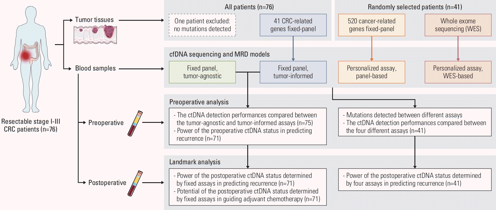
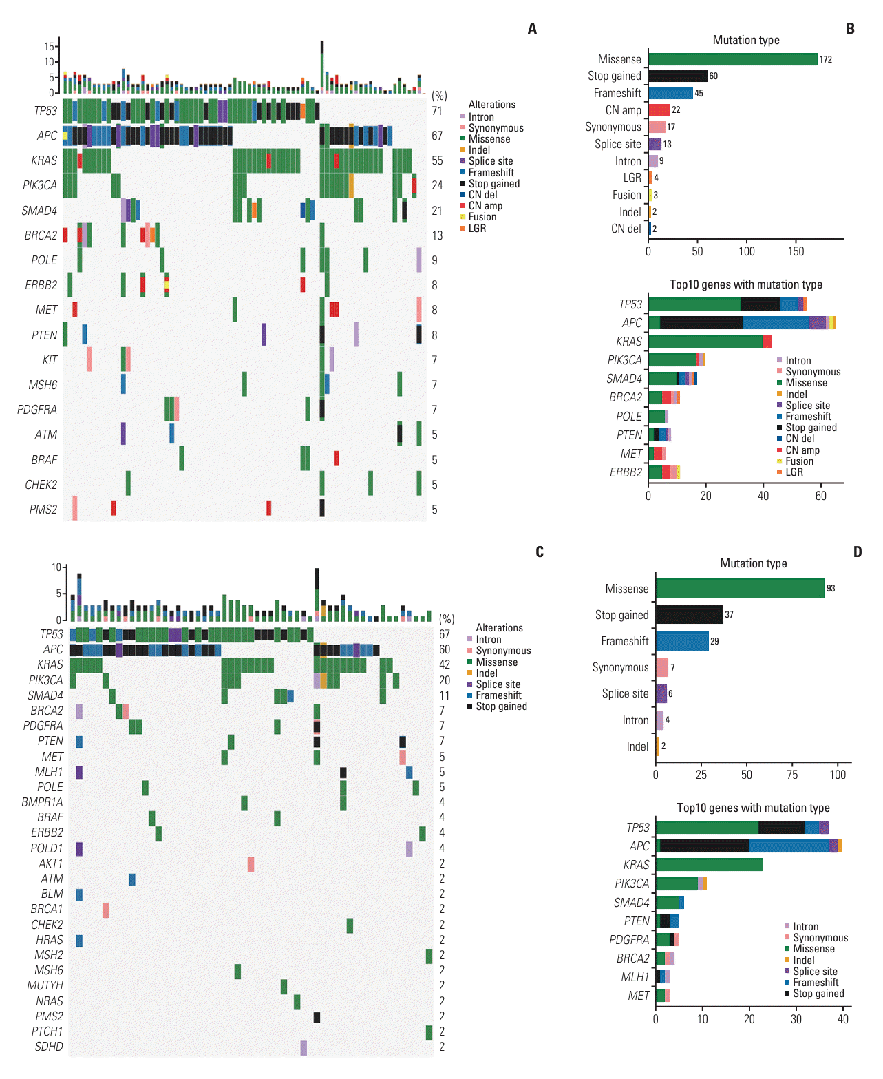
Fig. 1.
Study design. cfDNA, cell-free DNA; CRC, colorectal cancer; ctDNA, circulating tumor DNA; MRD, molecular residual disease.
crt-2023-1371f1.tif
Fig. 2.
Genomic alterations identified from tumor tissues. (A) Heatmap of genetic mutations identified from tumor tissues of 75 patients with detectable mutations. Different types of alterations are presented with colors as illustrated. (B) The most predominate mutation types and the top 10 mutant genes in tissue samples are presented as illustrated. (C) Heatmap of genetic mutations identified from 55 baseline circulating tumor DNA–positive patients defined by the tumor-informed fixed assay. (D) The most predominate mutation types and the top 10 mutant genes in blood samples are presented as illustrated.
crt-2023-1371f2.tif
Fig. 3.
Prognostic analysis of preoperative circulating tumor DNA (ctDNA). (A) Boxplots of ctDNA-positive rate determined by both tumor-agnostic fixed assay and tumor-informed fixed assay in patients with different stages and different tumor sites. (B) Kaplan-Meier plots of disease-free survival in patients stratified by preoperative ctDNA levels determined by the tumor-informed fixed assay. HR, hazard ratio.
crt-2023-1371f3.tif
Fig. 4.
Landmark prognostic analysis of postoperative circulating tumor DNA (ctDNA). (A) Kaplan-Meier plot of disease-free survival (DFS) analysis by postoperative ctDNA status determined by the tumor-informed fixed assay. (B) Boxplot of DFS status in postoperative ctDNA-positive and ctDNA-negative patients determined by the tumor-informed fixed assay. (C) Multivariabel analysis of factors associated with recurrence. ADT, adjuvant therapy; HR, hazard ratio; MRD, molecular residual disease. (D) The dynamic change of positive predictive value (PPV) and negative predictive value (NPV) over time by landmark ctDNA status. (E, F) Boxplots of preoperative ctDNA fraction (E) and time to relapse (F) in postoperative positive and relapsed (P-RE) patients and negative but relapsed (N-RE) patients.
crt-2023-1371f4.tif
Fig. 5.
Head-to-head comparison of different fixed and personalized panels in the sub-cohort. (A) Boxplot of preoperative circulating tumor DNA (ctDNA)–positive rate determined by all four assays. (B) Venn diagram of ctDNA-positive patients by different assays. (C) Boxplot of ctDNA fractions in positive patients specific determined by whole-exome sequencing (WES)–based personalized assay and commonly determined by all assays. (D) Positive variants within fixed panel in patients specific determined by WES-based personalized assay. (E, F) Kaplan-Meier plot of disease-free survival analysis by postoperative ctDNA status determined by WES-based personalized assay (E) and panel-based personalized assay (F). HR, hazard ratio.
crt-2023-1371f5.tif
Table 1.
Baseline characteristics
No. (%) (n=75)
Sex
 Female 40 (53.3)
 Male 35 (46.7)
Age (yr)
 Mean±SD 65.3±10.1
 Median (IQR) 67.0 (57.0-71.0)
Tumor site
 Left colon 31 (41.3)
 Right colon 18 (24.0)
 Rectum 23 (30.7)
 Other 3 (4.0)
TNM stage
 I 2 (2.7)
 II 42 (56.0)
 III 31 (41.3)
dMMR/pMMR
 dMMR 6 (8.0)
 pMMR 68 (90.7)
 Missing 1 (1.3)
Adjuvant
 No 29 (38.7)
 Yes 46 (61.3)
DFS status
 Disease-free 50 (66.7)
 Recurrence 21 (28.0)
 Missing 4 (5.3)
OS status
 Alive 59 (78.7)
 Dead 12 (16.0)
 Missing 4 (5.3)

DFS, disease-free survival; dMMR, deficient mismatch repair; IQR, interquartile range; OS, overall survival; pMMR, proficient mismatch repair; SD, standard deviation.

TOOLS
Similar articles