Journal List > Cancer Res Treat > v.56(4) > 1516088684

Ou, Wu, Wu, Fu, Zeng, Li, Li, Liu, Li, Li, Zhou, Zhuang, Cheng, Zhang, Wang, Qu, and Yan: Efficacy of Lenvatinib Combined with Anti–PD-1 Antibodies Plus Transcatheter Arterial Chemoembolization for Hepatocellular Carcinoma with Portal Vein Tumor Thrombus: A Retrospective, Multicenter Study

Abstract

Purpose

The prognosis of patients with hepatocellular carcinoma (HCC) and portal vein tumor thrombus (PVTT) is extremely poor, and systemic therapy is currently the mainstream treatment. This study aimed to assess the efficacy and safety of lenvatinib combined with anti–programmed cell death-1 antibodies and transcatheter arterial chemoembolization (triple therapy) in patients with HCC and PVTT.

Materials and Methods

This retrospective multicenter study included patients with HCC and PVTT who received triple therapy, were aged between 18 and 75 years, classified as Child-Pugh class A or B, and had at least one measurable lesion. The overall survival (OS), progression-free survival (PFS), objective response rates, and disease control rates were analyzed to assess efficacy. Treatment-related adverse events were analyzed to assess safety profiles.

Results

During a median follow-up of 11.23 months (range, 3.07 to 34.37 months), the median OS was greater than 24 months, and median PFS was 12.53 months. The 2-year OS rate was 54.9%. The objective response rate and disease control rate were 69.8% (74/106) and 84.0% (89/106), respectively; 20.8% (22/106) of the patients experienced grade 3/4 treatment-related adverse events and no treatment-related deaths occurred. The conversion rate to liver resection was 31.1% (33/106), with manageable postoperative complications. The median OS was not reached in the surgery group, but was 19.08 months in the non-surgery group. The median PFS in the surgery and non-surgery groups were 20.50 and 9.00 months, respectively.

Conclusion

Triple therapy showed promising survival benefits and high response rates in patients with HCC and PVTT, with manageable adverse effects.

Introduction

Hepatocellular carcinoma (HCC) is the most common primary liver cancer and the fourth-leading cause of cancer-related deaths worldwide [1]. Most patients diagnosed with HCC are already in intermediate-advanced stages, and approximately 40% of them are complicated with portal vein tumor thrombus (PVTT) and have lost the opportunity for radical resection [2]. These patients have an extremely poor prognosis, with a median survival time of 2.7-4.0 months without treatment [2].
According to current National Comprehensive Cancer Network (NCCN) guidelines, systemic therapy is the optimal treatment. The efficacy of sorafenib and lenvatinib as first-line therapies for advanced HCC remains poor. However, Asian guidelines suggest a multidisciplinary treatment for patients with HCC and PVTT, including transcatheter arterial chemoembolization (TACE), radiotherapy (RT), hepatic artery infusion chemotherapy (HAIC), and systemic therapy [3-5]. It is worth noting that TACE plus lenvatinib for the treatment of HCC with PVTT can improve prognosis, with a median overall survival (mOS) time of 16.4 months and median progression-free survival (mPFS) time of 8.40 months [6]. Moreover, TACE plus lenvatinib is more effective than TACE plus sorafenib [6,7]. Although these outcomes indicated that the TACE plus lenvatinib treatment regimen can significantly improve the prognosis of patients with HCC and PVTT, the treatment effects still remain unsatisfactory.
Recently, immune checkpoint inhibitors therapy, particularly antibodies targeting programmed cell death-1 (anti–PD-1 antibodies), has become an important regimen for improving the tumor response and survival time of patients with advanced HCC [8-10]. However, the efficacy of anti–PD-1 antibodies as monotherapy is unsatisfactory, with approximately 20% objective response rate (ORR) [11]. Triple therapy using anti–PD-1 antibodies in combination with tyrosine kinase inhibitors (TKIs) and TACE for advanced HCC has become a popular research topic. Some studies evaluating its efficacy showed an ORR of 56.1%-69.3% and mOS of 16.9-18.0 months, making it superior to TKIs plus locoregional therapy [12,13]. In our previous study, TACE combined with lenvatinib and anti–PD-1 antibodies showed satisfactory results for advanced HCC, with an ORR of 77.4% [14]. These studies demonstrated the application prospects of triple therapy in the treatment of advanced HCC. Furthermore, a previous study also evaluated the efficacy of triple therapy in HCC with PVTT, with an ORR of 48.7%, and the mOS and mPFS were 14.0 and 9.2 months, respectively [15]. However, the extent of the clinical benefits needs to be confirmed with further research and larger sample sizes.
Therefore, we conducted this study to further explore the efficacy and safety of triple therapy for HCC patients with PVTT.

Materials and Methods

1. Patients

This retrospective, multicenter study was conducted in patients with HCC and PVTT who received triple therapy between November 2018 and May 2022 at eight major cancer centers in China (Fujian Provincial Hospital, Affiliated Hospital of Guilin Medical University, First Affiliated Hospital of Xiamen University, Zhongshan Hospital of Xiamen University, Zhangzhou Affiliated Hospital of Fujian Medical University, Eastern Hepatobiliary Surgery Hospital, First Affiliated Hospital of Fujian Medical University, and Second Affiliated Hospital of Nanchang University). Baseline data, including demographic, clinical and pathological characteristics, and treatment-related outcomes, were retrospectively collected. Detailed radiological examinations (tumor number, maximum tumor size, extent of PVTT, and extrahepatic metastases), physical examination, liver function tests (alanine aminotransferase, aspartate aminotransferase [AST], total bilirubin, albumin, coagulation function tests [blood platelet count], tumor markers [α-fetoprotein, AFP]), and detection of hepatitis B surface antigen were performed at the initial visit.
The diagnosis of HCC with PVTT was based on histological examination of the tumor tissue or clinicoradiological criteria according to the American Association for the Study of Liver Diseases (AASLD) criteria [16]. The presence and extent of PVTT were evaluated using contrast-enhanced dynamic computed tomography (CT) or contrast-enhanced dynamic magnetic resonance imaging (MRI) findings. The extent of the PVTT was classified according to the Japanese Liver Cancer Study Group classification as follows: Vp0, no PVTT; Vp1, portal vein invasion distal to the second-order branches of the portal vein; Vp2, portal vein invasion of the second-order branches; Vp3, invasion of the first-order branches of the portal vein; Vp4, invasion of the main trunk [17].

2. Inclusion and exclusion criteria

The inclusion criteria were as follows: (1) diagnosis of HCC with PVTT treated with triple therapy; (2) Barcelona Clinic Liver Cancer (BCLC) C stage; (3) age between 18 and 75 years; (4) Child-Pugh class A or B; (5) Eastern Cooperative Oncology Group performance status (ECOG PS) score of 0-1; and (6) at least one measurable lesion according to the modified Response Evaluation Criteria in Solid Tumors (mRECIST). The exclusion criteria were as follows: (1) previous acceptance of other antitumor treatments; (2) treatment in conjunction with other treatment regimens, such as RT and chemotherapy; (3) history of other malignancies; and (4) incomplete data.

3. Procedure

All patients received lenvatinib (8 mg for bodyweight < 60 kg or 12 mg for bodyweight ≥ 60 kg) orally once daily and anti–PD-1 antibodies (sintilimab 200 mg, tislelizumab 200 mg, camrelizumab 200 mg, toripalimab 240 mg, pembrolizumab 200 mg, nivolumab 200 mg, or penpulimab 200 mg) intravenously once every 3 weeks. Moreover, TACE was performed every 4-6 weeks if there was an obvious hepatic arterial blood supply to the HCC according to contrast-enhanced dynamic CT or MRI. Lenvatinib and anti–PD-1 antibodies were stopped for 3 days before and after TACE. All the patients with active hepatitis B virus (HBV) infection received oral antiviral treatment.
TACE was performed via the right femoral artery under local anesthesia. After identifying the artery supplying the tumor using selective hepatic angiography, iodized oil and pirarubicin were mixed and injected into the selected tumor artery through a microcatheter. Subsequently, the feeding arteries were selectively embolized with gelatin sponge particles until complete arterial flow stasis was observed.

4. Efficacy and safety evaluation

Tumor responses, including complete response (CR), partial response (PR), stable disease (SD), progressive disease (PD), ORR, and disease control rate (DCR) were assessed according to the mRECIST criteria. ORR was defined as the sum of CR and PR rates. DCR was defined as the sum of CR, PR, and SD rates. Overall survival (OS) was defined as the time interval from the start date of treatment to the date of death or the most recent follow-up. Progression-free survival (PFS) was defined as the time interval from treatment initiation to the first radiologically confirmed PD, last follow-up, or death. Data were right censored at the last follow-up for living patients with no evidence of disease progression or death.
Conversion to resectable HCC was defined as follows: (1) R0 resection can be performed (PVTT in the main trunk (Vp4) regressed to the first-order (Vp3) or second-order branches (Vp2), or was completely inactivated); (2) adequate residual liver volume (40% for patients with liver cirrhosis or 30% for those without liver cirrhosis) is available postoperatively; (3) Child-Pugh class A; (4) ECOG PS score of 0-1; (5) no extrahepatic lesions or extrahepatic lesions achieved CR; and (6) no other hepatectomy contraindications.
Treatment safety was evaluated continuously based on clinical symptoms and laboratory indicators. Treatment-related adverse events (TRAEs) were assessed according to the National Cancer Institute Common Terminology Criteria for Adverse Events ver. 5.0. Postoperative complications were assessed using the Clavien-Dindo grade.

5. Follow-up

The patients were followed up every 4-8 weeks. Clinical, laboratory, and radiological data (contrast-enhanced dynamic CT or MRI) were collected at each time point.
If a patient met the criteria for surgical excision during the triple therapy, conversion surgery was recommended after obtaining informed consent. Lenvatinib was discontinued for 1 week, and anti–PD-1 antibodies for 3 weeks before and after surgery. They were then recommended to receive systemic therapy (Lenvatinib+anti–PD-1 antibodies) for 3-6 months as a postoperative adjuvant treatment. The other patients continued the previous treatment regimen until PD, intolerable toxicity, or withdrawal of consent (PD refers to at least a 20% increase in the sum of the longest diameter of target lesions, taking as reference the smallest sum longest diameter recorded since treatment started or the appearance of one or more new lesions according to the mRECIST criteria. Intolerable toxicity refers to TRAEs assessed as grade 3 or above according to the National Cancer Institute Common Terminology Criteria for Adverse Events ver. 5.0, such as abnormal liver function, abdominal pain, gastrointestinal bleeding events). Subsequent treatment, including TACE, radiofrequency ablation, or systemic therapy, depended on discussion by a multidisciplinary team and patient preference. All patients were followed up until death or the end date of January 2023.

6. Statistical analysis

Continuous data were expressed as mean (standard deviation) or median (range) according to the normality of the data, and categorical data were expressed as number (%). OS and PFS were calculated using the Kaplan-Meier method and compared using the log-rank test. Univariate and multivariate Cox regression analyses were used to evaluate the significance of potential variables associated with OS and PFS. Statistical significance was defined as a two-tailed p-value < 0.05. Statistical analyses were performed using IBM SPSS Statistics ver. 25.0 (IBM Corp., Armonk, NY) and R ver. 4.2.2 (https://www.R-project.org/).

Results

1. Patient characteristics

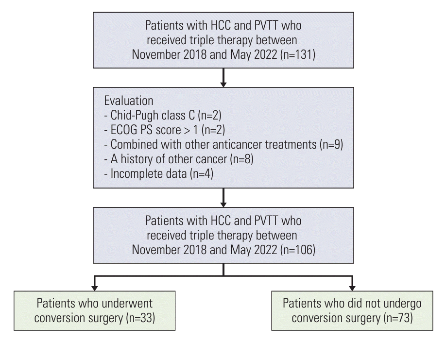
Between November 2018 and May 2022, 106 patients who received triple therapy at eight major cancer centers in China were included in this study (Fig. 1). The baseline demographics and characteristics of the patients diagnosed with HCC and PVTT are listed in Table 1. The mean age was 54.25 (11.25) years. Among them, 93 patients (87.7%) were males; the majority of patients (91.5%) had hepatitis B virus infection; 74 patients (69.8%) had an ECOG PS score of 0; 70 patients (66.0%) had baseline AFP ≥ 400 ng/mL, and 96 patients’ liver function (90.6%) was classified as Child-Pugh class A. Furthermore, 93 patients (87.7%) had maximum tumor size ≥ 5 cm, while 76 patients (71.7%) had multiple tumors. Extrahepatic metastases were observed in 15 patients (14.2%). With regard to the type of PVTT, Vp4 was presented in 41 patients (38.7%) and non-Vp4 was presented in 65 patients (61.3%) (Vp1 in 21 patients [19.8%], Vp2 in 13 patients [12.5%], Vp3 in 31 patients [29.2%]). Thirty-three patients (31.1%) reached the standard of conversion to resectable HCC after triple therapy and underwent conversion surgery.
Of 106 patients, 53 received camrelizumab, 27 received sintilimab, 10 received tislelizumab, nine received toripalimab, four received pembrolizumab, two received penpulimab, and one received nivolumab. The median cycle of the administering of anti–PD-1 antibodies was 13 (from 2 to 32). The median duration of treatment for lenvatinib was 270 days (from 60 to 1,030). And the median course of TACE that was performed in each patient was 2 (from 1 to 13).

2. Efficacy outcomes

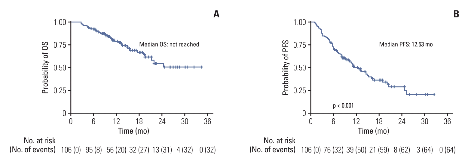
As of the data cutoff date of January 2023, the median follow-up time was 11.23 months (range, 3.07 to 34.37 months). Of the 106 patients, there were 17 patients (16.0%) with CR, 57 patients (53.8%) with PR, 15 patients (14.2%) with SD, and 17 patients (16.0%) with PD according to mRECIST criteria. The ORR and DCR were 69.8% and 84.0%, respectively (Table 2). The mOS was > 24 months, and the mPFS was 12.53 months (95% confidence interval [CI], 9.349 to 15.717). The 6-, 12-, 18-, and 24-month OS rates were 92.4%, 79.0%, 67.0%, and 54.9%, respectively. The 6-, 12-, 18-, and 24-month PFS rates were 72.6%, 50.2%, 36.7%, and 29.3%, respectively (Fig. 2).
In addition, we performed a subgroup analysis showing that the ORR and DCR of the Non-Vp4 group were 70.8% (46/65) and 83.1% (54/65), respectively, while those of the Vp4 group were 68.29% (28/41) and 85.37% (35/41), respectively. The mPFS was 11.53 months (95% CI, 7.650 to 15.410) in the non-Vp4 group and 14.50 months (95% CI, 9.664 to 19.336) in the Vp4 group. There was no statistically significant difference in mPFS between the two groups (p=0.802). The mOS of the non-Vp4 group was 24.37 months, but it was not reached in the Vp4 group. Similarly, there was no statistically significant difference in mOS between these two groups (p=0.677).

3. Risk factor analysis for OS and PFS

Univariate Cox regression analysis revealed that AFP (≥ 400 ng/mL) (hazard ratio [HR], 2.863; 95% CI, 1.176 to 6.969; p=0.021), tumor response with CR or PR (HR, 0.192; 95% CI, 0.094 to 0.391; p < 0.001), extrahepatic metastases (HR, 2.462; 95% CI, 1.137 to 5.330; p=0.022), and conversion surgery after triple therapy (HR, 0.095; 95% CI, 0.023 to 0.400; p=0.001) were associated with OS. In multivariate Cox regression analysis, CR or PR (HR, 0.385; 95% CI, 0.181 to 0.816; p=0.012) and conversion surgery (HR, 0.151; 95% CI, 0.033 to 0.683; p=0.014) were associated with OS. For PFS, age (≥ 65) (HR, 0.327; 95% CI, 0.131 to 0.817; p=0.017), sex (male) (HR, 0.485; 95% CI, 0.245 to 0.959; p=0.038), CR or PR (HR, 0.330; 95% CI, 0.196 to 0.556; p < 0.001), conversion surgery (HR, 0.415; 95% CI, 0.231 to 0.745; p=0.003) were the independent risk factors in univariate Cox regression analysis. Similarly, these factors were independent risk factors for PFS in the multivariate Cox regression analysis (Tables 3 and 4).

4. Outcomes of conversion surgery after successful conversion therapy with triple therapy

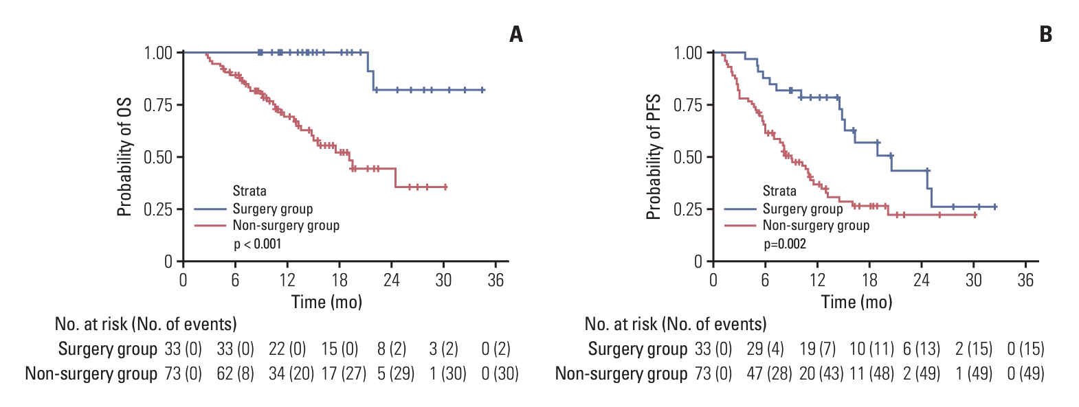
The characteristics of patients who could receive conversion surgery versus those who could not are shown in S1 Table. There were significant differences in AST levels (p=0.040), extrahepatic metastasis (p=0.005), and tumor response (p<0.001) between the two groups. The surgery group showed longer OS and PFS than the non-surgery group (Fig. 3). The mOS in the surgery group was not reached, whereas it was 19.08 months (95% CI, 13.636 to 24.524) in the non-surgery group. The mPFS rates in the surgery and non-surgery groups were 20.50 months (95% CI, 13.432 to 27.568) vs 9.00 months (95% CI, 6.241 to 11.759), respectively. Among the 33 patients who underwent conversion surgery, the median operation time and estimated intraoperative blood loss were 210 minutes (range, 150 to 310 minutes) and 300 ml (range, 100 to 6,000 mL), respectively. Clavien-Dindo grade IIIb or higher complications occurred in four patients, including pulmonary embolism (n=1) and postoperative hepatic function failure (n=3). The median postoperative hospital stay was 8 days (range, 6 to 22 days). During the follow-up period, 14 patients experienced postoperative recurrence. Intrahepatic recurrence was observed in 12 patients, extrahepatic recurrence in one patient, and both in one patient.

5. Safety outcomes

From 8 to 12 weeks after the initial treatment, we collected and analyzed the changes of liver function of 106 patients that was shown in S2 Table. There were 53 patients with a Child-Pugh score of 5, 31 patients with a score of 6, 14 patients with a score of 7, and eight patients with a score of 8. In terms of albumin-bilirubin (ALBI) grade, 55 patients had ALBI grade 1 (score ≤ –2.6), 49 patients had ALBI grade 2 (–2.6 < score ≤ –1.39), and two patients had ALBI grade 3 (score > –1.39). Compared to baseline data, 26 patients had varying degrees of liver function deterioration. Of these, the Child-Pugh class of 16 patients was changed from A to B. The ALBI grade of 16 patients changed from grade 1 to 2, and two patients changed from grade 2 to 3. Owning to the deterioration of liver function, four patients discontinued anti–PD-1 antibodies and six patients reduced the dose of lenvatinib. The mOS and mPFS of patients who had liver function deterioration from 8 to 12 weeks after triple therapy were 21.23 months (95% CI, 7.164 to 35.296) and 8.13 months (95% CI, 5.573 to 10.694), respectively.
Until January 2023, TRAEs occurred in 84 patients (79.2%), and 22 patients (20.8%) experienced grade 3/4 TRAEs (Table 5). Among these, the most frequent were abnormal liver function (60.4%), fever (30.2%), and fatigue (24.5%). The most frequent grade 3/4 TRAEs were gastrointestinal bleeding events (7.5%), thrombocytopenia (5.7%), and abnormal liver function 5 (4.7%). Immune-related adverse reactions occurred in two patients (1.9%). During the follow-up period, seven patients (6.6%) discontinued anti–PD-1 antibodies and lenvatinib, and nine patients (8.5%) reduced the dose of lenvatinib owing to grade 3/4 TRAEs, respectively. None of the patients experienced grade 5 TRAEs. All TRAEs were manageable during follow-up.

Discussion

Triple therapy has demonstrated significant survival benefits in patients with HCC and PVTT. During a median follow-up of 11.23 months (range, 3.07 to 34.37 months), the mOS was greater than 24 months, and the mPFS was 12.53 months. The 2-year OS rate was 54.9%, and it revealed a high ORR (69.8%) and DCR (84.0%) based on the mRECIST criteria. Regarding the safety of the triple therapy, 84 (79.2%) patients experienced at least one TRAEs, which may be due to the presence of PVTT, as the patients originally had poor liver function. However, most TRAEs were grade 1. Only 22 (20.8%) patients experienced grade 3/4 TRAEs and no treatment-related deaths occurred. The overall and severe adverse events rates were similar to those reported previously. The conversion rate to liver resection was 31.1%, with manageable postoperative complications. Conversion surgery after successful conversion therapy with triple therapy could improve the prognosis of patients with HCC and PVTT.
HCC with PVTT has an extremely poor prognosis and a high recurrence rate. This may be because PVTT can easily cause intrahepatic spread of the tumor and block blood flow to the liver, leading to further deterioration of liver function [18]. Therefore, it is difficult for a single treatment regimen to exert a significant effect. Currently, the combination of multiple programs is becoming a trend in the treatment of patients with advanced HCC. In the phase III IMbrave150 trial, the PD-L1 inhibitor atezolizumab combined with antiangiogenic bevacizumab significantly improved OS and PFS compared to sorafenib, making it a first-line immunotherapy regimen currently approved by the U.S. Food and Drug Administration for the treatment of unresectable or metastatic HCC [19]. A phase II trial of camrelizumab in combination with apatinib in patients with advanced HCC showed an ORR of 34.4% [20]. These studies demonstrated the efficacy of combination therapy. Although the regimen of lenvatinib plus pembrolizumab did not meet prespecified significance for improving OS and PFS versus lenvatinib plus placebo in LEAP002, the mOS and mPFS were still 21.2 months and 8.2 months in lenvatinib plus pembrolizumab group, respectively [21]. Moreover, in the Asian subgroup of LEAP002, the mOS reached 26.3 months in lenvatinib plus pembrolizumab group. It is known that most HCC patients in Asia are infected with HBV. This may indicate that the regimen of lenvatinib plus anti–PD-1 antibodies is more effective for HBV-infected HCC patients. In addition, subgroup analyses of LEAP002 showed that patients with extrahepatic metastasis or PVTT had a greater OS benefit from lenvatinib plus pembrolizumab therapy. Therefore, the efficacy of lenvatinib plus anti–PD-1 antibodies in the treatment of advanced HCC is worthy of further exploration. Moreover, TACE therapy was used in addition to lenvatinib plus anti–PD-1 antibodies in this study. Recently, many studies have shown that triple therapy has shown good efficacy in advanced HCC [12-14,22-24]. So we performed this study to explore the efficacy of triple therapy for patients with HCC and PVTT.
Guidelines in Western countries do not advocate TACE for the treatment of patients with HCC and PVTT because of concerns about disruption of the hepatic artery supply, which may lead to ischemia-related liver failure after TACE [25]. However, this concept has gradually evolved with advances in medical technology and further research. TACE can superselectively insert a catheter into the tumor blood supply target artery and inject an appropriate amount of embolic agent at an appropriate speed to block the target artery and cause ischemic necrosis of the tumor tissue. Lee et al. [26] published a prospective study showing that patients with PVTT may benefit from TACE therapy when their liver function is good (Child-Pugh A) and adequate collateral circulation has been established. A controlled experiment confirmed that patients in the TACE group had significantly better survival than those in the conservative treatment group (mOS, 8.67 vs. 1.4 months; p < 0.001) for different extents of PVTT [27]. All the above studies have confirmed the efficacy and safety of TACE in the treatment of patients with PVTT. Recently, the combination of locoregional and systemic therapy has achieved good curative effect in patients with HCC and PVTT. Two randomized controlled studies had shown that HAIC combined with sorafenib had better efficacy, with a mOS of 13.4-16.3 months [28,29]. A controlled study from Korea showed that radiotherapy combined with lenvatinib achieved a 6-month PFS rate of 67.2% [30]. In addition, TACE plus lenvatinib in the treatment of HCC with PVTT demonstrated a survival benefit, with a mOS of 16.4 months [6], which suggests the feasibility and advantage of TACE combined with lenvatinib for HCC with PVTT.
Li et al. [12] conducted a real-world study in China and showed that the triple therapy for the treatment of HCC achieved a mOS of 18.0 months and an ORR of 69.3%. A retrospective cohort study found that advanced HCC patients in the triple therapy group had prolonged OS (median, 16.9 vs. 12.1 months; p=0.009) and higher ORR (56.1% vs. 32.5%, p=0.033) than those in dual therapy (Lenvatinib+TACE) group [13]. Our previous studies have also suggested that triple therapy has a good survival benefit in patients with initially unresectable HCC [14,22]. As for HCC with PVTT, Zou et al. [23,24] conducted two studies showing that triple therapy achieved a mOS of 21.7-23.5 months and an ORR of 38.57%-41.25%. However, the number of existing studies and cases is limited. Thus, in the present study, we further assessed the efficacy of triple therapy, which achieved a high ORR of 69.8% and DCR of 84.0%. The mOS was beyond 24 months and mPFS was 12.53 months. This indicates that triple therapy can significantly improve the prognosis of patients with HCC and PVTT.
Conversion surgery refers to surgery for patients with initially unresectable HCC after successful conversion therapy that meets the resectable criteria mentioned in the Materials and Methods. It could also be called salvage surgery, which has been widely reported to result in a better prognosis in patients with initially unresectable HCC [22,31-33]. Based on previous research, the achievement of imaging remission after conversion therapy does not mean achievement of pathological remission. Most cases with imaging remission will progress in about 1 year, even if the medication is continued [19,34] Therefore, conversion surgery is of great significance and can eliminate the potential residual tumor cells and prolong the OS. Zhu et al. [31] demonstrated that surgical resection after successful conversion therapy with TKIs and anti–PD-1 antibodies was effective and safe with careful preparation and patient assessment [31]. A retrospective cohort study demonstrated that patients with BCLC C stage HCC undergoing conversion surgery after receiving TKIs and anti–PD-1 antibodies had a 6- and 12-month recurrence-free survival (RFS) rates of 77.0% and 64.8%, respectively, while the 6-, 12-, 24-, and 36-month OS rates were 98.4%, 93.4%, 76.8%, and 69.8%, respectively [32]. Another multicenter study of patients undergoing conversion surgery after treatment with TACE combined with TKIs and anti–PD-1 antibodies had 1- and 2-year RFS rates of 68.2% and 61.8%, 1- and 2-year OS rates of 92.2% and 87.3%, respectively, and the perioperative complications were manageable with appropriate medical interventions [33]. Our previous study also confirmed that conversion surgery for initially unresectable HCC converted by triple therapy resulted in a good prognosis. The 1- and 2-year OS rates could reach 97.1% and 94.4%, respectively [22]. Conversion surgery not only prolongs survival, but also reduces acquired drug resistance due to tumor heterogeneity during conversion treatment. In this study, we found that patients who underwent conversion surgery after conversion by triple therapy had better survival benefits and that conversion surgery was an independent prognostic factor for OS and PFS. Particularly, patients who accepted conversion surgery had longer mPFS (20.50 vs. 9.00 months), meaning that radical resection after triple therapy can provide a chance of cancer-free survival for advanced HCC patients, reduce the duration of continuous medication, the large costs associated with long-term medication use and occurrence of TRAEs.
In multivariate Cox regression analysis, tumor with objective response (CR or PR) was an independent prognostic factor for both PFS and OS, indicating that patients with objective response had better survival outcomes after triple therapy. A subanalysis of the REFLECT study showed that patients with objective response had a better OS than those without objective response (mOS, 21.6 vs. 11.9 months) [35]. An exploratory analysis of IMbrave150 indicated that objective response was an independent prognostic factor for OS [36]. These results all suggested that objective response was associated with better survival benefits. It may be a strong predictor of OS in HCC patients after conversion therapy. In this study, the high ORR (69.8%) associated with triple therapy shows the effectiveness of this therapeutic regimen. Moreover, our study revealed that tumor size, tumor number, extrahepatic metastasis, and Vp4 or non-Vp4 status were not significantly correlated with OS or PFS. In subgroup analysis, there was no significant difference in OS and PFS between Vp4 and non-Vp4 groups. This may be due to the synergistic effect of the triple therapy, which also works well in patients with a high tumor burden or main portal vein thrombus. However, the sample size of this study is limited. More studies with large sample sizes are needed to further explore this outcome.
The present study has several limitations. First, this was a retrospective study with a limited sample size and a relatively short follow-up time, leading to inevitable selection bias and relatively insufficient medical evidence. However, to the best of our knowledge, this is the largest series of reported cases of triple therapy for patients with HCC and PVTT. Second, there was no control group; therefore, it was impossible to compare the efficacy and safety of this triple therapy with those of other combined therapeutic approaches. Third, inconsistencies in the types of anti–PD-1 antibodies used may have influenced the interpretation of treatment outcomes. Fourth, HBV-related cases accounted for a relatively high proportion of the cases. Whether triple therapy can be applied to HCC due to other causes requires further investigation. Thus, more prospective and well-designed randomized controlled trials are required to determine the benefits of triple therapy in patients with HCC and PVTT.
In conclusion, lenvatinib combined with anti–PD-1 antibodies plus TACE showed promising survival benefits and high response rates in the treatment of patients with HCC and PVTT, with manageable adverse effects. Patients who underwent conversion surgery after successful conversion therapy had better prognoses. Therefore, triple therapy may be the ideal treatment regimen for patients with HCC and PVTT.

Electronic Supplementary Material

Supplementary materials are available at Cancer Research and Treatment website (https://www.e-crt.org).

Notes

Ethical Statement

This study was conducted in accordance with the World Medical Association Declaration of Helsinki and with approval from the Institutional Review Board (IRB) of Fujian Provincial Hospital, approval number: K2024-02-006. The requirement of informed consent has been waived because of the retrospective nature of this study.

Author Contributions

Conceived and designed the analysis: Qu X, Wu J (Junyi Wu), Wu J (Jiayi Wu), Qu S, Yan M.

Collected the data: Qu X, Wu J (Junyi Wu), Wu J (Jiayi Wu), Fu Y, Zeng Z, Li S, Li Y, Liu D, Li H, Li B, Zhou J, Zhuang S, Cheng S, Zhang Z, Wang K, Qu S, Yan M.

Contributed data or analysis tools: Qu X, Wu J (Junyi Wu), Wu J (Jiayi Wu), Fu Y, Zeng Z, Li S, Li Y, Liu D, Li H, Qu S, Yan M.

Performed the analysis: Qu X, Wu J (Junyi Wu), Wu J (Jiayi Wu), Fu Y, Zeng Z, Li S, Li Y, Liu D, Li H, Qu S, Yan M.

Wrote the paper: Qu X, Wu J (Junyi Wu), Wu J (Jiayi Wu), Qu S, Yan M.

Conflicts of Interest

Conflict of interest relevant to this article was not reported.

ACKNOWLEDGMENTS

This study was funded by the Natural Science Foundation of Fujian Province (Grant number: 2022J011021) and the Medical Innovation Project of Health and Family Planning Commission of Fujian Province (Grant number: 2022CXA002). The authors thank all the staff of the participating hospitals for their efforts, as well as all of the patients for their participation.

References

1. Llovet JM, Kelley RK, Villanueva A, Singal AG, Pikarsky E, Roayaie S, et al. Hepatocellular carcinoma. Nat Rev Dis Primers. 2021; 7:6.
crossref
2. Cheng S, Chen M, Cai J, Sun J, Guo R, Bi X, et al. Chinese expert consensus on multidisciplinary diagnosis and treatment of hepatocellular carcinoma with portal vein tumor thrombus (2018 Edition). Liver Cancer. 2020; 9:28–40.
crossref
3. Zhou J, Sun H, Wang Z, Cong W, Wang J, Zeng M, et al. Guidelines for the diagnosis and treatment of hepatocellular carcinoma (2019 edition). Liver Cancer. 2020; 9:682–720.
crossref
4. Korean Liver Cancer Association; National Cancer Center. 2018 Korean Liver Cancer Association-National Cancer Center Korea practice guidelines for the management of hepatocellular carcinoma. Gut Liver. 2019; 13:227–99.
5. Sun J, Guo R, Bi X, Wu M, Tang Z, Lau WY, et al. Guidelines for diagnosis and treatment of hepatocellular carcinoma with portal vein tumor thrombus in China (2021 edition). Liver Cancer. 2022; 11:315–28.
6. Yang B, Jie L, Yang T, Chen M, Gao Y, Zhang T, et al. TACE plus lenvatinib versus TACE plus sorafenib for unresectable hepatocellular carcinoma with portal vein tumor thrombus: a prospective cohort study. Front Oncol. 2021; 11:821599.
crossref
7. Ding X, Sun W, Li W, Shen Y, Guo X, Teng Y, et al. Transarterial chemoembolization plus lenvatinib versus transarterial chemoembolization plus sorafenib as first-line treatment for hepatocellular carcinoma with portal vein tumor thrombus: a prospective randomized study. Cancer. 2021; 127:3782–93.
crossref
8. Rimassa L, Pressiani T, Merle P. Systemic treatment options in hepatocellular carcinoma. Liver Cancer. 2019; 8:427–46.
crossref
9. Finn RS, Zhu AX, Farah W, Almasri J, Zaiem F, Prokop LJ, et al. Therapies for advanced stage hepatocellular carcinoma with macrovascular invasion or metastatic disease: a systematic review and meta-analysis. Hepatology. 2018; 67:422–35.
crossref
10. Finn RS, Zhu AX. Evolution of systemic therapy for hepatocellular carcinoma. Hepatology. 2021; 73 Suppl 1:150–7.
crossref
11. Lewis S, Cedillo MA, Lee KM, Bane O, Hectors S, Ma W, et al. Comparative assessment of standard and immune response criteria for evaluation of response to PD-1 monotherapy in unresectable HCC. Abdom Radiol (NY). 2022; 47:969–80.
crossref
12. Li X, Fu Z, Chen X, Cao K, Zhong J, Liu L, et al. Efficacy and safety of lenvatinib combined with PD-1 inhibitors plus TACE for unresectable hepatocellular carcinoma patients in China real-world. Front Oncol. 2022; 12:950266.
crossref
13. Cai M, Huang W, Huang J, Shi W, Guo Y, Liang L, et al. Transarterial chemoembolization combined with lenvatinib plus PD-1 inhibitor for advanced hepatocellular carcinoma: a retrospective cohort study. Front Immunol. 2022; 13:848387.
crossref
14. Wu JY, Yin ZY, Bai YN, Chen YF, Zhou SQ, Wang SJ, et al. Lenvatinib combined with anti-PD-1 antibodies plus transcatheter arterial chemoembolization for unresectable hepatocellular carcinoma: a multicenter retrospective study. J Hepatocell Carcinoma. 2021; 8:1233–40.
crossref
15. Feng JK, Liu ZH, Fu ZG, Chai ZT, Sun JX, Wang K, et al. Efficacy and safety of transarterial chemoembolization plus antiangiogenic-targeted therapy and immune checkpoint inhibitors for unresectable hepatocellular carcinoma with portal vein tumor thrombus in the real world. Front Oncol. 2022; 12:954203.
16. Marrero JA, Kulik LM, Sirlin CB, Zhu AX, Finn RS, Abecassis MM, et al. Diagnosis, staging, and management of hepatocellular carcinoma: 2018 practice guidance by the American Association for the Study of Liver Diseases. Hepatology. 2018; 68:723–50.
crossref
17. Kudo M, Izumi N, Kubo S, Kokudo N, Sakamoto M, Shiina S, et al. Report of the 20th nationwide follow-up survey of primary liver cancer in Japan. Hepatol Res. 2020; 50:15–46.
crossref
18. Yoon SM, Lim YS, Won HJ, Kim JH, Kim KM, Lee HC, et al. Radiotherapy plus transarterial chemoembolization for hepatocellular carcinoma invading the portal vein: long-term patient outcomes. Int J Radiat Oncol Biol Phys. 2012; 82:2004–11.
crossref
19. Galle PR, Finn RS, Qin S, Ikeda M, Zhu AX, Kim TY, et al. Patient-reported outcomes with atezolizumab plus bevacizumab versus sorafenib in patients with unresectable hepatocellular carcinoma (IMbrave150): an open-label, randomised, phase 3 trial. Lancet Oncol. 2021; 22:991–1001.
crossref
20. Xu J, Shen J, Gu S, Zhang Y, Wu L, Wu J, et al. Camrelizumab in combination with apatinib in patients with advanced hepatocellular carcinoma (RESCUE): a nonrandomized, open-label, phase II trial. Clin Cancer Res. 2021; 27:1003–11.
crossref
21. Llovet JM, Kudo M, Merle P, Meyer T, Qin S, Ikeda M, et al. Lenvatinib plus pembrolizumab versus lenvatinib plus placebo for advanced hepatocellular carcinoma (LEAP-002): a randomised, double-blind, phase 3 trial. Lancet Oncol. 2023; 24:1399–410.
22. Wu JY, Zhang ZB, Zhou JY, Ke JP, Bai YN, Chen YF, et al. Outcomes of salvage surgery for initially unresectable hepatocellular carcinoma converted by transcatheter arterial chemoembolization combined with lenvatinib plus anti-PD-1 antibodies: a multicenter retrospective study. Liver Cancer. 2023; 12:229–37.
crossref
23. Zou X, Xu Q, You R, Yin G. Evaluating the benefits of TACE combined with lenvatinib plus PD-1 inhibitor for hepatocellular carcinoma with portal vein tumor thrombus. Adv Ther. 2023; 40:1686–704.
crossref
24. Zou X, Xu Q, You R, Yin G. Correlation and efficacy of TACE combined with lenvatinib plus PD-1 inhibitor in the treatment of hepatocellular carcinoma with portal vein tumor thrombus based on immunological features. Cancer Med. 2023; 12:11315–33.
25. Yamada R, Sato M, Kawabata M, Nakatsuka H, Nakamura K, Takashima S. Hepatic artery embolization in 120 patients with unresectable hepatoma. Radiology. 1983; 148:397–401.
crossref
26. Lee HS, Kim JS, Choi IJ, Chung JW, Park JH, Kim CY. The safety and efficacy of transcatheter arterial chemoembolization in the treatment of patients with hepatocellular carcinoma and main portal vein obstruction: a prospective controlled study. Cancer. 1997; 79:2087–94.
crossref
27. Niu ZJ, Ma YL, Kang P, Ou SQ, Meng ZB, Li ZK, et al. Transarterial chemoembolization compared with conservative treatment for advanced hepatocellular carcinoma with portal vein tumor thrombus: using a new classification. Med Oncol. 2012; 29:2992–7.
crossref
28. Zheng K, Zhu X, Fu S, Cao G, Li WQ, Xu L, et al. Sorafenib plus hepatic arterial infusion chemotherapy versus sorafenib for hepatocellular carcinoma with major portal vein tumor thrombosis: a randomized trial. Radiology. 2022; 303:455–64.
crossref
29. He M, Li Q, Zou R, Shen J, Fang W, Tan G, et al. Sorafenib plus hepatic arterial infusion of oxaliplatin, fluorouracil, and leucovorin vs sorafenib alone for hepatocellular carcinoma with portal vein invasion: a randomized clinical trial. JAMA Oncol. 2019; 5:953–60.
crossref
30. Yu JI, Kang W, Yoo GS, Goh MJ, Sinn DH, Gwak GY, et al. Safety and efficacy of liver-directed radiotherapy in combination with lenvatinib for hepatocelluar carcinoma with macroscopic tumor thrombosis. Front Oncol. 2022; 12:888755.
crossref
31. Zhu XD, Huang C, Shen YH, Ji Y, Ge NL, Qu XD, et al. Downstaging and resection of initially unresectable hepatocellular carcinoma with tyrosine kinase inhibitor and anti-PD-1 antibody combinations. Liver Cancer. 2021; 10:320–9.
crossref
32. Pan Y, Yang L, Cao Y, Jun H, Tang H, Zhang W, et al. Factors influencing the prognosis patients with Barcelona Clinic Liver Cancer stage C hepatocellular carcinoma undergoing salvage surgery after conversion therapy. Transl Cancer Res. 2023; 12:1852–62.
crossref
33. Lin KY, Lin ZW, Chen QJ, Luo LP, Zhang JX, Chen JH, et al. Perioperative safety, oncologic outcome, and risk factors of salvage liver resection for initially unresectable hepatocellular carcinoma converted by transarterial chemoembolization plus tyrosine kinase inhibitor and anti-PD-1 antibody: a retrospective multicenter study of 83 patients. Hepatol Int. 2023; 17:1477–89.
crossref
34. Finn RS, Ikeda M, Zhu AX, Sung MW, Baron AD, Kudo M, et al. Phase Ib study of lenvatinib plus pembrolizumab in patients with unresectable hepatocellular carcinoma. J Clin Oncol. 2020; 38:2960–70.
crossref
35. Kudo M, Finn RS, Qin S, Han KH, Ikeda K, Cheng AL, et al. Overall survival and objective response in advanced unresectable hepatocellular carcinoma: a subanalysis of the REFLECT study. J Hepatol. 2023; 78:133–41.
crossref
36. Ducreux M, Zhu AX, Cheng AL, Galle PR, Ikeda M, Nicholas A, et al. IMbrave150: exploratory analysis to examine the association between treatment response and overall survival (OS) in patients (pts) with unresectable hepatocellular carcinoma (HCC) treated with atezolizumab (atezo) + bevacizumab (bev) versus sorafenib (sor). J Clin Oncol. 2021; 39(15 Suppl):4071.
crossref

Fig. 1.
Flowchart of patient selection. ECOG PS, Eastern Cooperative Oncology Group performance status; HCC, hepatocellular carcinoma; PVTT, portal vein tumor thrombus.
crt-2023-1165f1.tif
Fig. 2.
Kaplan-Meier estimates of overall survival (OS) (A) and progression-free survival (PFS) (B).
crt-2023-1165f2.tif
Fig. 3.
Kaplan-Meier estimates of overall survival (OS) (A) and progression-free survival (PFS) (B) in surgery group and non-surgery group.
crt-2023-1165f3.tif
Table 1.
Baseline demographic and clinical characteristics of patients
Characteristic No. (%) (n=106)
Age (yr)
 < 65 89 (84.0)
 ≥ 65 17 (16.0)
ECOG PS score
 0 74 (69.8)
 1 32 (30.2)
Sex
 Female 13 (12.3)
 Male 93 (87.7)
Platelet (×109/L)
 < 100 12 (11.3)
 ≥ 100 94 (88.7)
ALT (IU/L)
 < 40 48 (45.3)
 ≥ 40 58 (54.7)
AST (IU/L)
 < 40 22 (20.8)
 ≥ 40 84 (79.2)
Total bilirubin (μmol/L)
 < 34 101 (95.3)
 ≥ 34 5 (4.7)
Albumin (g/L)
 < 35 24 (22.6)
 ≥ 35 82 (77.4)
ALBI grade
 1 46 (43.4)
 2 60 (56.6)
AFP (ng/mL)
 < 400 36 (34.0)
 ≥ 400 70 (66.0)
HBsAg
 Negative 9 (8.5)
 Positive 97 (91.5)
Child-Pugh
 A 96 (90.6)
 B 10 (9.4)
Maximum tumor size (cm)
 < 5 13 (12.3)
 ≥ 5 93 (87.7)
Tumor number
 Solitary 30 (28.3)
 Multiple 76 (71.7)
Extent of PVTT
 Non-VP4 65 (61.3)
 VP4 41 (38.7)
Extrahepatic metastases
 Yes 15 (14.2)
 No 91 (85.8)
mRECIST
 CR or PR 74 (69.8)
 SD or PD 32 (30.2)
Conversion surgery
 Yes 33 (31.1)
 No 73 (68.9)

AFP, α-fetoprotein; ALBI, albumin-bilirubin; ALT, alanine aminotransferase; AST, aspartate aminotransferase; CR, complete response; ECOG PS, Eastern Cooperative Oncology Group performance status; HBsAg, hepatitis B surface antigen; mRECIST, modified Response Evaluation Criteria in Solid Tumors; PD, progressive disease; PR, partial response; PVTT, portal vein tumor thrombus; SD, stable disease.

Table 2.
Best tumor response to triple therapy
Best response Triple therapy (n=106)
Complete response 17 (16.0)
Partial response 57 (53.8)
Stable disease 15 (14.2)
Progressive disease 17 (16.0)
Objective response rate 74 (69.8)
Disease control rate 89 (84.0)

Values are presented as number (%).

Table 3.
Univariate and multivariate analyses of the prognostic factors for overall survival in all patients
Variable Univariate analysis
Multivariate analysis
Hazard ratio 95% CI p-value Hazard ratio 95% CI p-value
Age (yr)
 ≥ 65 vs. < 65 0.700 0.245-1.999 0.505
Sex
 Male vs. female 0.534 0.204-1.398 0.202
ECOG PS
 1 vs. 0 1.083 0.522-2.249 0.831
AFP (ng/mL)
 ≥ 400 vs. < 400 2.863 1.176-6.969 0.021 2.326 0.934-5.794 0.070
HBsAg
 Positive vs. negative 1.004 0.306-3.302 0.994
PLT (×109/L)
 ≥ 100 vs. < 100 2.398 0.572-10.050 0.232
Tbil (μmol/L)
 ≥ 34 vs. < 34 1.763 0.531-5.858 0.355
Albumin (g/L)
 ≥ 35 vs. < 35 1.212 0.498-2.949 0.672
ALT (IU/L)
 ≥ 40 vs. < 40 0.899 0.449-1.802 0.765
AST (IU/L)
 ≥ 40 vs. < 40 1.522 0.652-3.551 0.331
ALBI grade
 ≥ 2 vs. 1 0.925 0.458-1.867 0.828
Child-Pugh
 B vs. A 0.754 0.180-3.162 0.699
Tumor number
 Multiple vs. solitary 1.139 0.524-2.475 0.742
Maximum tumor size (cm)
 ≥ 5 vs. < 5 1.588 0.483-5.217 0.446
Portal vein thrombus
 VP4 vs. non-VP4 1.161 0.575-2.341 0.677
mRECIST
 CR or PR vs. SD or PD 0.192 0.094-0.391 < 0.001 0.385 0.181-0.816 0.013
Extrahepatic metastases
 Yes vs. no 2.462 1.137-5.330 0.022 1.200 0.543-2.652 0.653
Conversion surgery
 Yes vs. no 0.095 0.023-0.400 0.001 0.151 0.033-0.683 0.014

AFP, α-fetoprotein; ALBI, albumin-bilirubin; ALT, alanine aminotransferase; AST, aspartate aminotransferase; CI, confidence interval; CR, complete response; ECOG PS, Eastern Cooperative Oncology Group performance status; HBsAg, hepatitis B surface antigen; mRECIST, modified Response Evaluation Criteria in Solid Tumors; PD, progressive disease; PLT, platelet; PR, partial response; SD, stable disease; Tbil, total bilirubin.

Table 4.
Univariate and multivariate analyses of the prognostic factors for progression-free survival in all patients
Variable Univariate analysis
Multivariate analysis
Hazard ratio 95% CI p-value Hazard ratio 95% CI p-value
Age (yr)
 ≥ 65 vs. < 65 0.327 0.131-0.817 0.017 0.354 1.141-0.886 0.027
Sex
 Male vs. female 0.485 0.245-0.959 0.038 0.454 0.224-0.921 0.029
ECOG PS
 1 vs. 0 1.024 0.606-1.728 0.930
AFP (ng/mL)
 ≥ 400 vs. < 400 1.406 0.833-2.373 0.202
HBsAg
 Positive vs. negative 0.573 0.260-1.264 0.168
PLT (×109/L)
 ≥ 100 vs. < 100 1.088 0.495-2.392 0.834
Tbil (μmol/L)
 ≥ 34 vs. < 34 1.329 0.480-3.680 0.584
Albumin (g/L)
 ≥ 35 vs. < 35 1.081 0.587-1.990 0.802
ALT (IU/L)
 ≥ 40 vs. < 40 0.913 0.558-1.493 0.717
AST (IU/L)
 ≥ 40 vs. < 40 1.146 0.640-2.054 0.647
ALBI grade
 ≥ 2 vs. 1 0.947 0.576-1.557 0.830
Child-Pugh
 B vs. A 1.114 0.479-2.593 0.802
Tumor number
 Multiple vs. solitary 1.406 0.797-2.481 0.240
Maximum tumor size (cm)
 ≥ 5 vs. < 5 1.208 0.574-2.541 0.618
Portal vein thrombus
 VP4 vs. non-VP4 0.938 0.567-1.551 0.802
mRECIST
 CR or PR vs. SD or PD 0.330 0.196-0.556 < 0.001 0.446 0.251-0.792 0.006
Extrahepatic metastases
 Yes vs. no 1.844 0.978-3.473 0.058
Conversion surgery
 Yes vs. no 0.415 0.231-0.745 0.003 0.490 0.256-0.939 0.032

AFP, α-fetoprotein; ALBI, albumin-bilirubin; ALT, alanine aminotransferase; AST, aspartate aminotransferase; CI, confidence interval; CR, complete response; ECOG PS, Eastern Cooperative Oncology Group performance status; HBsAg, hepatitis B surface antigen; mRECIST, modified Response Evaluation Criteria in Solid Tumors; PD, progressive disease; PLT, platelet; PR, partial response; SD, stable disease; Tbil, total bilirubin.

Table 5.
Summary of adverse events
Adverse events Any grades Grade 3/4
Total 84 (79.2) 22 (20.8)
Abnormal liver function 64 (60.4) 5 (4.7)
Fever 32 (30.2) 3 (2.8)
Fatigue 26 (24.5)
Decreased appetite 24 (22.6)
Hypertension 21 (19.8) 2 (1.9)
Hypothyroidism 17 (16.3)
Hand-foot skin reaction 15 (14.2)
Thrombocytopenia 14 (13.2) 6 (5.7)
Skin rash 12 (11.3)
Weight decreased 11 (10.3)
Diarrhea 11 (10.3)
Abdominal pain 10 (9.4) 4 (3.8)
Proteinuria 10 (9.4) 2 (1.9)
Anemia 9 (8.4)
Gastrointestinal bleeding events 8 (7.5) 8 (7.5)
Immune-related adverse reactions 2 (1.9) 2 (1.9)

Values are presented as number (%).

TOOLS
Similar articles