Journal List > J Liver Cancer > v.24(2) > 1516088664

Bae, Jang, Mortensen, Weber, Kim, and Høyer: Recent update of proton beam therapy for hepatocellular carcinoma: a systematic review and meta-analysis

Abstract

Backgrounds/Aims

Although access to proton beam therapy (PBT) is limited worldwide, its use for the treatment of hepatocellular carcinoma (HCC) is gradually increasing with the expansion of new facilities. Therefore, we conducted a systematic review and metaanalysis to investigate the updated evidence of PBT for HCC.

Methods

The MEDLINE, EMBASE, Cochrane Library, and Web of Science databases were systematically searched for studies that enrolled patients with liver-confined HCC that were treated with PBT for a cure up to February 2024.

Results

A total of 1,858 HCC patients receiving PBT from 22 studies between 2004 and 2023 were selected for this meta-analysis. The median proportion of Child-Pugh class A was 86% (range, 41-100), and the median tumor size was 3.6 cm (range, 1.2-9.0). The median total dose ranged from 55 GyE to 76 GyE (median, 69). The pooled rates of 3- and 5-year local progression-free survival after PBT were 88% (95% confidence interval [CI], 85-91) and 86% (95% CI, 82-90), respectively. The pooled 3- and 5-year overall rates were 60% (95% CI, 54-66) and 46% (95% CI, 38-54), respectively. The pooled rates of grade 3 hepatic toxicity, classic radiationinduced liver disease (RILD), and non-classic RILD were 1%, 2%, and 1%, respectively.

Conclusions

The current study supports PBT for HCC and demonstrates favorable long-term survival and low hepatic toxicities compared with other published studies on other radiotherapy modalities. However, further studies are needed to identify the subgroups that will benefit from PBT.

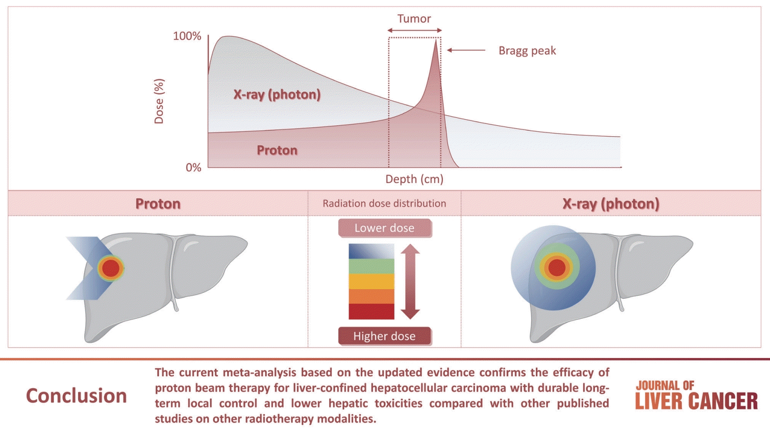
GRAPHICAL ABSTRACT

INTRODUCTION

Proton beam therapy (PBT) is a type of charged particle therapy (CPT) with unique physical properties, consisting of a finite range in tissues and a zero dose beyond the end of their path, the so-called Bragg peak. PBT has a dose-distribution profile which results in better sparing of healthy tissues compared to that with photon therapy in the low-medium dose areas.1 Many in-vitro and in-vivo studies were conducted to determine the biological effect of proton relative to photon irradiation (relative biological effectiveness [RBE]), and an average RBE of 1.1 is currently used, implying that proton therapy is 10% more biologically effective compared to photon therapy.2 Since Robert Wilson proposed the use of accelerated protons for use in clinical setting in 1946, PBT was firstly applied for a cancer patient in 1954 and extended for the treatment of hepatocellular carcinoma (HCC) in the 1980s.3-5 Although the access to PBT has been limited, the number of treatment facilities and patients receiving PBT has rapidly increased in recent years. Over 100 PBT centers are in operation worldwide and over 350,000 patients have been treated with PBT as of February 2024.6
HCC is the most common primary liver cancer and one of the leading causes of cancer-related deaths worldwide.7 Because most HCCs develop in patients with liver cirrhosis from various cause, including chronic viral hepatitis, alcoholic and/or nonalcoholic fatty liver disease, the treatment decision in HCC is affected by both tumor stage and patient-related factors (e.g., performance status, underlying liver disease, and liver function).8,9 Therefore, several treatment guidelines are used for the treatment of HCC, reflecting different stating system for HCC and various etiology across different regions of the world.10-16 Historically, the role of external beam radiotherapy (EBRT) for HCC has been limited to palliative treatments. Advances of radiotherapy (RT) techniques including 3-dimensional conformal RT (3DCRT), intensity-modulated RT (IMRT), stereotactic body RT (SBRT), and CPT together with improved understanding of tumor biology and the tolerance dose of normal and cirrhotic liver, however, lead to the expansion of the role of EBRT to curative treatments.17 RT, mainly 3DCRT, IMRT or SBRT, is considered as an alternative treatment option for HCC that is deemed inoperable or unsuitable for other liver-directed therapies (LDT) in some guidelines; however, there are few recommendations on the use of PBT in treatment of HCC.11,13,14,16 The National Comprehensive Cancer Network guidelines recommend that PBT is an acceptable option for hypofractionated RT for intrahepatic lesions and may be appropriate in specific situations.16 Recent updated Korea guideline suggests that PBT is not inferior to radiofrequency ablation (RFA) in treating recurrent or residual HCCs ≤3 cm in size as the level of evidence of A2.14,18
With the increasing number of PBT facilities, many clinical studies on the use of PBT in treatment of HCC have been recently published. Therefore, we conducted a systematic review and meta-analysis to investigate the updated evidence of PBT for HCC.

METHODS

A systematic literature search was performed according to the preferred reporting items for systematic reviews and meta-analyses (PRISMA) guidelines.19

Study selection

The following inclusion criteria for selection of studies were used: 1) prospective or retrospective studies including patients with liver-confined HCC treated with PBT for a cure, 2) inclusion of ≥10 patients, and 3) reporting of at least one endpoint of interest in terms of survivals and/or toxicities. Survival was indirectly estimated using descriptive plots in the absence of numerical data. If there was an overlap of patients among the studies, we selected the study with the following criteria: 1) the study with the largest number of patients, 2) the study that reported quantified data for PBT outcomes, and 3) the most recent ones. However, studies from a single center were independently categorized if they were reported in distinct periods. We excluded studies in which PBT was applied to 1) pediatric patients, 2) patients with distant metastases, or 3) patients with a history of prior RT to the liver.

Search strategy

The population, intervention, comparison, and outcome (PICO) model was used to outline the initial literature search (Supplementary Table 1). The search strategy was developed and reviewed by all authors in cooperation with a professional librarian at Soonchunhyang University College of Medicine, Bucheon. The MEDLINE, EMBASE, Cochrane Library, and Web of Science databases were systematically searched for articles published through February 2024. Full-text articles on humans, published in English between 1975 and February 2024, were identified. In addition, the reference lists of relevant studies and review articles were manually reviewed, allowing for the identification of 18 additional studies that were not identified in the original literature search. A total of 6,365 articles were identified, and two authors (SH Bae and WI Jang) independently screened the article titles, abstracts, and full texts as necessary. Conflicts were resolved through discussion with a third author (MS Kim).

Data extraction and quality assessment

Data from the included studies were independently extracted by two authors (SH Bae and WI Jang). Data included 1) patient and tumor characteristics, 2) details of PBT, 3) survivals, and 4) treatment related toxicities ≥grade 3. The PBT doses are described in gray equivalents (GyE=proton physical dose [in gray]×RBE [1.1]). Because various fractionation schemes were used among studies, total doses were converted to the biologically equivalent dose (BED, GyE10) using a linear quadratic model with α/β of 10. Survival rates at 1-, 3-, and 5-year were obtained. Hepatic, gastrointestinal (GI) toxicity, and skin toxicities were assessed. Severe toxicity was mainly defined by crude rates of grade ≥3 toxicity by the common terminology criteria for adverse events. Classic radiation-induced liver disease (RILD) and non-classic RILD were independently assessed for hepatic toxicity.
Because most studies were retrospective, the Newcastle-Ottawa scale (NOS) was employed to assess the quality of the included studies.20 Studies with over seven stars were categorized as high quality, and studies with a score of 4-6 stars were categorized as medium quality, respectively.

Statistical analysis

Meta-analysis was performed using the DerSimonian-Laird random-effects model. Given the variation in PBT indication, different study periods among studies, and different etiologies according to country, we adopted the random-effects model but reported both estimations in the tables.21 The Higgins’ (I2) statistic was used to assess the heterogeneity in the results of individual studies, and an I2>50% was considered as the threshold indicating significant heterogeneity.22 Funnel plots were used to assess the publication bias, and Egger’s regression tests were used to quantitatively analyze the symmetry of the funnel plots. If the funnel plot was symmetrical or P-value was >0.05 in Egger’s test, then the null hypothesis of no publication bias was accepted. For comparisons between subgroups, a Q test based on analysis of variance and a random-effects model were used. A P<0.05 was considered statistically significant. Statistical analyses were conducted using Rex Excel-based statistical analysis software ver. 3.6.0 (RexSoft, Seoul, Korea; http://rexsoft.org).

RESULTS

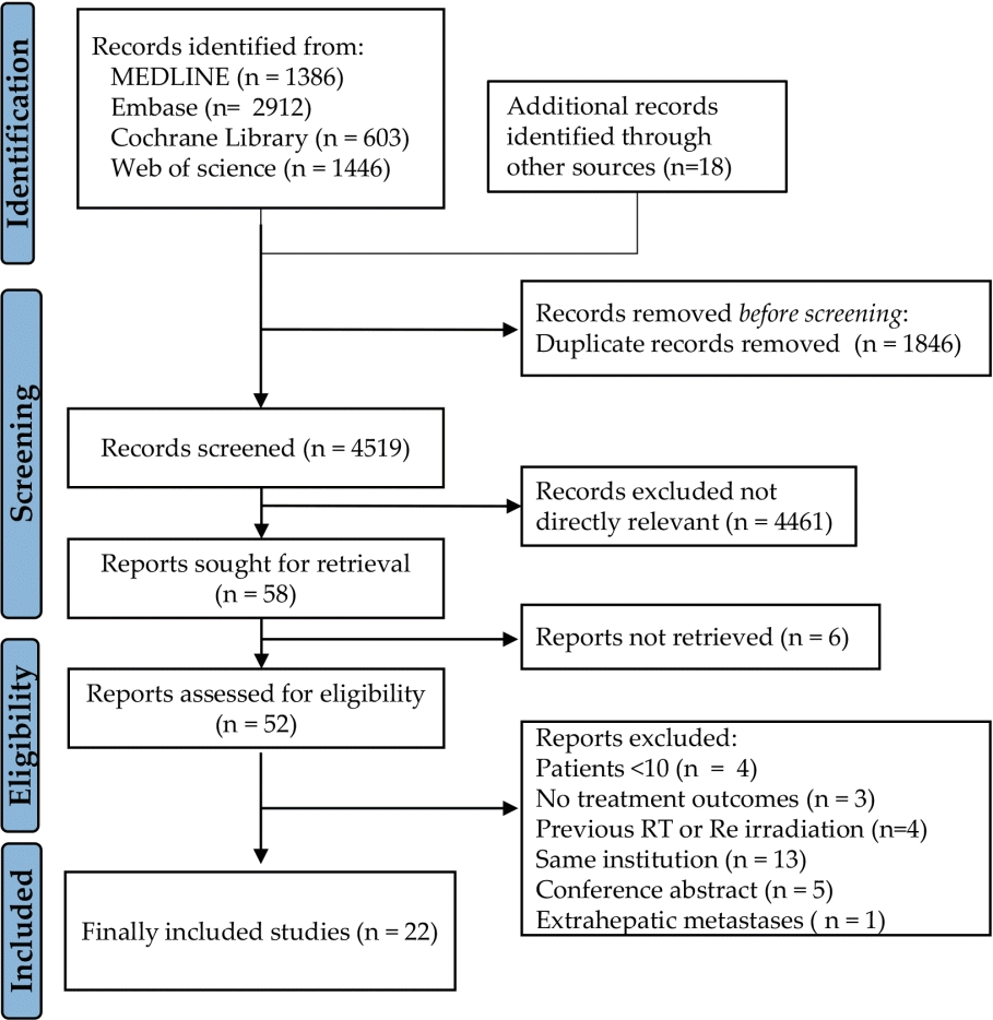
We retrieved 6,347 studies from the initial database searches and 18 from additional sources. After multilayered screening, this systematic review and meta-analysis included 22 eligible studies published between 2004 and 2023, comprising 1,858 HCC patients treated with PBT (Fig. 1).18,23-43 Because six studies from two institutions might have had the possibility of including overlapping patients among the studies, we emailed both the corresponding authors and confirmed that each study was conducted with independent cohorts.18,27,28,33,37,38

Studies’ characteristics

An overview of the study and treatment details of our study population is presented in Tables 1 and 2. Five studies were prospective and the remaining studies were retrospective. Sixteen studies were single-arm trials and six studies were comparative trials. The indications for PBT and the quality of each study according to NOS are presented in Supplementary Table 2.
The median age across the studies was 69.5 years (range, 55-82), and the proportion of patients with Eastern Cooperative Oncology Group performance status of 0 or 1 was 97% (range, 68-100). The proportion of patients with Child-Pugh (CP) class A was between 41% and 100% (median, 86). The median tumor size was 3.6 cm (range, 1.2-9.0), and 0-75% of the patients (median, 22) had multiple HCCs in the liver. Portal vein tumor thrombosis (PVTT) was present in 0-100% of patients (median, 10.5). The median total dose ranged from 55 to 76 GyE (median, 69) and the median BED values ranged from 68.8 to 109.6 GyE10 (median, 101.1). Concurrent treatment during PBT was applied in three studies, where sorafenib was used in 3-22% of patients.33,34,38 Four studies reported that between 1% and 37% of patients ultimately received liver transplantation after PBT.25,29,37,43

Progression and survival estimates

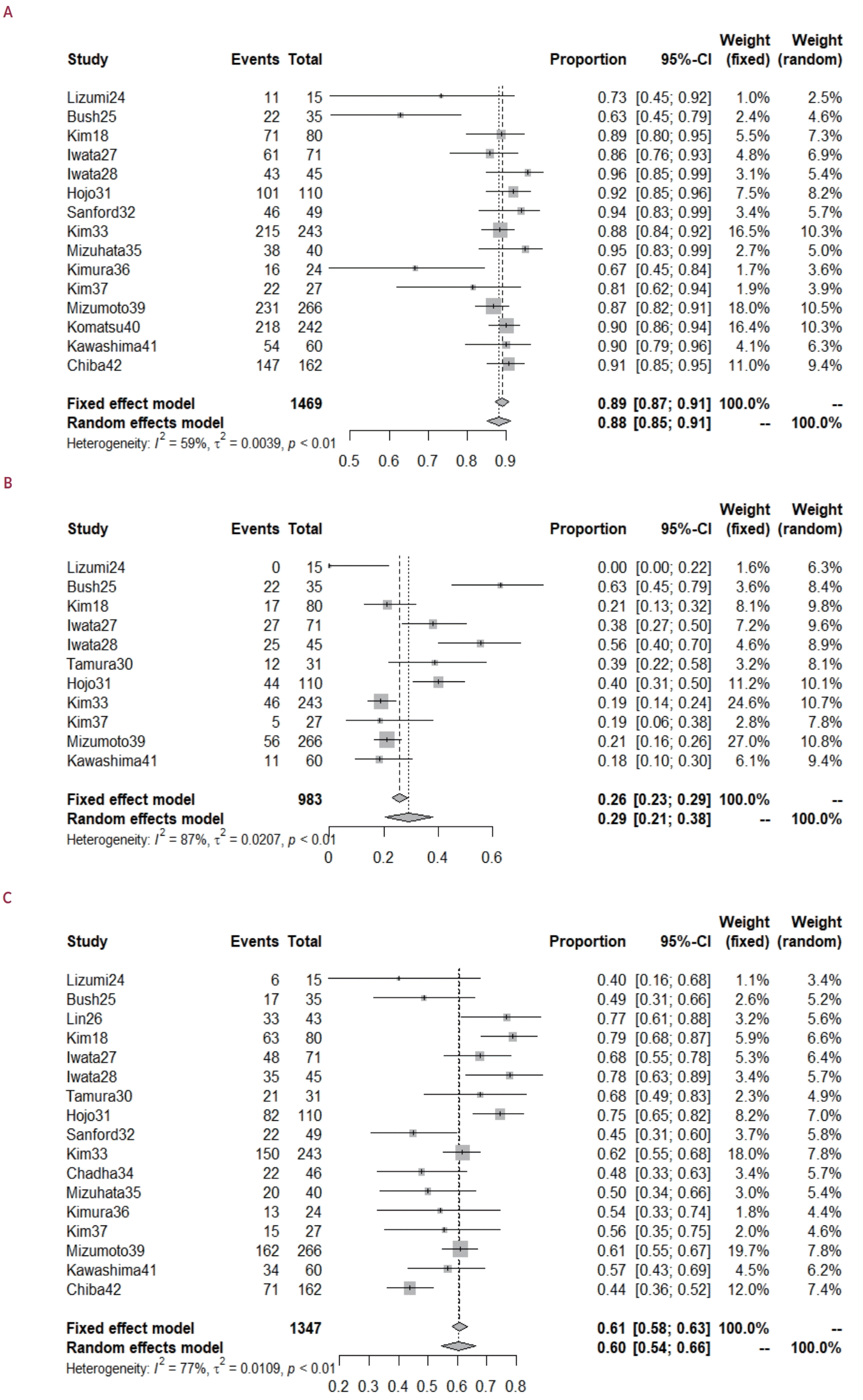
The median follow-up period was 13-56 months (median, 31). The median 3- and 5-year local progression-free survival (LPFS) rates were 89% (range, 62-95) and 88% (range, 64-94), respectively. The median 3- and 5-year progression-free survival (PFS) rates were 21% (range, 0-63) and 19% (range, 0-56), respectively. The median 3- and 5-year overall survival (OS) rates were 56% (range, 40-79) and 45% (range, 23-70), respectively. Using random effects analysis, the pooled 3- and 5-year LPFS rates were 88% (95% confidence interval [CI], 85-91) and 86% (95% CI, 82-90), respectively (Table 3, Fig. 2). The pooled 3- and 5-year OS rates were 60% (95% CI, 54-66) and 46% (95% CI, 38-54), respectively. Significant heterogeneity in survival estimates was present among the studies; however, there was no publication bias, as shown in Table 3 and Supplementary Fig. 1.
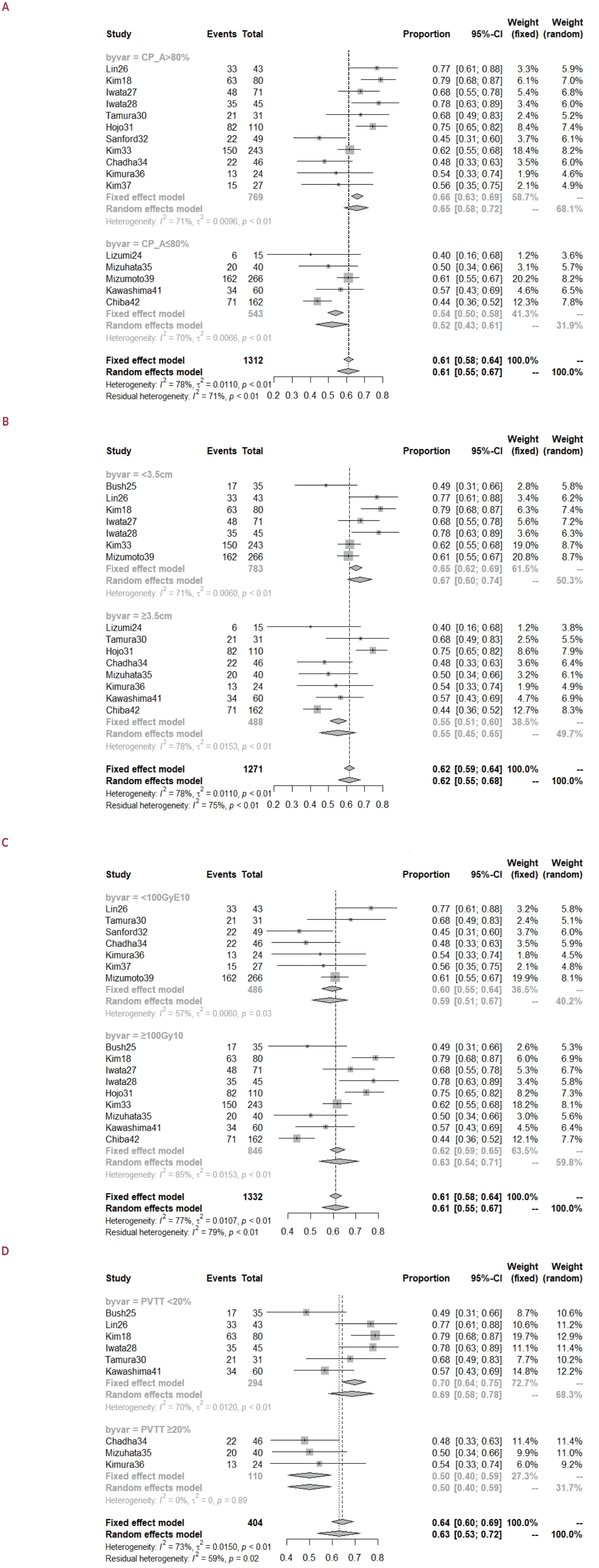
In the subgroup comparison, no significant factors affected the LPFS. The proportion of patients with CP-A >80% was the only significant favorable factor for PFS (Supplementary Table 3). The proportion of patients with PVTT <20% was the only consistently favorable factor for the 1-5-year OS with statistical significance (Table 4, Fig. 3). The proportion of patients with CP-A >80% was a significantly favorable factor for the 3- and 5-year OS. The median tumor size <3.5 cm and median BED >100 GyE10 were statistically favorable factors for the 1-year OS; however, statistical significance was not maintained for the 5-year OS.

Toxicities

Table 5 reveals the overall incidence of severe toxicities after PBT for HCC: hepatic toxicity ≥grade 3 occurred in 0-13%; classic RILD in 0-18%; non-classic RILD in 0-13%; GI toxicity ≥grade 3 in 0-7%; and skin toxicity ≥grade 3 in 0-8%, respectively. The pooled rates of hepatic toxicity, classic RILD, and non-classic RILD were 1% (95% CI, 0-2), 2% (95% CI, 0-5), and 1% (95% CI, 0-3), respectively (Supplementary Fig. 2).

DISCUSSION

To the best of our knowledge, this is the largest up-to-date systematic review and meta-analysis focusing on treatment outcomes after PBT for liver-confined HCC. Previous meta-analysis of CPT for HCC reported that the pooled rate of LPFS at the longest duration of follow-up was 86% (95% CI, 83-88), and the pooled rates of 3- and 5-year OS rates were 59% (95% CI, 51-66) and 37% (95% CI, 31-43), respectively.44 Our systematic review and meta-analysis reported that the pooled rates of LPFS and OS after PBT were 88% (95% CI, 85-91) and 60% (95% CI, 54-66) at 3 years and 86% (95% CI, 82-90) and 46% (95% CI, 38-54) at 5 years, respectively. The current meta-analysis, based on updated evidence, confirmed the efficacy of PBT with durable long-term local control and favorable survival.
Through a comprehensive systematic review, we found two randomized phase 3 trials which compared PBT with other LDT. The National Cancer Center in Korea conducted a noninferiority trial comparing PBT to RFA in patients with recurrent/residual HCC (tumor size <3 cm, number ≤2).18 Passive scattering PBT was applied with 66 GyE in 10 fractions. The primary endpoint was 2-year LPFS and the study met this endpoint with 95% in PBT (n=80) vs. 84% in RFA (n=56) (difference of 10.9%; 90% CI, 1.8-20.0), respectively. The 2-year OS rate (secondary endpoint) was 89% in PBT and 93% in RFA, respectively (hazard ratio [HR], 1.19; 95% CI, 0.62-2.27, P=0.60). Two recent retrospective studies comparing PBT with RFA for untreated HCC also showed no significant differences in treatment outcomes.45,46 These data support the idea that PBT can be an effective local treatment modality for small HCC lesions. The other phase 3 trial was launched by the Linda University Medical Center, comparing PBT to transarterial chemoembolization (TACE) in patients with newly diagnosed and previously untreated HCC meeting Milan or San Francisco transplant criteria.25 Passive scattering PBT was applied with 70.2 GyE in 15 fractions. The primary endpoint was 2-year OS, which was found to be 68% (n=36; 95% CI, 54-86) in PBT vs. 65% (n=40; 95% CI, 52-83) in TACE, respectively, indicating no major difference between the groups (P=0.80). However, LPFS (HR, 5.64; 95% CI, 1.78-17.9; P<0.05) and PFS (HR, 3.62; 95% CI, 1.62-8.05; P<0.05) as the secondary endpoints were significantly improved in the PBT arm and this result was consistent across multiple SBRT studies. The TRENDY, a randomized phase 2 trial comparing SBRT with TACE, showed that the median local control was 12 months for TACE and >40 months (not reached) for SBRT (P=0.08) and the median OS was 36.8 months for TACE vs. 44.1 months for SBRT (P=0.36).47 Another phase 3 trial comparing salvage SBRT after incomplete TACE vs. exclusive transarterial embolization (TAE)/TACE reported that the 1-year LPFS rate was 84% in the SBRT arm vs. 23% in TAE/TACE arm; however, the OS was not different.48 A few retrospective studies showed superior local control rates after SBRT compared with TACE but without survival benefit.49,50 These studies indicate that PBT might be an effective alternative to TACE for inoperable HCC, however, further prospective studies will be needed to confirm survival benefit.
Systemic therapy is the standard treatment for advanced HCC. The median OS in patients treated with first-line sorafenib, after the approval in 2007, was 10 to 12 months as demonstrated in several phase 3 trials.51,52 In 2018, lenvatinib showed non-inferiority to sorafenib in the REFLECT trial.53 In 2020, IMbrave150, a global phase 3 randomized trial, demonstrated that atezolizumab plus bevacizumab had a superior OS to sorafenib (median OS, 19.2 months; 1-year OS rate, 67% vs. median OS, 13.4 months; 1-year OS rate, 56%).54,55 Another phase 3 study, the HIMALAYA study showed the efficacy of tremelimumab plus durvalumab (median OS, 16.4 months; 3-year OS rate, 31%).56,57 The successful development of effective systemic treatments has improved OS for advanced HCC; however, the overall prognosis, especially in HCC with macrovascular invasion (MVI), remains unsatisfactory. One randomized trial comparing TACE plus RT against sorafenib for HCC patients with MVI demonstrated that the TACE-RT group had a higher 12-week PFS rate (87% vs. 34%, P<0.05), and longer OS (55 weeks vs. 43 weeks, P=0.04) compared to that with sorafenib.58 A multinational retrospective study in Asia reported that LDT (such as RFA, TACE, and hepatic arterial infusion chemotherapy) plus RT had longer OS than that with sorafenib (10.6 months vs. 4.2 months, P<0.05) in 1,035 HCC patients with PVTT.59 These studies on RT for HCC with PVTT, compared with the previously used regimen of the tyrosine kinase inhibitor (sorafenib), showed that the OS rates seem lower with novel combined regimens. However, RT could probably still be an effective LDT in these patients considering that the median OS in patients with MVI vs. Vp4 (main trunk and/or contralateral PV) PVTT from the IMbrave150 study were 14.2 months and 7.6 months despite of the use of atezolizumab plus bevacizumab.55,60 One retrospective study using PBT for HCC with PVTT reported a median OS of 13.2 months and 2-year OS rate of 33%, respectively.38 Another study on HCC with Vp3 (the first branch of the PV) or Vp4 PVTT reported a median OS of 22 months and 5-year OS rate of 21%, respectively.61 Our meta-analysis showed the pooled 3- and 5-year OS rates in group, where the proportion of patients with PVTT ≥20% were 50% (95% CI, 40-59) and 22% (95% CI, 11-37), respectively. Although PVTT leads to both intrahepatic and distant tumor spread, PVTT itself is a local disease, and PBT might play a role as a potent LDT in selected patients with PVTT. In addition, PBT can enhance the antitumor effects when combined with systemic treatment. Recently, a case report showed promising results; however, further clinical studies are required to confirm the efficacy of combined treatment for HCC with PVTT.62
Despite the increasing use of RT for HCC treatment, the optimal RT dose remains controversial. The American Society for Radiation Oncology clinical practice guideline recommends a wide range of fractionation regimens for HCC (BED of 59-180 Gy10 in 3-35 fractions).17 Recent expert consensus recommend a BED ≥80 Gy10 within ≤6 weeks to ablate HCC tumor.63 Kim et al.33 conducted risk-adaptive PBT using 3 fractionation regimens (50 GyE [BED, 75 GyE10] vs. 60 GyE [BED, 96 GyE10] vs. 66 GyE [BED, 109.6 GyE10] in 10 fractions) according to the proximity to the GI organ. Depending on the dose regimen, the 5-year LRFS and OS rates were 55%, 95%, and 92% (P<0.05), and 17%, 39%, and 68%, respectively (P<0.05). The BED ≥80 GyE10 was found to be a statistically favorable factor for both LPFS and OS. The Japanese Society for Radiation Oncology stipulated the PBT protocol for HCC based on tumor location.64 The recommended BED is about 91.2 GyE10 in 38 fractions (adjacent to GI organ), 96.6 GyE10 in 22 fractions (adjacent to porta hepatis) and 109.6 GyE10 in 10 fractions (peripheral). Their prospective registry study found that the local recurrence rates did not differ significantly according to the distance from the GI organ <1 vs. 1-2 cm (P=0.63); however, the rates were significantly lower for distances from the GI tract >2 cm vs. <1 cm (P<0.05).65 Similar results were revealed for the OS rates. The authors suggested that the local recurrence rates for tumors adjacent to luminal GI organs were high, most likely because of an insufficient tumor dose (<90 GyE10). The need for a high dose was supported by the University of Texas MD Anderson Cancer Center study that reported that a high dose (BED ≥90 GyE10) was an independent predictor of improved OS.34 The National Cancer Database study for T1-2N0 HCC patients receiving PBT or SBRT showed that both PBT (HR, 0.48; 95% CI, 0.29-0.78) and BED ≥100 Gy10 (HR, 0.61; 95% CI, 0.38-0.98) were independent predictors for longer survival rates.66 A retrospective study from Taiwan, using PBT or SBRT for HCC >5 cm, reported that BED ≥75 Gy10 independently predicted better LPFS (P=0.03) and PBT significantly correlated with superior LPFS, PFS, and OS outcomes (P<0.05).67 A systematic review on CPT for HCC described that dose escalation seems to improve survival at 1-3 years, while it does not influence survival at 5 years (median BED, 96.4 GyE10; range, 68.8-122.5).68 In line with these findings, the present meta-analysis shows durable long-term local control of 86% at 5-year, but with the cut-off point of median BED of 100 GyE10, high doses did not result in improved survival outcomes. This might be due to inclusion of studies which prescribed relatively higher PBT dose for a cure (median BED, 101.1 GyE10; range, 68.8-109.6). We presented the distribution of the median BED according to treatment outcomes in Supplementary Fig. 3. In the future, we expect that the optimal dose-fractionation regimens for PBT will be individualized using a personalized approach based on a panel of patient- and tumor-related predictive factors.
PBT has the potential to widen the therapeutic window by allowing ablative doses to be delivered to tumors while limiting exposure to the normal liver, thereby reducing the risk of hepatic toxicity.69 PBT has evolved with the introduction of the pencil-beam scanning method and respiratory and image guidance.1 These technologies provide highly conformal and precise planning of delivery and delivery of proton doses to liver tumors while sparing the surrounding normal liver tissue. Our meta-analysis showed that the pooled rate of hepatic toxicity was as low as ≤2% after PBT. Only a few studies have evaluated the possible differences in hepatic toxicity between proton and photon therapies. Sanford et al.32 compared PBT and photon RT in 133 patients with unresectable HCC. PBT decreased the risk of non-classic RILD (odds ratio, 0.26; P=0.03; 95% CI, 0.08-0.86). The development of non-classic RILD at 3 months was significantly associated with worse OS. The mean liver doses were similar between the groups. The authors suggested that the Bragg peak phenomenon inherent in PBT, which eliminates the low-dose bath distal to the target beam path, was more strongly associated with liver toxicity than the mean liver dose, which has historically been used to evaluate the photon-RT treatment plans. A multicenter study including eastern and western patients found that the unirradiated liver volume (the absolute liver volume receiving <1 GyE)/standard liver volume (the liver volume calculating body surface area), not mean liver dose, was an independent predictor of RILD regardless of countries.70 A study from Japan reported that classic RILD was not observed and non-classic RILD was not different between PBT and photon-SBRT for HCC ≤5 cm.23 Another study for large HCC >5 cm showed that classic RILD was uncommon and non-classic RILD had significantly lower incidence in PBT.67 When authors performed additional photon plans for the 105 patients who received PBT using same fractionation regimen, photon plans demonstrated significantly higher mean liver dose compared with actually delivered PBT plans, and 69% of them had mean liver dose exceeding 28 Gy, which necessitated the target dose de-escalation. An ongoing phase 3 trial (NCT03186898) comparing PBT and photon-RT is expected to provide high-level evidence supporting the use of PBT for HCC.
This study has several limitations. First, most studies were retrospective in nature, although two phase 3 randomized studies were included. In addition, the study designs, inclusion criteria, patient characteristics, fractionation regimens, and endpoint definitions were heterogeneous and inconsistently reported. This may affect the interpretation of the results. Second, PBT operates in a limited number of facilities worldwide. Some institutions reported consecutive cohorts, although we excluded studies with overlapping patients, which may have introduced a potential selection bias and limited the generalizability of the results. Lastly, we found several prognostic factors affecting survival outcomes in the subgroup analysis, but could not draw specific cut-off points to define the subgroup that benefited from PBT in HCC in the absence of individual patient data.
In conclusion, the current systematic review and meta-analysis of 22 studies comprising recently updated evidence demonstrates the efficacy and safety of PBT for HCC. One phase 3 trial supported PBT as a non-inferior local modality treatment compared to RFA for small HCC, with level 1 evidence. Another phase 3 trial failed to show a survival benefit of PBT compared to TACE for inoperable HCC, but suggested its efficacy as an LDT. An ongoing phase 3 trial comparing PBT with photon-RT will clarify the role of PBT over photon. Although the present meta-analysis demonstrates favorable local control of HCC and provides important information on prognostic factors and doses of PBT for HCC, there is a need for prospective studies in this area.

Notes

Conflicts of Interest

Sun Hyun Bae is an editorial board member of Journal of Liver Cancer, and was not involved in the review process of this article. Otherwise, the authors have no conflicts of interest to disclose.

Ethics Statement

Not applicable.

Funding Statement

This work was supported by a grant from the Korea Institute of Radiological and Medical Sciences (KIRAMS) funded by the Ministry of Science and ICT (MSIT), Republic of Korea (No. 50572-2024). This work was also supported by the Soonchunhyang University Research Fund. No funding bodies had any role in study design, data collection and analysis, decision to publish, or preparation of the manuscript.

Data Availability

Not applicable.

Author Contributions

Conceptualization: SHB, WIJ, MSK, HRM, BW, MH

Data curation: SHB, WIJ, MSK

Formal analysis: SHB

Supervision: MH

Writing - original draft: SHB, WIJ

Writing - review & editing: SHB, WIJ, MSK, HRM, BW, MH

All authors approved the final version of the manuscript.

References

1. Dionisi F, Scartoni D, Fracchiolla F, Giacomelli I, Siniscalchi B, Goanta L, et al. Proton therapy in the treatment of hepatocellular carcinoma. Front Oncol. 2022; 12:959552.
crossref
2. Mohan R, Grosshans D. Proton therapy - present and future. Adv Drug Deliv Rev. 2017; 109:26–44.
crossref
3. Wilson RR. Radiological use of fast protons. Radiology. 1946; 47:487–491.
crossref
4. Lawrence JH, Tobias CA, Born JL, McCombs RK, Roberts JE, Anger HO, et al. Pituitary irradiation with high-energy proton beams: a preliminary report. Cancer Res. 1958; 18:121–134.
5. Tsujii H, Tsuji H, Inada T, Maruhashi A, Hayakawa Y, Takada Y, et al. Clinical results of fractionated proton therapy. Int J Radiat Oncol Biol Phys. 1993; 25:49–60.
crossref
6. Particle Therapy Co-Operative Group (PTCOG). Facilities in operation [Internet]. PTCOG; [cited 2024 Apr 25]. Available from: https://www.ptcog.site/index.php/facilities-in-operation-public.
7. Sung H, Ferlay J, Siegel RL, Laversanne M, Soerjomataram I, Jemal A, et al. Global cancer statistics 2020: GLOBOCAN estimates of incidence and mortality worldwide for 36 cancers in 185 countries. CA Cancer J Clin. 2021; 71:209–249.
crossref
8. Cao G, Liu J, Liu M. Global, regional, and national trends in incidence and mortality of primary liver cancer and its underlying etiologies from 1990 to 2019: results from the global burden of disease study 2019. J Epidemiol Glob Health. 2023; 13:344–360.
crossref
9. Vitale A, Cabibbo G, Iavarone M, Viganò L, Pinato DJ, Ponziani FR, et al. Personalised management of patients with hepatocellular carcinoma: a multiparametric therapeutic hierarchy concept. Lancet Oncol. 2023; 24:e312–e322.
10. Reig M, Forner A, Rimola J, Ferrer-Fàbrega J, Burrel M, Garcia-Criado Á, et al. BCLC strategy for prognosis prediction and treatment recommendation: the 2022 update. J Hepatol. 2022; 76:681–693.
11. Singal AG, Llovet JM, Yarchoan M, Mehta N, Heimbach JK, Dawson LA, et al. AASLD practice guidance on prevention, diagnosis, and treatment of hepatocellular carcinoma. Hepatology. 2023; 78:1922–1965.
crossref
12. European Association for the Study of the Liver. EASL clinical practice guidelines: management of hepatocellular carcinoma. J Hepatol. 2018; 69:182–236.
13. Vogel A, Martinelli E; ESMO Guidelines Committee. Updated treatment recommendations for hepatocellular carcinoma (HCC) from the ESMO clinical practice guidelines. Ann Oncol. 2021; 32:801–805.
crossref
14. Korean Liver Cancer Association (KLCA); National Cancer Center (NCC) Korea. 2022 KLCA-NCC Korea practice guidelines for the management of hepatocellular carcinoma. J Liver Cancer. 2023; 23:1–120.
15. Hasegawa K, Takemura N, Yamashita T, Watadani T, Kaibori M, Kubo S, et al. Clinical practice guidelines for hepatocellular carcinoma: The Japan Society of Hepatology 2021 version (5th JSH-HCC guidelines). Hepatol Res. 2023; 53:383–390.
crossref
16. National Comprehensive Cancer Network (NCCN). NCCN guidelines version 1. 2014, hepatobiliary cancers [Internet]. Plymouth Meeting (US): NCCN; [cited 2024 Apr 25]. Available from: https://www.nccn.org/professionals/physician_gls/pdf/hcc.pdf.
17. Apisarnthanarax S, Barry A, Cao M, Czito B, DeMatteo R, Drinane M, et al. External beam radiation therapy for primary liver cancers: an ASTRO clinical practice guideline. Pract Radiat Oncol. 2022; 12:28–51.
crossref
18. Kim TH, Koh YH, Kim BH, Kim MJ, Lee JH, Park B, et al. Proton beam radiotherapy vs. radiofrequency ablation for recurrent hepatocellular carcinoma: a randomized phase III trial. J Hepatol. 2021; 74:603–612.
19. Page MJ, McKenzie JE, Bossuyt PM, Boutron I, Hoffmann TC, Mulrow CD, et al. The PRISMA 2020 statement: an updated guideline for reporting systematic reviews. BMJ. 2021; 372:n71.
20. Wells GA, Shea B, O’Connell D, Peterson J, Welch V, Losos M, et al. The Newcastle-Ottawa scale (NOS) for assessing the quality of nonrandomised studies in meta-analyses [Internet]. Ottawa (CA): Ottawa Hospital Research Institute; [cited 2021 Nov 22]. Available from: https://www.ohri.ca/programs/clinical_epidemiology/oxford.asp.
crossref
21. Raudenbush SW. Analyzing effect sizes: random-effects models. In : Cooper H, Hedges LV, Valentine JC, editors. The handbook of research synthesis and meta-analysis. 2nd ed. New York: Russell Sage Foundation;2009. p. 295–316.
22. Higgins JP, Thompson SG, Deeks JJ, Altman DG. Measuring inconsistency in meta-analyses. BMJ. 2003; 327:557–560.
crossref
23. Uchinami Y, Katoh N, Abo D, Morita R, Taguchi H, Fujita Y, et al. Study of hepatic toxicity in small liver tumors after photon or proton therapy based on factors predicting the benefits of proton. Br J Radiol. 2023; 96:20220720.
24. Iizumi T, Okumura T, Hasegawa N, Ishige K, Fukuda K, Seo E, et al. Proton beam therapy for hepatocellular carcinoma with bile duct invasion. BMC Gastroenterol. 2023; 23:267.
25. Bush DA, Volk M, Smith JC, Reeves ME, Sanghvi S, Slater JD, et al. Proton beam radiotherapy versus transarterial chemoembolization for hepatocellular carcinoma: results of a randomized clinical trial. Cancer. 2023; 129:3554–3563.
26. Lin SY, Chen CM, Huang BS, Lai YC, Pan KT, Lin SM, et al. A preliminary study of hepatocellular carcinoma post proton beam therapy using MRI as an early prediction of treatment effectiveness. PLoS One. 2021; 16:e0249003.
27. Iwata H, Ogino H, Hattori Y, Nakajima K, Nomura K, Hayashi K, et al. Image-guided proton therapy for elderly patients with hepatocellular carcinoma: high local control and quality of life preservation. Cancers (Basel). 2021; 13:219.
28. Iwata H, Ogino H, Hattori Y, Nakajima K, Nomura K, Hashimoto S, et al. A phase 2 study of image-guided proton therapy for operable or ablation-treatable primary hepatocellular carcinoma. Int J Radiat Oncol Biol Phys. 2021; 111:117–126.
29. Yoo GS, Yu JI, Park HC, Hyun D, Jeong WK, Lim HY, et al. Do biliary complications after proton beam therapy for perihilar hepatocellular carcinoma matter? Cancers (Basel). 2020; 12:2395.
30. Tamura S, Okamura Y, Sugiura T, Ito T, Yamamoto Y, Ashida R, et al. A comparison of the outcomes between surgical resection and proton beam therapy for single primary hepatocellular carcinoma. Surg Today. 2020; 50:369–378.
crossref
31. Hojo H, Raturi V, Nakamura N, Arahira S, Akita T, Mitsunaga S, et al. Impact of proton beam irradiation of an anatomic subsegment of the liver for hepatocellular carcinoma. Pract Radiat Oncol. 2020; 10:e264–e271.
crossref
32. Sanford NN, Pursley J, Noe B, Yeap BY, Goyal L, Clark JW, et al. Protons versus photons for unresectable hepatocellular carcinoma: liver decompensation and overall survival. Int J Radiat Oncol Biol Phys. 2019; 105:64–72.
crossref
33. Kim TH, Park JW, Kim BH, Kim H, Moon SH, Kim SS, et al. Does riskadapted proton beam therapy have a role as a complementary or alternative therapeutic option for hepatocellular carcinoma? Cancers (Basel). 2019; 11:230.
34. Chadha AS, Gunther JR, Hsieh CE, Aliru M, Mahadevan LS, Venkatesulu BP, et al. Proton beam therapy outcomes for localized unresectable hepatocellular carcinoma. Radiother Oncol. 2019; 133:54–61.
crossref
35. Mizuhata M, Takamatsu S, Shibata S, Bou S, Sato Y, Kawamura M, et al. Respiratory-gated proton beam therapy for hepatocellular carcinoma adjacent to the gastrointestinal tract without fiducial markers. Cancers (Basel). 2018; 10:58.
crossref
36. Kimura K, Nakamura T, Ono T, Azami Y, Suzuki M, Wada H, et al. Clinical results of proton beam therapy for hepatocellular carcinoma over 5 cm. Hepatol Res. 2017; 47:1368–1374.
crossref
37. Kim TH, Park JW, Kim YJ, Kim BH, Woo SM, Moon SH, et al. Phase I dose-escalation study of proton beam therapy for inoperable hepatocellular carcinoma. Cancer Res Treat. 2015; 47:34–45.
crossref
38. Lee SU, Park JW, Kim TH, Kim YJ, Woo SM, Koh YH, et al. Effectiveness and safety of proton beam therapy for advanced hepatocellular carcinoma with portal vein tumor thrombosis. Strahlenther Onkol. 2014; 190:806–814.
39. Mizumoto M, Okumura T, Hashimoto T, Fukuda K, Oshiro Y, Fukumitsu N, et al. Proton beam therapy for hepatocellular carcinoma: a comparison of three treatment protocols. Int J Radiat Oncol Biol Phys. 2011; 81:1039–1045.
crossref
40. Komatsu S, Fukumoto T, Demizu Y, Miyawaki D, Terashima K, Sasaki R, et al. Clinical results and risk factors of proton and carbon ion therapy for hepatocellular carcinoma. Cancer. 2011; 117:4890–4904.
crossref
41. Kawashima M, Kohno R, Nakachi K, Nishio T, Mitsunaga S, Ikeda M, et al. Dose-volume histogram analysis of the safety of proton beam therapy for unresectable hepatocellular carcinoma. Int J Radiat Oncol Biol Phys. 2011; 79:1479–1486.
crossref
42. Chiba T, Tokuuye K, Matsuzaki Y, Sugahara S, Chuganji Y, Kagei K, et al. Proton beam therapy for hepatocellular carcinoma: a retrospective review of 162 patients. Clin Cancer Res. 2005; 11:3799–3805.
crossref
43. Bush DA, Hillebrand DJ, Slater JM, Slater JD. High-dose proton beam radiotherapy of hepatocellular carcinoma: preliminary results of a phase II trial. Gastroenterology. 2004; 127 Suppl 1:S189–S193.
44. Qi WX, Fu S, Zhang Q, Guo XM. Charged particle therapy versus photon therapy for patients with hepatocellular carcinoma: a systematic review and meta-analysis. Radiother Oncol. 2015; 114:289–295.
crossref
45. Sekino Y, Tateishi R, Fukumitsu N, Okumura T, Maruo K, Iizumi T, et al. Proton beam therapy versus radiofrequency ablation for patients with treatment-naïve single hepatocellular carcinoma: a propensity score analysis. Liver Cancer. 2022; 12:297–308.
crossref
46. Seo SH, Yu JI, Park HC, Yoo GS, Choi MS, Rhim H, et al. Proton beam radiotherapy as a curative alternative to radiofrequency ablation for newly diagnosed hepatocellular carcinoma. Anticancer Res. 2024; 44:2219–2230.
crossref
47. Romero AM, van der Holt B, Willemssen FEJA, de Man RA, Heijmen BJM, Habraken S, et al. Transarterial chemoembolization with drugeluting beads versus stereotactic body radiation therapy for hepatocellular carcinoma: outcomes from a multicenter, randomized, phase 2 trial (the TRENDY trial). Int J Radiat Oncol Biol Phys. 2023; 117:45–52.
crossref
48. Comito T, Loi M, Franzese C, Clerici E, Franceschini D, Badalamenti M, et al. Stereotactic radiotherapy after incomplete transarterial (chemo-) embolization (TAE/TACE) versus exclusive TAE or TACE for treatment of inoperable HCC: a phase III trial (NCT02323360). Curr Oncol. 2022; 29:8802–8813.
49. Sapir E, Tao Y, Schipper MJ, Bazzi L, Novelli PM, Devlin P, et al. Stereotactic body radiation therapy as an alternative to transarterial chemoembolization for hepatocellular carcinoma. Int J Radiat Oncol Biol Phys. 2018; 100:122–130.
crossref
50. Su TS, Liang P, Zhou Y, Huang Y, Cheng T, Qu S, et al. Stereotactic body radiation therapy vs. transarterial chemoembolization in inoperable Barcelona Clinic Liver Cancer stage A hepatocellular carcinoma: a retrospective, propensity-matched analysis. Front Oncol. 2020; 10:347.
crossref
51. Llovet JM, Ricci S, Mazzaferro V, Hilgard P, Gane E, Blanc JF, et al. Sorafenib in advanced hepatocellular carcinoma. N Engl J Med. 2008; 359:378–390.
crossref
52. Cheng AL, Kang YK, Chen Z, Tsao CJ, Qin S, Kim JS, et al. Efficacy and safety of sorafenib in patients in the Asia-Pacific region with advanced hepatocellular carcinoma: a phase III randomised, double-blind, placebo-controlled trial. Lancet Oncol. 2009; 10:25–34.
crossref
53. Kudo M, Finn RS, Qin S, Han KH, Ikeda K, Piscaglia F, et al. Lenvatinib versus sorafenib in first-line treatment of patients with unresectable hepatocellular carcinoma: a randomised phase 3 non-inferiority trial. Lancet. 2018; 391:1163–1173.
54. Finn RS, Qin S, Ikeda M, Galle PR, Ducreux M, Kim TY, et al. Atezolizumab plus bevacizumab in unresectable hepatocellular carcinoma. N Engl J Med. 2020; 382:1894–1905.
crossref
55. Cheng AL, Qin S, Ikeda M, Galle PR, Ducreux M, Kim TY, et al. Updated efficacy and safety data from IMbrave150: atezolizumab plus bevacizumab vs. sorafenib for unresectable hepatocellular carcinoma. J Hepatol. 2022; 76:862–873.
crossref
56. Abou-Alfa GK, Lau G, Kudo M, Chan SL, Kelley RK, Furuse J, et al. Tremelimumab plus durvalumab in unresectable hepatocellular carcinoma. NEJM Evid. 2022; 1:EVIDoa2100070.
crossref
57. Sangro B, Chan SL, Kelley RK, Lau G, Kudo M, Sukeepaisarnjaroen W, et al. Four-year overall survival update from the phase III HIMALAYA study of tremelimumab plus durvalumab in unresectable hepatocellular carcinoma. Ann Oncol. 2024; 35:448–457.
58. Yoon SM, Ryoo BY, Lee SJ, Kim JH, Shin JH, An JH, et al. Efficacy and safety of transarterial chemoembolization plus external beam radiotherapy vs sorafenib in hepatocellular carcinoma with macroscopic vascular invasion: a randomized clinical trial. JAMA Oncol. 2018; 4:661–669.
59. Kim J, Cheng JC, Nam TK, Kim JH, Jang BK, Huang WY, et al. Efficacy of liver-directed combined radiotherapy in locally advanced hepatocellular carcinoma with portal vein tumor thrombosis. Cancers (Basel). 2023; 15:3164.
crossref
60. Breder VV, Vogel A, Merle P, Finn RS, Galle PR, Zhu AX, et al. IMbrave150: exploratory efficacy and safety results of hepatocellular carcinoma (HCC) patients (pts) with main trunk and/or contralateral portal vein invasion (Vp4) treated with atezolizumab (atezo) + bevacizumab (bev) versus sorafenib (sor) in a global Ph III study. J Clin Oncol. 2021; 39 Suppl 15:4073.
crossref
61. Sugahara S, Nakayama H, Fukuda K, Mizumoto M, Tokita M, Abei M, et al. Proton-beam therapy for hepatocellular carcinoma associated with portal vein tumor thrombosis. Strahlenther Onkol. 2009; 185:782–788.
crossref
62. Komatsu S, Terashima K, Ishihara N, Matsuo Y, Kido M, Yanagimoto H, et al. Novel concept of “sequential particle radiotherapy” with atezolizumab plus bevacizumab for hepatocellular carcinoma with portal vein tumor thrombus. Surg Today. 2024; 54:972–976.
crossref
63. Yanagihara TK, Tepper JE, Moon AM, Barry A, Molla M, Seong J, et al. Defining minimum treatment parameters of ablative radiation therapy in patients with hepatocellular carcinoma: an expert consensus. Pract Radiat Oncol. 2024; 14:134–145.
64. Japanese Society for Radiation Oncology (JASTRO). English translation of JASTRO treatment policy of proton beam therapy. Ver 1.0 at 2016 May [Internet]. Tokyo (JP): JASTRO; [cited 2023 Oct 23]. Available from: https://www.jastro.or.jp/en/news/proton_guideline_jastro_7_13_2017-2_cmarkandwatermark.pdf.
65. Mizumoto M, Ogino H, Okumura T, Terashima K, Murakami M, Ogino T, et al. Proton beam therapy for hepatocellular carcinoma: multicenter prospective registry study in Japan. Int J Radiat Oncol Biol Phys. 2024; 118:725–733.
crossref
66. Hasan S, Abel S, Verma V, Webster P, Arscott WT, Wegner RE, et al. Proton beam therapy versus stereotactic body radiotherapy for hepatocellular carcinoma: practice patterns, outcomes, and the effect of biologically effective dose escalation. J Gastrointest Oncol. 2019; 10:999–1009.
crossref
67. Hsieh RC, Lee CH, Huang HC, Wu SW, Chou CY, Hung SP, et al. Clinical and dosimetric results of proton or photon radiation therapy for large (>5 cm) hepatocellular carcinoma: a retrospective analysis. Int J Radiat Oncol Biol Phys. 2024; 118:712–724.
crossref
68. Spychalski P, Kobiela J, Antoszewska M, Błażyńska-Spychalska A, Jereczek-Fossa BA, Høyer M. Patient specific outcomes of charged particle therapy for hepatocellular carcinoma - a systematic review and quantitative analysis. Radiother Oncol. 2019; 132:127–134.
crossref
69. Zaki P, Chuong MD, Schaub SK, Lo SS, Ibrahim M, Apisarnthanarax S. Proton beam therapy and photon-based magnetic resonance imageguided radiation therapy: the next frontiers of radiation therapy for hepatocellular carcinoma. Technol Cancer Res Treat. 2023; 22:15330338231206335.
70. Hsieh CE, Venkatesulu BP, Lee CH, Hung SP, Wong PF, Aithala SP, et al. Predictors of radiation-induced liver disease in eastern and western patients with hepatocellular carcinoma undergoing proton beam therapy. Int J Radiat Oncol Biol Phys. 2019; 105:73–86.
crossref

Figure 1.
Preferred reporting items for systematic reviews and meta-analyses (PRISMA) diagram of study selection process. RT, radiotherapy.
jlc-2024-06-26f1.tif
Figure 2.
Forest plot of survivals. (A) Three-year local progression-free survival. (B) Three-year progression-free survival. (C) Three-year overall survival. CI, confidence interval.
jlc-2024-06-26f2.tif
Figure 3.
Forest plot of 3-year overall survival on subgroup analysis. (A) The proportion of Child-Pugh (CP) A. (B) Median tumor size. (C) Median total dose of biologically equivalent dose (GyE10). (D) The proportion of portal vein tumor thrombosis (PVTT). CI, confidence interval.
jlc-2024-06-26f3.tif
jlc-2024-06-26f4.tif
Table 1.
Study details for hepatocellular carcinoma treated with proton beam therapy
Study Nation Study type Study period Number of patients Age (years) ECOG PS (%)
Hepatitis (%)
CP class (%)
BCLC stage (%)
Tumor size (cm) Multiplicity (%) PVTT (%)
0 1 2 3 HBV HCV Alcoholic A B C 0 A B C D
Uchinami et al., 202323 Japan R/SC/CT 2015-2021 41 69 (44-88) NR 22 27 NR 100 0 0 NR 2.6 (1.9-3.6) 27 NR
Lizumi et al., 202324 Japan R/SC/ST 2009-2018 15 71 (58-90) 40 60 13 53 NR 53 47 NR 4.0 (1.5-8.0) 27 NR
Bush et al., 202325 USA P3/MC/CT 2008-2017 35 62 (53-70)* 3 65 32 0 0 69 34 NR 0 86 11 3 0 3.3 (2.0-4.5) 17 0
Lin et al., 202126 Taiwan R/SC/ST 2014-2017 43 71 (48-85) 51 44 5 0 53 35 5 93 7 0 NR 3.1 (1.1-17.1) 23 19
Kim et al., 202118 Korea P3/SC/CT 2013-2017 80 61 (40-82) 91 9 0 0 84 NR NR 96 4 0 6 54 35 5 0 1.2 (1.0-2.9) 6 0
Iwata et al., 202127 Japan R/SC/ST 2013-2019 71 82 (80-96) 62 28 6 4 10 39 7 90 10 0 10 63 2 21 4 3.2 (0.8-11.1) NR NR
Iwata et al., 202128 Japan P2/SC/ST 2013-2016 45 68 (36-80) 93 7 0 0 35 29 7 91 9 0 27 67 0 6 0 2.5 (1.0-10.0) 0 0
Yoo et al., 202029 Korea R/SC/ST 2016-2017 167 62 (35-91) 55 45 0 78 9 5 89 9 2 66 34 NR NR NR
Tamura et al., 202030 Japan R/SC/CT 2003-2017 31 72 (51-84) 65 32 3 0 13 58 NR 94 6 0 NR 3.5 (1.0-9.0) 0 0
Hojo et al., 202031 Japan R/SC/ST 2008-2015 110 74 (48-90) 65 35 0 25 45 NR 86 14 0 NR 4.3 (0.8-15.0) 23 NR
Sanford et al., 201932 USA R/SC/CT 2008-2017 49 65 (60-74) 47 49 4 12 49 NR 83 17 0 NR NR 49 NR
Kim et al., 201933 Korea R/SC/ST 2012-2017 243 61 (24-92) 98 2 0 0 77 8 7 94 6 0 0 40 35 25 0 2.2 (1.0-17.0) NR NR
Chadha et al., 201934 USA R/SC/ST 2007-2016 46 72 (52-90) 44 54 2 0 4 28 52 83 17 0 NR 6.0 (1.5-21.0) 22 20
Mizuhata et al., 201835 Japan R/SC/ST 2011-2015 40 72 (38-87) 95 5 0 12 38 25 70 30 0 NR 3.7 (1.1-12.4) 75 30
Kimura et al., 201736 Japan R/SC/ST 2008-2015 24 73 (49-89) 67 33 0 0 29 25 17 100 0 0 NR 9.0 (5.0-18.0) 0 83
Kim et al., 201537 Korea P1/SC/ST 2007-2010 27 NR (51-78) 78 22 0 0 56 30 7 89 11 0 0 48 37 15 0 NR (1.3-7.0) NR NR
Lee et al., 201438 Korea R/SC/ST 2008-2011 27 55 (42-70) 67 33 0 0 74 15 NR 67 33 0 0 0 0 100 0 7.0 (3.0-16.0) NR 100
Mizumoto et al., 201139 Japan R/SC/ST 2001-2007 266 70 (26-88) 60 38 2 0 12 75 NR 76 23 1 NR 3.4 (0.6-13.0) 53 NR
Komatsu et al., 201140 Japan R/SC/CT 2001-2009 242 NR 71 24 4 1 12 67 NR 76 23 1 4 34 13 47 2 NR 12 NR
Kawashima et al., 201141 Japan R/SC/ST 1999-2007 60 70 (48-92) 95 5 0 7 83 NR 78 22 0 NR 4.5 (2.0-9.0) 15 2
Chiba et al., 200542 Japan R/SC/ST 1985-1998 162 63 (41-84) 37 49 13 1 11 81 1 56 38 6 NR 3.8 (1.5-14.5) 51 NR
Bush et al., 200443 USA P2/SC/ST 1998-2003 34 65 (47-86) NR NR 41 41 NR 5.6 (1.5-10.0) 6 NR

ECOG PS, Eastern Cooperative Oncology Group performance status; HBV, hepatitis B virus; HCV, hepatitis C virus; CP, Child-Pugh; BCLC, Barcelona Clinic Liver Cancer Stage; PVTT, portal vein tumor thrombosis; R, retrospective study; SC, single center; CT, comparative trial; NR, not reported; ST, single-arm trial; P3, prospective phase 3 study; MC, multicenter; P2, prospective phase 2 study; P1, prospective phase 1 study.

* Mean value (standard deviation);

Median (interquartile range);

Mean value.

Table 2.
Treatment outcomes for hepatocellular carcinoma treated with proton beam therapy
Study Previous treatment (%) Median total dose (GyE) Number of fx Median BED (GyE10) Median fu (months) LPFS (%)
PFS (%)
OS (%)
1-yr 3-yr 5-yr 1-yr 3-yr 5-yr 1-yr 3-yr 5-yr
Uchinami et al., 202323 - 72.6 (66.0-76.0) 10-22 99.0 - - - - - - - - - -
Iizumi et al., 202324 53 - - - 23 (8-54) 93 75 - 73 0 - 80 40 -
Bush et al., 202325 0 70.2 15 103.1 30 89 62 - 76 63 - 81 49 42
Lin et al., 202126 84 72.6 (66.0-72.6) 10-22 96.6 40 (9-62) - - 93 74 - 56 88 76 63
Kim et al., 202118 95 66.0 10 109.6 52 (46-60) 95 88 75 54 21 11 98 79 65
Iwata et al., 202127 35 66.0 (66.0 or 72.6) 10 or 22 109.6 33 (9-68) 97 86 70 72 38 25 93 68 38
Iwata et al., 202128 - 66.0 (66.0 or 72.6) 10 or 22 109.6 53 (10-75) 99 95 92 83 55 40 98 79 70
Yoo et al., 202029 - - - - 14 (1-29) 93 - - - - - 95 - -
Tamura et al., 202030 - 73.5 (47.6-78.4) 10-38 99.2 56 (22-82) - - - 69 39 31 92 69 51
Hojo et al., 202031 - 76.0 20 104.9 37 (1-100) 97 92 89 70 40 28 94 74 69
Sanford et al., 201932 24 58.1 (30.0-67.5) 5-16 80.5 14 - 93 93 - - - 69 44 -
Kim et al., 201933 79 66.0 (50.0, 60.0, or 66.0) 10 109.6 32 (2-68) - 89 88 - 19 12 - 62 48
Chadha et al., 201934 54 67.5 (24.0-91.0) 6-25 97.9 15 (1-60) 95 - - 74 - - 73 47 -
Mizuhata et al., 201835 90 76.0 20-38 104.9 20 (1-72) 100 94 94 70 - - 86 51 23
Kimura et al., 201736 - 72.6 (60.8-85.8) 10-35 96.6 18 (3-64) 95 66 - - - - 70 52 -
Kim et al., 201537 96 66.0 (60.0, 66.0, or 72.0) 20-24 85.8 31 (5-63) - 80 64 - 17 0 - 56 42
Lee et al., 201438 78 55.0 (50.0-66.0) 20-22 68.8 13 (2-52) 71 - - 11 - - 56 - -
Mizumoto et al., 201139 63 72.6 (66.0, 72.6, or 77.0) 10-35 96.6 - 98 87 81 56 21 12 87 61 48
Komatsu et al., 201140 47 66.0 (52.8-84.0) 4-38 109.6 31 - 90 90 - - - - - 38
Kawashima et al., 201141 60 76.0 (60.0, 65.0, or 76.0) 10-26 104.9 - 97 90 86 -18 4 83 56 25
Chiba et al., 200542 72 72.0 (50.0-84.0) 10-24 104.4 32 (3-133) 97 91 87 - - - 81 44 24
Bush et al., 200443 - 63.0 15 89.5 20 - - - - - - - - -

fx, fraction; median BED, median total dose of biologically effective dose, using a linear quadratic model with alpha/beta ratios of 10 for tumor; fu, follow-up; yr, years; LPFS, local progression-free survival; PFS, progression-free survival; OS, overall survival.

Table 3.
Pooled rates of treatment outcomes
Group Cohorts Patients heterogeneity* I2 (%) Egger’s test* Fixed event rate Random event rate
LPFS
 1-year LPFS 14 1,148 0.0031 58.34 0.0993 0.97 (0.95-0.98) 0.96 (0.94-0.98)
 3-year LPFS 15 1,469 0.0019 59.15 0.1683 0.89 (0.87-0.91) 0.88 (0.85-0.91)
 5-year LPFS 13 1,438 <0.0001 73.78 0.8187 0.86 (0.84-0.88) 0.86 (0.82-0.90)
PFS
 1-year PFS 12 809 <0.0001 82.91 0.5067 0.64 (0.60-0.67) 0.65 (0.56-0.74)
 3-year PFS 11 983 <0.0001 87.22 0.3211 0.26 (0.23-0.29) 0.29 (0.21-0.38)
 5-year PFS 10 976 <0.0001 90.22 0.3193 0.16 (0.14-0.18) 0.19 (0.11-0.28)
OS
 1-year OS 17 1,271 <0.0001 81.02 0.1802 0.88 (0.87-0.90) 0.86 (0.81-0.91)
 3-year OS 17 1,347 <0.0001 76.74 0.8238 0.61 (0.58-0.63) 0.60 (0.54-0.66)
 5-year OS 14 1,455 <0.0001 89.09 0.7089 0.45 (0.42-0.47) 0.46 (0.38-0.54)

Values are presented as number (95% confidence interval).

LPFS, local progression-free survival; PFS, progression-free survival; OS, overall survival.

* P-value.

Table 4.
Subgroup meta-analysis affecting overall survival
Group Cohorts Patients heterogeneity* I2 (%) Egger’s test* Fixed event rate Random event rate P-value between groups
1-year
 CP_A >80% 10 666 <0.0001 81.49 0.0694 0.92 (0.90-0.94) 0.90 (0.83-0.95) 0.0536
 CP_A ≤80% 6 570 0.0152 64.45 0.1765 0.84 (0.81-0.87) 0.81 (0.74-0.87)
 mSize <3.5 cm 6 540 0.0037 71.35 0.5529 0.91 (0.88-0.93) 0.92 (0.86-0.96) 0.0264
 mSize ≥3.5 cm 9 515 <0.0001 75.82 0.2988 0.84 (0.81-0.87) 0.82 (0.74-0.89)
 Median BED ≥100 GyE10 8 603 <0.0001 78.26 0.7585 0.90 (0.87-0.92) 0.90 (0.84-0.95) 0.0279
 Median BED <100 GyE10 7 486 0.0002 77.45 0.1681 0.83 (0.79-0.86) 0.79 (0.69-0.87)
 PVTT <20% 6 294 0.0054 69.80 0.4145 0.92 (0.89-0.95) 0.91 (0.84-0.97) 0.0039
 PVTT ≥20% 4 137 0.0740 56.73 0.3961 0.74 (0.66-0.81) 0.73 (0.60-0.84)
3-year
 CP_A >80% 11 769 0.0001 71.44 0.7695 0.66 (0.63-0.69) 0.65 (0.58-0.72) 0.0216
 CP_A ≤80% 5 543 0.0095 70.12 0.4854 0.54 (0.50-0.58) 0.52 (0.43-0.61)
 mSize <3.5 cm 7 783 0.0024 70.55 0.2785 0.65 (0.62-0.69) 0.67 (0.60-0.74) 0.0488
 mSize ≥3.5 cm 8 488 <0.0001 77.54 0.9648 0.55 (0.51-0.60) 0.55 (0.45-0.65)
 Median BED ≥100 GyE10 9 846 <0.0001 84.51 0.7053 0.62 (0.59-0.65) 0.63 (0.54-0.71) 0.4894
 Median BED <100 GyE10 7 486 0.0314 56.67 0.7397 0.60 (0.55-0.64) 0.59 (0.51-0.67)
 PVTT <20% 6 294 0.0052 69.95 0.4491 0.70 (0.64-0.75) 0.69 (0.58-0.78) 0.0095
 PVTT ≥20% 3 110 0.8862 0.00 0.1256 0.50 (0.40-0.59) 0.50 (0.40-0.59)
5-year
 CP_A >80% 8 650 <0.0001 78.98 0.6883 0.55 (0.51-0.59) 0.56 (0.47-0.65) 0.0009
 CP_A ≤80% 5 770 <0.0001 88.58 0.2195 0.36 (0.33-0.40) 0.32 (0.22-0.43)
 mSize <3.5 cm 7 783 0.0006 74.51 0.3558 0.51 (0.47-0.54) 0.53 (0.45-0.61) 0.1924
 mSize ≥3.5 cm 5 403 <0.0001 94.36 0.9484 0.38 (0.33-0.42) 0.38 (0.18-0.60)
 Median BED ≥100 GyE10 10 1,088 <0.0001 91.87 0.7846 0.43 (0.40-0.46) 0.44 (0.33-0.55) 0.3464
 Median BED <100 GyE10 4 367 0.2563 25.89 0.7088 0.50 (0.44-0.55) 0.50 (0.43-0.58)
 PVTT <20% 6 294 <0.0001 85.19 0.8998 0.53 (0.48-0.59) 0.53 (0.38-0.68) 0.0043
 PVTT ≥20% 1 40 1.0000 - - 0.22 (0.11-0.37) 0.22 (0.11-0.37)

Values are presented as number (95% confidence interval).

CP, Child-Pugh; mSize, median tumor size; median BED, median total dose of biologically effective dose using a linear quadratic model with alpha/beta ratios of 10 for tumor; PVTT, portal vein tumor thrombosis.

* P-value;

The proportion of patients with CP class of A was >80% among the whole patients;

The proportion of patients with PVTT was <20% among the whole patients.

Table 5.
Severe toxicity after proton beam therapy for hepatocellular carcinoma
Study Toxicity criteria Hepatic toxicity ≥Gr 3 (%) Classic RILD (%) Non-classic RILD (%) GI toxicity ≥Gr 3 (%) Skin toxicity ≥Gr 3 (%)
Uchinami et al., 202323 CTCAE - 0 5 - -
Iizumi et al., 202324 CTCAE 4.0 7 - - 7 0
Bush et al., 202325 CTCAE 5.0 11 - - 6 0
Lin et al., 202126 CTCAE 4.0 0 - 0 0 2
Kim et al., 202118 CTCAE 3.0 0 0 0 0 0
Iwata et al., 202127 CTCAE 4.0 0 0 0 0 1
Iwata et al., 202128 CTCAE v4.0 0 2 0 0 0
Yoo et al., 202029 CTCAE 5.0 4 6 1 1 0
Tamura et al., 202030 CTCAE 5.0 0 - - 3 -
Hojo et al., 202031 CTCAE 4.0 2 - 4 - -
Sanford et al., 201932 Not specify - - 8 - -
Kim et al., 201933 CTCAE v4.0 0 - 0 0 -
Chadha et al., 201934 CTCAE v4.03 13 0 9 2 2
Mizuhata et al., 201835 CTCAE v4.0 3 - - 3 0
Kimura et al., 201736 CTCAE v4.0 0 - 13 0 8
Kim et al., 201537 CTCAE v3.0 0 0 0 0 0
Lee et al., 201438 CTCAE v3.0 0 0 0 0 -
Mizumoto et al., 201139 CTCAE v3.0 + RTOG/EORTC 0 - - 1 1
Komatsu et al., 201140 CTCAE v2.0 1 2 - 0 2
Kawashima et al., 201141 CTCAE v3.0 - 18 - 3 -
Chiba et al., 200542 RTOG/EORTC - 11 - 1 -
Bush et al., 200443 CTCAE v2.0 - 0 - 0 0
Pooled rates
Group Cohorts Patients I2 (%) I2 (%) Egger’s test* Fixed event rate (95% CI) Random event rate (95% CI)
Hepatic toxicity ≥Gr 3 17 1,512 <0.0001 67.12 0.0898 0.00 (0.00-0.01) 0.01 (0.00-0.02)
Classic RILD 12 999 <0.0001 80.22 0.3300 0.03 (0.02-0.04) 0.02 (0.00-0.05)
Non-classic RILD 13 970 0.0002 67.62 0.0709 0.01 (0.00-0.01) 0.01 (0.00-0.03)
GI toxicity ≥Gr 3 19 1,658 0.6106 0.00 0.0477 0.00 (0.00-0.01) 0.00 (0.00-0.01)
Skin toxicity ≥Gr 3 14 1,135 0.4488 0.00 0.6103 0.00 (0.00-0.01) 0.00 (0.00-0.01)

Gr, grade; RILD, radiation-induced liver disease; GI, gastrointestinal; CTCAE, common terminology criteria for adverse events; RTOG/EORTC, Radiation Therapy Oncology Group/European Organization for Research and Treatment of Cancer; CI, confidence interval.

* P-value.

TOOLS
Similar articles