Journal List > J Liver Cancer > v.24(2) > 1516088663

Hwang, Chun, Chie, and Lee: Assessment of real-time US-CT/MR-guided percutaneous gold fiducial marker implementation in malignant hepatic tumors for stereotactic body radiation therapy

Abstract

Backgrounds/Aims

This study explored the initial institutional experience of using gold fiducial markers for stereotactic body radiotherapy (SBRT) in treating malignant hepatic tumors using real-time ultrasound-computed tomography (CT)/magnetic resonance (MR) imaging fusion-guided percutaneous placement.

Methods

From May 2021 to August 2023, 19 patients with 25 liver tumors that were invisible on pre-contrast CT received fiducial markers following these guidelines. Postprocedural scans were used to confirm their placement. We assessed technical and clinical success rates and monitored complications. The implantation of fiducial markers facilitating adequate treatment prior to SBRT, which was achieved in 96% of the cases (24 of 25 tumors), was considered technical success. Clinical success was the successful completion of SBRT without evidence of marker displacement and was achieved in 88% of cases (22 of 25 tumors). Complications included one major subcapsular hematoma and marker migration into the right atrium in two cases, which prevented SBRT.

Results

Among the treated tumors, 20 of 24 (83.3%) showed a complete response, three of 24 (12.5%) remained stable, and one of 24 (4.2%) progressed during an average 11.7-month follow-up (range, 2-32 months).

Conclusions

This study confirms that percutaneous gold fiducial marker placement using real-time CT/MR guidance is effective and safe for SBRT in hepatic tumors, but warns of marker migration risks, especially near the hepatic veins and in subcapsular locations. Using fewer markers than traditionally recommended-typically two per patient, the outcomes were still satisfactory, particularly given the increased risk of migration when markers were placed near major hepatic veins.

GRAPHICAL ABSTRACT

INTRODUCTION

Stereotactic body radiotherapy (SBRT) has emerged as an effective alternative, providing precise, high-dose radiation with minimal impact on adjacent tissues. Demonstrating local control rates over 80% of lesions,1-4 SBRT is increasingly recognized as a viable option for primary liver cancers and oligometastatic lesions, especially for small liver tumors located in areas difficult to address by ablation.5-7 Despite the lack of phase III studies, phase II studies and retrospective analyses cite SBRT’s promising outcomes in early-stage hepatocellular carcinoma (HCC), leading to its endorsement by the American Association for the Study of Liver Diseases (AASLD) and the Korean Association for the Study of the Liver-National Cancer Center (KASL-NCC) as a second-line treatment.8,9 Advancements in radiation therapy, notably in SBRT, have necessitated accurate tumor localization and tracking.10 Cone-beam computed tomography (CBCT) guidance is a valuable tool in SBRT for delivering precise radiation doses to targeted tumors while sparing the surrounding healthy tissue. However, several limitations associated with CBCT guidance in the context of SBRT, including limited soft tissue contrast and susceptibility to artifacts, such as scatter, beam hardening, and motion artifacts,11,12 hinder accurate delineation of the target tumor. The use of fiducial markers is crucial for overcoming these issues.13 These markers, which have a spherical appearance and are sized less than 2.0 mm in diameter, comprise 99.99% pure gold, and were chosen for their biocompatibility and radiopacity.14-16 Placed near the tumor, under image guidance from either computed tomography (CT)17 or ultrasound (US),18 fiducial markers act as effective surrogates for monitoring tumor position, enabling real-time tracking, and thereby enhancing the precision of radiation delivery.19 Moreover, their use has markedly reduced setup errors in liver SBRT, surpassing traditional methods such as uncorrected alignment, vertebral alignment, and three-dimensional diaphragm-based setups.20 The strategic placement of these markers prior to radiotherapy is instrumental in monitoring and compensating for respiratory motion.19 Although the recent introduction of magnetic resonance (MR)-guided SBRT has marked notable advancements in the field, offering superior imaging capabilities for enhanced tumor and organ-at-risk visualization, its accessibility remains limited.10
To further enhance precision, real-time CT and MR imaging (MRI)-US fusion guidance have emerged as significant advancements in fiducial marker placement. Traditional methods, such as B-mode US or CT guidance, although dependable, sometimes fail to achieve accurate lesion localization owing to limited lesion visibility or the lack of comprehensive real-time imaging capabilities.18,21,22 In contrast, the fusion of CT or MR with US effectively merges the high resolution and soft-tissue contrast of CT/MR with the real-time capabilities of US.23 This technique is particularly advantageous for dynamically changing anatomical regions such as the liver or pancreas and is crucial for accurately targeting tumors near vital structures5 or tumors that are poorly visible or inconspicuous on US.
This study aimed to evaluate the technical success and identify potential complications associated with the use of real-time CT/MR-US fusion guidance for placing gold fiducial markers, which is a critical step in SBRT planning for HCC treatment.

METHODS

Study population

From May 2021 to August 2023, 19 consecutive patients (14 men and five women; mean age, 67.9 years [range, 42-80]) with 25 hepatic tumors were referred to the department of radiology for fiducial marker implantation for SBRT. This retrospective analysis was approved by the Institutional Review Board (IRB) of Seoul National University Hospital (IRB No. 2403-029-1518). The requirement for informed consent was waived due to the retrospective nature of this study. The inclusion criteria were as follows: 1) patients aged between 20 and 85 years, 2) those with HCC, which was not discernible on pre-contrast CT and poorly depicted on US, 3) those who planned to receive curative-intent SBRT, and 4) those whose liver function was categorized as Child-Pugh class A or B. The exclusion criteria were as follows: 1) presence of multiple tumors >3, 2) largest tumor size exceeding 5 cm, 3) tumors with macrovascular invasion and/or distant metastasis, and 4) coagulation disorder, platelet count <50,000 mm3 or international normalized ratio greater than 1.5.

Fiducial marker implantation

All fiducial marker implantations were performed by a single operator with 26 years of experience in radiological interventions, including hepatic tumor radiofrequency ablation. The surgeon was assisted by either a resident or a clinical fellow. Prior to the procedure, a thorough review of the patient’s diagnostic imaging findings, such as CT or MRI, was conducted to determine the optimal percutaneous needle approach for fiducial insertion. The procedure began with preparation of the skin entry site, which was meticulously disinfected with betadine to maintain sterility. Subsequently, local anesthesia was administered, typically using 10 mL of 1% lidocaine, to ensure patient comfort. The fiducial insertion was performed under real-time multimodality US fusion guidance (Easy Fusion; Samsung Medison, Seoul, Korea), allowing for precise placement of the markers and optimal targeting during subsequent SBRT.
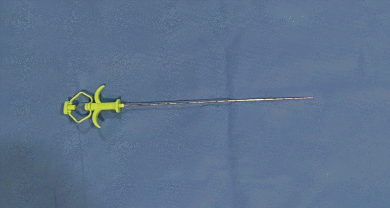
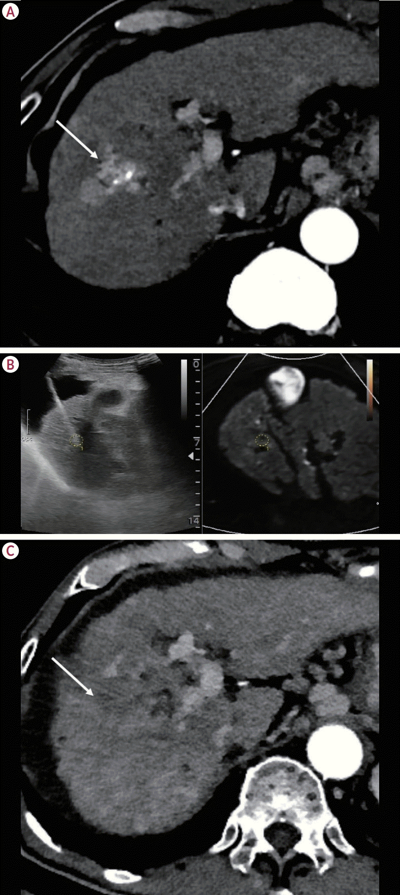
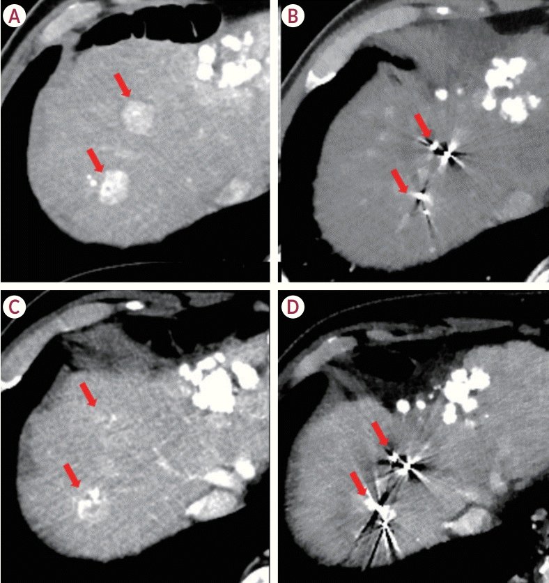
Under the guidance of real-time multimodality US fusion, the radiologist placed one to four gold fiducial markers from a Fiducial Marker Kit (Smart G. Medical, Incheon, Korea) on and around the target, using 18-gauge introducer needles with a convex probe (2-5 MHz) during transabdominal ultrasonography. Each fiducial marker measured 0.9×3.0 mm in diameter and length, respectively (Fig. 1). When performing fiducial implantation, it is advisable to maintain a minimum spacing of 20 mm and a minimum angle of 15° between the fiducials. Additionally, the fiducials should not be more than 50-60 mm away from the target. In most cases, the standard practice involves installing two fiducial markers. Typically, one marker is placed in the upper portion and the other in the lower portion of the lesion. If the lesion is small (1 cm or less) and located in the central portion of the liver, a single fiducial marker is placed at the center of the tumor. When the lesion has an irregular shape and is close to vital structures, such as the portal vein or hepatic vein, three to four markers are placed around the periphery of the lesion (Fig. 2).

Delivery of SBRT

Fiducial markers are typically implanted approximately 2 weeks before the planning CT scan, with a 1-week period following implantation dedicated to scan preparation. This standard practice, although not formalized in a specific protocol, is designed to minimize marker movement, allow inflammation to subside, and enhance imaging and treatment planning accuracy. Subsequently, four-dimensional planning CT scans were performed to account for respiratory motion before treatment began. Techniques such as abdominal compression are used to minimize the effects of motion. The internal target volume was determined by summing the volumes of the gross tumors observed in each respiratory phase, which were then expanded by a margin of 4-10 mm to define the planning target volume. SBRT treatments were delivered using a TrueBeam STX linear accelerator (Varian Medical Systems, Palo Alto, CA, USA), typically over 4-5 sessions, occurring 2-3 times weekly and spaced every other day. The entire treatment process generally spans approximately 10 days, with the total duration from the procedure to the end of radiation therapy ranging from 17 to 32 days (median, 22). Respiratory monitoring during treatment is not feasible. Instead, cone-beam CT scans are used before each SBRT session to ensure that the fiducial marker positions align with those on the planning CT. This method compensates for respiratory motion during both the treatment planning and execution phases, thereby effectively guiding the treatment.

Assessment of technical and clinical success rates and clinical outcomes

Technical success was defined as the implantation of fiducial markers that facilitated adequate treatment prior to SBRT. Clinical success was defined as the successful completion of SBRT without marker displacement. The marker positions were verified using contrast-enhanced liver CT images obtained immediately after placement. Furthermore, radiation therapy planning CT images were acquired before the initiation of radiotherapy and served as a reference for the initial positioning of markers in or around the tumor.
To ascertain the potential displacement of the fiducial markers during SBRT, cone-beam CT scans were systematically performed at each treatment session. This protocol facilitates the ongoing monitoring of marker stability, which is a critical factor in the precision of radiotherapy delivery. A retrospective analysis was implemented to rigorously evaluate the stability of these markers. This analysis entailed a comparative assessment of the fiducial positions as recorded in the cone-beam CT scans obtained during the final session of radiotherapy relative to their positions in the initial radiotherapy planning CT scans. The focus of this assessment was on two fiducials for which the centers were meticulously recorded. Displacement was quantitatively defined as any variation in the positional coordinates (X for left-right, Y for superior-inferior, and Z for anterior-posterior) of these fiducials between the initial planning and final cone-beam CT scans. Using the Varian Eclipse program (Varian Medical Systems), the fiducial markers identified on the planning CT were contoured and their positions on the cone-beam CT acquired during SBRT were pinpointed. Subsequently, these positions were converted into X-, Y-, and Z-coordinates to precisely measure the distances between the fiducials on the planning CT and those on the cone-beam CT acquired during treatment, further defining the observed displacement and ensuring treatment accuracy.
To assess clinical outcomes following SBRT, all patients were monitored for serum markers, either alpha-fetoprotein or protein induced by vitamin K absence or antagonist-II, along with regular imaging tests. The initial follow-up, conducted 1 month after SBRT, involved contrast-enhanced CT or MRI. Patients who continued SBRT treatment subsequently underwent regular contrast-enhanced CT or MRI scans every 3 months to monitor their progress.
Evaluation of local tumor response was based on the modified response evaluation criteria in solid tumors criteria.24 Complete response was defined as the total disappearance of arterial contrast enhancement in all target lesions, indicating complete local tumor control. A partial response was identified by a reduction in the total diameter of the target lesion by 30% or more in arterial contrast enhancement. Stable disease was defined as a lesion diameter change of less than 30% decrease or less than 20% increase. Progressive disease was defined by a target lesion diameter increase of 20% or more.

Complications

Immediately following the procedure, contrast-enhanced liver CT was performed to determine whether any complications, such as pneumothorax or bleeding, had occurred. Major and minor procedure-related complications were documented during and after the implantation. Complications were graded according to the Clavien-Dindo classification,25 where complications of grade IIIa or higher were classified as major, and others were considered as minor.

Statistical analysis

Statistical analyses were performed using the SPSS program (version 27; IBM, Armonk, NY, USA) with p-values <0.05 considered to indicate statistical significance.

RESULTS

Information on patient demographics and procedures involved in the placement of fiducial markers is depicted in Table 1. Among the 19 patients with 25 tumors, 15 were male (78.9%) and four were female (21.1%). The average age was 67.9 years (standard deviation, 8.6) and ranged from 42 to 80 years. Of the 25 tumors, 22 (88.0%) were HCC, and the remaining three (12.0%) were liver metastases. The average tumor size was 1.47 cm. Each patient had at least one radio-opaque marker implanted near the liver lesion, with a median of two markers per patient (range, 1-4). The average number of fiducial markers implanted per tumor was 1.9, with a standard deviation of 0.53. Of these markers, 15 (34.9%) were inserted directly into the tumors and 28 (65.1%) were placed adjacent to them.

Technical success assessment

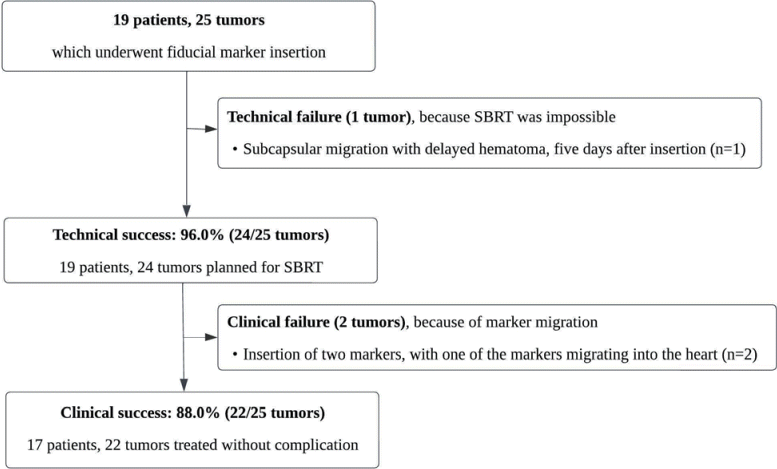
As shown in Fig. 3, we achieved a technical success rate of 96.0% (24 of 25 tumors). A total of 43 fiducial markers were inserted into 25 tumors. When more than two fiducial markers were inserted, they were placed with a minimum spacing of 20 mm and a minimum angle of 15° between them. Furthermore, the fiducials were positioned no more than 50-60 mm away from the target, a standard that was met in 100% of cases. Placement was confirmed in real time using sono-guided imaging.
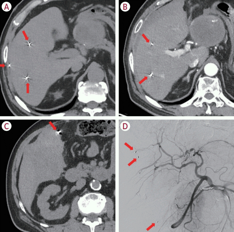
Technical failure occurred in one patient who experienced a delayed hematoma 5 days after the insertion procedure. A 66-year-old man with three HCCs had three fiducial markers implanted into each tumor. Five days after the insertion of the fiducial markers and prior to SBRT, the patient experienced right flank pain and sought emergency care. A CT tomography revealed that two of the fiducial markers were correctly positioned; however, one had migrated to the subcapsular region, resulting in the formation of a delayed hematoma. Transarterial embolization was immediately performed to control bleeding (Fig. 4). Bleeding was successfully managed, and the patient underwent SBRT for the remaining two tumors without further complications. This was the only major complication noted (one of 19 patients, 5.3%).

Clinical success assessment

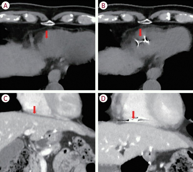
Of the 25 tumors, 22 (88.0%) achieved clinical success, marked by completion of SBRT without evidence of marker displacement. Clinical failure occurred in two hepatic tumors of two patients due to the migration of fiducial markers, necessitating the decision to withhold SBRT treatment for these specific instances. In these two instances, the fiducial markers migrated into the heart and were detected on follow-up CT scans conducted after marker insertion and before the SBRT procedure (Fig. 5). The median time from fiducial marker implantation to the start of SBRT was 13 days, ranging from 8 to 25 days. In terms of SBRT treatment specifics, the median radiation dose delivered was 50 Gy (range, 50-60) administered in 4-10 fractions. The median internal target volume measured 5.1 cm³ (range, 0.9-33.5), and the median planning target volume was 23.1 cm³ (range, 9.3-114.7).
In the context of radiotherapy, we analyzed 19 patients treated for 25 tumors. Initially, one patient with only one fiducial marker was excluded from the displacement calculations because it was impossible to perform stereotactic calculations, although radiotherapy was still successfully administered. Furthermore, two other patients were excluded because their fiducials migrated into the heart, making displacement calculations unfeasible; however, they also received successful treatment. Consequently, the remaining 16 patients were analyzed. Among these 16 patients, five had two tumors each. In three of these patients, the tumors were located in the same hepatic segment and were sufficiently close to be considered together in one treatment session. However, the tumors in the other two patients were in different regions and had to be treated in separate sessions. Thus, data from 16 patients and 18 treatment sessions are included in Table 2, summarizing the positional changes between cone-beam CT and pretreatment planning CT. Positional changes in the fiducials were assessed in three directions, X (left-right), Y (superior-inferior), and Z (anterior-posterior). The observed movements were minimal, with average displacements of 0.09 mm in the X direction, 0.30 mm in the Y direction, and 0.23 mm in the Z direction, respectively, reflecting the high stability of the markers during treatment.
Over a mean follow-up period of 11.7 months (range, 2-32), we observed a complete response post-SBRT in 20 tumors (83.3%) (Fig. 6); three tumors (12.5%) maintained stable disease, and one tumor showed progressive disease (4.2%), as depicted in Table 3.

DISCUSSION

Our study demonstrated the effectiveness of real-time CT/MRI-US fusion technology in placing gold fiducial markers for SBRT, achieving a technical success rate of 96.0% (24 of 25 tumors) and a clinical success rate of 88.0% (22 of 25 tumors). Despite using fewer markers than traditionally recommended, with a median of two markers per patient (range, 1-4) and an average of 1.9 per tumor, the outcomes were satisfactory. This suggests that a reduced number of markers, compared with the conventional recommendation of at least to 3-4 fiducials with defined distances and angles, may suffice, especially considering the increased risk of marker migration near major hepatic veins.
Advancements in stereotactic radiosurgery now enable frameless real-time tumor tracking with 1 mm accuracy,14,16,26,27 significantly enhancing the role of fiducial markers in treating hepatic tumors. Our methodology not only improves localization accuracy but also incorporates high-resolution imaging from contrast-enhanced CT or MR with real-time B-mode US, which enhances lesion visibility, particularly in challenging conditions such as cirrhosis.28
We observed minimal marker displacement, with average movements of 0.09 mm in the X direction, 0.30 mm in the Y direction, and 0.23 mm in the Z direction. The planning target volume margins were set between 4-10 mm, adequately compensating for both inter-fractional and intra-fractional movements. The maximum displacements observed in this study were approximately 3 mm with standard deviations of 1.5 mm in the X direction, 1.1 mm in the Y direction, and 1.1 mm in the Z direction, respectively. This suggests that the margins used sufficiently covered the typical range of movement (up to three standard deviations), ensuring accurate targeting and effective treatment coverage. Our approach, which leverages advancements in stereotactic radiosurgery for frameless, real-time tumor tracking with 1 mm accuracy, significantly improves the localization accuracy.
In our study, integrating real-time CT/MRI-US fusion guidance during the placement of gold fiducial markers represented a significant advancement in tumor visualization and localization of SBRT in hepatic tumors. This approach is especially advantageous because lesions are not always detectable using sonography alone, mostly due to the highly heterogeneous parenchyma found in conditions, such as cirrhosis in patients with HCC or because of their proximity to intrahepatic vital structures.5,23 This innovative technique enhances lesion visibility within the liver by combining high-resolution images obtained using contrast-enhanced CT or MR with real-time B-mode ultrasonography.
Percutaneous fiducial marker placement is a recognized method for treating malignant hepatic tumors; however, it carries risks such as marker migration, subsequent bleeding,29,30 cardiac embolization, pneumothorax, and other complications that require careful procedure execution. In our study, the major complication rate was 5.3%, primarily delayed subcapsular hematoma, which was effectively treated with transarterial embolization. The elimination of viable hypervascular tumor tissue was confirmed using post-embolization imaging. These findings are consistent with the results of a similar study by Kothary et al.,31 who reported a complication rate of 5%.
Two minor complications were noted, including fiducial marker migration into the right atrium, which were both asymptomatic and detectable on pre-SBRT CT. Initially placed near the middle and right hepatic veins, the markers later migrated to the right atrium. This type of migration has not yet been reported in the literature. Although these incidents did not result in clinical symptoms, they highlighted the importance of caution when positioning markers near hepatic veins, especially centrally. Based on our findings, we recommend careful marker placement near the large hepatic veins or the liver capsule to minimize the risk of migration.
Our study has certain limitations. First, the small sample size may affect the generalizability of our results to a broader patient population. Additionally, the specific timeframe of the study and the absence of extended follow-up periods limited our ability to fully ascertain the long-term implications of gold fiducial marker placement on SBRT outcomes. Another limitation was marker migration, especially in the right atrium. It is conceivable that a larger and more diverse cohort may exhibit different frequencies and varieties of complications, thereby offering a more comprehensive understanding of the risks associated with this treatment. Finally, our focus on the percutaneous method, without directly comparing it with other techniques, may limit the scope of our findings to the broader context of HCC treatment.
In conclusion, the percutaneous placement of gold fiducial markers under real-time CT/MRI guidance is a safe and effective method for enhancing the precision and outcomes of SBRT for hepatic tumors. Despite the insertion of fewer markers than conventionally recommended, treatment outcomes were satisfactory. However, instances of marker migration underscore the need for caution, particularly when inserting markers near hepatic veins and subcapsular tumor areas. These findings emphasize the importance of ongoing evaluation and refinement of this technique.

Notes

Conflicts of Interest

The authors have no conflicts of interest to disclose.

Ethics Statement

This retrospective analysis was approved by the Institutional Review Board (IRB) of the Seoul National University Hospital (IRB No. 2403-029-1518). The requirement for informed consent was waived due to the retrospective nature of this study.

Funding Statement

This study was supported by the Scientific Research Fund of the Korean Liver Cancer Association 2020.

Data Availability

The datasets used and/or analyzed during the current study are available from the corresponding author on reasonable request.

Author Contributions

Conceptualization: JML

Data curation: SH, SC

Formal analysis: SH, SC

Investigation: JML

Methodology: JML

Project administration: EKC, JML

Resources: EKC, JML

Supervision: EKC, JML

Visualization: SH

Writing - original draft: SH, JML

Writing - review & editing: SC, EKC

References

1. Jackson WC, Tao Y, Mendiratta-Lala M, Bazzi L, Wahl DR, Schipper MJ, et al. Comparison of stereotactic body radiation therapy and radiofrequency ablation in the treatment of intrahepatic metastases. Int J Radiat Oncol Biol Phys. 2018; 100:950–958.
crossref
2. Lee MT, Kim JJ, Dinniwell R, Brierley J, Lockwood G, Wong R, et al. Phase I study of individualized stereotactic body radiotherapy of liver metastases. J Clin Oncol. 2009; 27:1585–1591.
crossref
3. Rusthoven KE, Kavanagh BD, Cardenes H, Stieber VW, Burri SH, Feigenberg SJ, et al. Multi-institutional phase I/II trial of stereotactic body radiation therapy for liver metastases. J Clin Oncol. 2009; 27:1572–1578.
crossref
4. Wahl DR, Stenmark MH, Tao Y, Pollom EL, Caoili EM, Lawrence TS, et al. Outcomes after stereotactic body radiotherapy or radiofrequency ablation for hepatocellular carcinoma. J Clin Oncol. 2016; 34:452–459.
5. Kim JH, Lee JY, Yu SJ, Lee DH, Joo I, Yoon JH, et al. Fusion imagingguided radiofrequency ablation with artificial ascites or pleural effusion in patients with hepatocellular carcinomas: the feasibility rate and midterm outcome. Int J Hyperthermia. 2023; 40:2213424.
crossref
6. Vouche M, Habib A, Ward TJ, Kim E, Kulik L, Ganger D, et al. Unresectable solitary hepatocellular carcinoma not amenable to radiofrequency ablation: multicenter radiology-pathology correlation and survival of radiation segmentectomy. Hepatology. 2014; 60:192–201.
crossref
7. Rim CH, Lee JS, Kim SY, Seong J. Comparison of radiofrequency ablation and ablative external radiotherapy for the treatment of intrahepatic malignancies: a hybrid meta-analysis. JHEP Rep. 2022; 5:100594.
crossref
8. Heimbach JK, Kulik LM, Finn RS, Sirlin CB, Abecassis MM, Roberts LR, et al. AASLD guidelines for the treatment of hepatocellular carcinoma. Hepatology. 2018; 67:358–380.
crossref
9. Korean Liver Cancer Association (KLCA); National Cancer Center (NCC) Korea. 2022 KLCA-NCC Korea practice guidelines for the management of hepatocellular carcinoma. J Liver Cancer. 2023; 23:1–120.
10. Weykamp F, Hoegen P, Klüter S, Spindeldreier CK, König L, Seidensaal K, et al. Magnetic resonance-guided stereotactic body radiotherapy of liver tumors: initial clinical experience and patient-reported outcomes. Front Oncol. 2021; 11:610637.
11. Jaffray DA, Siewerdsen JH, Wong JW, Martinez AA. Flat-panel conebeam computed tomography for image-guided radiation therapy. Int J Radiat Oncol Biol Phys. 2002; 53:1337–1349.
crossref
12. Lam KL, Ten Haken RK, McShan DL, Thornton AF Jr. Automated determination of patient setup errors in radiation therapy using spherical radio-opaque markers. Med Phys. 1993; 20:1145–1152.
crossref
13. Goodman KA, Kavanagh BD. Stereotactic body radiotherapy for liver metastases. Semin Radiat Oncol. 2017; 27:240–246.
crossref
14. Chang SD, Main W, Martin DP, Gibbs IC, Heilbrun MP. An analysis of the accuracy of the CyberKnife: a robotic frameless stereotactic radiosurgical system. Neurosurgery. 2003; 52:140–146. discussion 146-147.
crossref
15. Shirato H, Harada T, Harabayashi T, Hida K, Endo H, Kitamura K, et al. Feasibility of insertion/implantation of 2.0-mm-diameter gold internal fiducial markers for precise setup and real-time tumor tracking in radiotherapy. Int J Radiat Oncol Biol Phys. 2003; 56:240–247.
16. Chang SD, Adler JR. Robotics and radiosurgery--the cyberknife. Stereotact Funct Neurosurg. 2001; 76:204–208.
17. Moskalenko M, Jones BL, Mueller A, Lewis S, Shiao JC, Zakem SJ, et al. Fiducial markers allow accurate and reproducible delivery of liver stereotactic body radiation therapy. Curr Oncol. 2023; 30:5054–5061.
crossref
18. Oldrini G, Taste-George H, Renard-Oldrini S, Baumann AS, Marchesi V, Troufléau P, et al. Implantation of fiducial markers in the liver for stereotactic body radiation therapy: feasibility and results. Diagn Interv Imaging. 2015; 96:589–592.
crossref
19. Ohta K, Shimohira M, Iwata H, Hashizume T, Ogino H, Miyakawa A, et al. Percutaneous fiducial marker placement under CT fluoroscopic guidance for stereotactic body radiotherapy of the lung: an initial experience. J Radiat Res. 2013; 54:957–961.
crossref
20. Bertholet J, Worm E, Høyer M, Poulsen P. Cone beam CT-based set-up strategies with and without rotational correction for stereotactic body radiation therapy in the liver. Acta Oncol. 2017; 56:860–866.
21. Sotiropoulou E, Stathochristopoulou I, Stathopoulos K, Verigos K, Salvaras N, Thanos L. CT-guided fiducial placement for cyberknife stereotactic radiosurgery: an initial experience. Cardiovasc Intervent Radiol. 2010; 33:586–589.
crossref
22. Anstadt EJ, Shumway R, Colasanto J, Grew D. Single community-based institutional series of stereotactic body radiation therapy (SBRT) for treatment of liver metastases. J Gastrointest Oncol. 2019; 10:330–338.
crossref
23. Han S, Lee JM, Lee DH, Yoon JH, Chang W. Utility of real-time CT/MRI-US automatic fusion system based on vascular matching in percutaneous radiofrequency ablation for hepatocellular carcinomas: a prospective study. Cardiovasc Intervent Radiol. 2021; 44:1579–1596.
crossref
24. Lencioni R, Llovet JM. Modified RECIST (mRECIST) assessment for hepatocellular carcinoma. Semin Liver Dis. 2010; 30:52–60.
crossref
25. Clavien PA, Barkun J, de Oliveira ML, Vauthey JN, Dindo D, Schulick RD, et al. The Clavien-Dindo classification of surgical complications: fiveyear experience. Ann Surg. 2009; 250:187–196.
26. Backlund EO, Johansson L, Sarby B. Studies on craniopharyngiomas. II. Treatment by stereotaxis and radiosurgery. Acta Chir Scand. 1972; 138:749–759.
27. Lohr F, Debus J, Frank C, Herfarth K, Pastyr O, Rhein B, et al. Noninvasive patient fixation for extracranial stereotactic radiotherapy. Int J Radiat Oncol Biol Phys. 1999; 45:521–527.
crossref
28. Miura H, Doi Y, Nakao M, Ozawa S, Kenjo M, Nagata Y. Interfractional liver positional motion under exhaled breath holding based on cone beam computed tomography. In Vivo. 2023; 37:1822–1827.
crossref
29. Kim JH, Hong SS, Kim JH, Park HJ, Chang YW, Chang AR, et al. Safety and efficacy of ultrasound-guided fiducial marker implantation for CyberKnife radiation therapy. Korean J Radiol. 2012; 13:307–313.
crossref
30. Ohta K, Shimohira M, Murai T, Nishimura J, Iwata H, Ogino H, et al. Percutaneous fiducial marker placement prior to stereotactic body radiotherapy for malignant liver tumors: an initial experience. J Radiat Res. 2016; 57:174–177.
31. Kothary N, Heit JJ, Louie JD, Kuo WT, Loo BW Jr, Koong A, et al. Safety and efficacy of percutaneous fiducial marker implantation for image-guided radiation therapy. J Vasc Interv Radiol. 2009; 20:235–239.
crossref

Figure 1.
Gold fiducial marker, 18-gauge syringe, and plunger from a Fiducial Marker Kit (Smart G. Medical, Incheon, Korea).
jlc-2024-06-03f1.tif
Figure 2.
Achievement of technical success and clinical success in fiducial marker insertion for a 74-year-old man with hepatocellular carcinoma (HCC) and hepatitis B-related liver cirrhosis. (A) An arterial phase CT scan displays a 3.0-cm hyperenhancing HCC (arrow) in segment VIII of the liver. (B) A real-time US-MR fusion image illustrates the target tumor, and the fiducial marker insertion is demonstrated. (C) Complete response (arrow) was achieved with no local tumor progression observed in the 12-month follow-up CT. The background hepatic parenchyma exhibits heterogeneous attenuation due to changes post-radiation therapy. CT, computed tomography; US, ultrasound; MR, magnetic resonance.
jlc-2024-06-03f2.tif
Figure 3.
Flow diagram of technical success and clinical success. SBRT, stereotactic body radiotherapy.
jlc-2024-06-03f3.tif
Figure 4.
Fiducial marker migration with delayed hematoma formation. (A) A 66-year-old man with three hepatocellular carcinomas (HCCs) had three fiducial markers implanted into each tumor. Five days post-procedure, the patient experienced right flank pain and sought emergency medical care. (B) A CT scan showed that two of the fiducial markers were properly positioned. (C) However, one marker had migrated to the subcapsular region, accompanied by a delayed hematoma. (D) Transarterial embolization was performed to control the associated bleeding. Three fiducial markers are indicated with red arrows. After the embolization, there was no contrast leakage observed during contrast enhancement. CT, computed tomography.
jlc-2024-06-03f4.tif
Figure 5.
Migration of a fiducial marker into the heart in an 80-year-old man. (A) Non-contrast enhanced axial CT scan before marker insertion showed no abnormal foreign material (arrow) in the right atrium. (B) The fiducial marker, which had migrated into the right atrium, was observed on the non-contrast phase of follow-up axial CT image (arrow), taken after marker placement and before the initiation of stereotactic body radiation therapy. (C) Contrast-enhanced coronal CT scan before marker insertion showed no abnormal foreign material (arrow) in the right atrium. (D) The fiducial marker, which had migrated into the right atrium, was observed on the portal phase of follow-up coronal CT image (arrow), taken after marker placement and before the initiation of stereotactic body radiation therapy. CT, computed tomography.
jlc-2024-06-03f5.tif
Figure 6.
Achievement of complete response in fiducial marker insertion for a 72-year-old man with hepatocellular carcinoma (HCC) and hepatitis B-related liver cirrhosis. (A) An arterial phase CT scan displays two 1.1-cm hyperenhancing HCCs (arrows) in segments VII and VIII of the liver. (B) A complete response (arrows) was achieved with no local tumor progression observed in the 12-month follow-up CT scan. Fiducial markers inserted on the peripheral aspect of the tumor show metallic artifact. (C) A portal phase CT scan displays two HCCs (arrows) showing washout in segments VII and VIII of the liver. (D) A complete response (arrows) was achieved with no local tumor progression observed in the 12-month follow-up CT scan. Fiducial markers inserted on the peripheral aspect of the tumor show metallic artifact. CT, computed tomography.
jlc-2024-06-03f6.tif
jlc-2024-06-03f7.tif
Table 1.
Participants characteristics
Characteristic Value
Age (years) 67.9±8.6
Sex
 Men 15 (78.9)
 Women 4 (21.1)
Tumor characteristics
 HCC 22 (88.0)
 Metastasis to liver 3 (12.0)
Mean tumor size (cm) 1.47±0.60
Tumor location
 Right anterior section 13 (53.0)
 Right posterior section 7 (28.0)
 Left medial section 2 (8.0)
 Left lateral section 3 (12.0)
Mean number of fiducials per tumor 1.9±0.53
Fiducial marker placement
 Inside the tumor 15 (34.9)
 Near the tumor 28 (65.1)

Values are presented as mean±standard deviation or number (%).

HCC, hepatocellular carcinoma.

Table 2.
A comprehensive summary of the positional changes between cone-beam CT and pre-treatment planning CT across a total of 18 treatment sessions
Treatment session Patient information Region information LR* (cm) SI (cm) AP (cm)
1 Patient 1 0.02 -0.11 -0.10
2 Patient 2 0.25 -0.19 0.15
3 Patient 3 -0.27 0.20 0.09
4 Patient 4 0.11 -0.01 0.01
5 Patient 5 Region 1 -0.10 0.20 0.11
6 Patient 5 Region 2 -0.03 -0.18 0.07
7 Patient 6 -0.10 0.10 -0.03
8 Patient 7 Region 1 0.03 -0.09 -0.02
9 Patient 7 Region 2 0.20 -0.02 0.01
10 Patient 8 -0.36 -0.12 0.19
11 Patient 9 -0.13 -0.19 0.13
12 Patient 10 -0.07 -0.10 0.07
13 Patient 11 -0.01 -0.01 -0.02
14 Patient 12 0.01 0.00 0.04
15 Patient 13 0.03 0.00 0.02
16 Patient 14 0.29 -0.12 -0.34
17 Patient 15 -0.03 0.00 -0.01
18 Patient 16   -0.01 0.10 0.05

CT, computed tomography; LR, left-right; SI, superior-inferior; AP, anterior-posterior.

* Positive value means left, negative value means right;

Positive value means superior, negative value means inferior;

Positive value means anterior, negative value means posterior.

Table 3.
Comparative analysis of stereotactic body radiation therapy following fiducial marker placement in 24 hepatic tumors from 19 patients
Treated tumors (n=24) Complete response (n=20) Partial response (n=0) Stable disease (n=3) Progressive diseas (n=1) p-value*
Tumor characteristics 0.732
 HCC 17 (85.0) 3 (100.0) 1 (100.0)
 Metastasis to liver 3 (15.0) 0 (0.0) 0 (0.0)
Mean tumor size (cm) 1.35±0.474 1.90±0.265 3.00±0.00 0.041
Tumor location 0.479
 Right anterior section 9 (45.0) 2 (66.7) 1 (100.0)
 Right posterior section 7 (35.0) 0 (0.0) 0 (0.0)
 Left medial section 2 (10.0) 0 (0.0) 0 (0.0)
 Left lateral section 2 (10.0) 1 (33.3) 0 (0.0)
Mean number of fiducials per tumor 1.8±0.410 2.33±0.577 3.00±0.00 0.030
Fiducial marker placement
 Inside the tumor 0.80±0.834 0.67±0.577 0.00±0.00 0.571
 Near the tumor 1.00±0.918 1.67±0.577 3.00±0.00 0.101

Values are presented as number (%) or mean±standard deviation.

HCC, hepatocellular carcinoma.

* p-values less than 0.05 indicate statistical significance.

TOOLS
Similar articles