Journal List > J Liver Cancer > v.24(2) > 1516088661

Song, Goh, Kang, Sinn, Gwak, Paik, Lee, and Choi: Role of transarterial chemoembolization for hepatocellular carcinoma with extrahepatic metastases in the era of advancing systemic therapy

Abstract

Backgrounds/Aims

Systemic therapy is the current standard treatment for hepatocellular carcinoma (HCC) with extrahepatic metastasis (EHM). However, some patients with HCC and EHM undergo transarterial chemoembolization (TACE) to manage intrahepatic tumors. Herein, we aimed to explore the appropriateness of TACE in patients with HCC and EHM in an era of advanced systemic therapy.

Methods

This study analyzed 248 consecutive patients with HCC and EHM (median age, 58.5 years; male, 83.5%; Child-Pugh A, 88.7%) who received TACE or systemic therapy (83 sorafenib, 49 lenvatinib, 28 immunotherapy-based) between January 2018 and January 2021.

Results

Among the patients, 196 deaths were recorded during a median follow-up of 8.9 months. Patients who received systemic therapy had a higher albumin-bilirubin grade, elevated tumor markers, an increased number of intrahepatic tumors, larger-sized tumors, and more frequent portal vein invasion than those who underwent TACE. TACE was associated with longer median overall survival (OS) than sorafenib (15.1 vs. 4.7 months; 95% confidence interval [CI], 11.1-22.2 vs. 3.7-7.3; hazard ratio [HR], 1.97; P<0.001). After adjustment for potential confounders, TACE was associated with statistically similar survival outcomes to those of lenvatinib (median OS, 8.0 months; 95% CI, 6.5-11.0; HR, 1.21; P=0.411) and immunotherapies (median OS, 14.3 months; 95% CI, 9.5-27.0; HR, 1.01; P=0.973), demonstrating survival benefits equivalent to these treatments.

Conclusions

In patients with HCC and EHM, TACE can provide a survival benefit comparable to that of newer systemic therapies. Accordingly, TACE remains a valuable option in this era of new systemic therapies.

GRAPHICAL ABSTRACT

INTRODUCTION

Hepatocellular carcinoma (HCC) is considered a substantial global health burden and the third leading cause of cancer-related deaths.1 The development of extrahepatic metastasis (EHM) in patients with HCC often signifies an advanced disease stage. Currently, the standard treatment for patients with HCC and EHM is systemic therapy.2 Prior to 2017, sorafenib was the only systemic treatment option. During this period, the role of transarterial chemoembolization (TACE) in managing HCC with EHM has been clearly defined and widely acknowledged, especially given the limited systemic options and therapeutic efficacy.3,4 A study conducted in Asia reported that repeated TACE with or without systemic chemotherapy could provide considerable survival benefits in patients with metastatic HCC and well-preserved liver function.3 Likewise, in a study conducted in Germany, Bettinger et al.4 highlighted that TACE, both alone and in combination with sorafenib, may improve the overall survival (OS) of patients with metastatic HCC. However, newer systemic agents, including newer tyrosine kinase inhibitors (TKIs) such as lenvatinib and novel immunotherapies (IO), have substantially evolved the treatment paradigm for advanced HCC.5,6 In 2017, lenvatinib was established as a non-inferior alternative to sorafenib, demonstrating notable improvements in the progression-free survival (PFS) and objective response rate (ORR).7,8 Following lenvatinib, several IO agents have shown promising results.9-12 Importantly, these newer agents have changed the treatment landscape of advanced HCC.13 With these newer agents eliciting promising results in HCC therapy, their adoption in earlier disease stages has increased. Consequently, research has notably shifted focus, with several randomized studies comparing these newer systemic agents to TACE in the intermediate stage of HCC.14 However, comparative studies in cases of HCC with EHM remain scarce.
In patients with advanced HCC, mortality is typically attributed to liver failure due to intrahepatic disease progression rather than the EHM itself, highlighting the importance of effective intrahepatic tumor control,15,16 and TACE is well-known for its efficacy in this respect. Thus, evaluating the role of TACE in the context of newer treatments for patients with EHM will offer insights into its relevance and effectiveness in the current therapeutic landscape.
In the current study, we compared the effectiveness of TACE with advanced systemic therapies in managing HCC with EHM and explored whether TACE retains its significance in the contemporary therapeutic milieu for HCC.

METHODS

Study population

This retrospective cohort study was performed at the Samsung Medical Center, a high-volume liver cancer center in Seoul, Korea, using a prospectively maintained HCC registry from January 2018 to January 2021. This registry proactively compiled data on baseline clinical characteristics, tumor characteristics, and initial treatment approaches for all newly diagnosed patients with HCC aged ≥18 years treated at the center. A diagnosis of HCC was established histologically or clinically according to the regional HCC guidelines.17 A total of 248 consecutive patients with EHM at the time of diagnosis and initially treated with systemic therapy or TACE were selected. This study adhered to the principles of the Declaration of Helsinki and was approved by the Ethics Committee of the Samsung Medical Center (Institutional Review Board [IRB] number 2024-02-075). The IRB waived the requirement for informed patient consent because the retrospective nature of the study relied on existing administrative and clinical data.

Treatment and follow-up

The systemic therapy cohort comprised 160 patients who were subdivided according to the specific agent received. Eighty-three patients were treated with sorafenib, initiated at a dosage of 400 mg twice daily and adjusted based on tolerability and adverse effects. Forty-nine patients received lenvatinib, with dosage determined by body weight: 12 mg daily for patients weighing ≥60 kg and 8 mg daily for those <60 kg. The IO subgroup (n=28) comprised patients receiving the following treatments: nivolumab monotherapy administered at 240 mg biweekly, four doses of combination therapy with nivolumab (1 mg/kg) and ipilimumab (3 mg/kg) followed by nivolumab maintenance (n=2), atezolizumab (1,200 mg) combined with bevacizumab (15 mg/kg) every 3 weeks (n=9), and clinical trials (n=17). Patients were monitored through regular clinical assessments, including imaging every 8-12 weeks, to evaluate treatment response, along with laboratory tests to monitor liver function and adverse events. The follow-up procedures were standardized for the cohort, with adjustments performed for individual patients based on treatment response and tolerability.
Conventional TACE was performed with intra-arterial injection of doxorubicin hydrochloride (Adriamycin; Dong-A Pharmaceutical, Seoul, Korea) and iodized oil (Lipiodol; Guerbet, Villepinte, France) following a femoral approach, a celiac angiogram, and superselection of the tumor feeder at the segmental or subsegmental artery level with a micro-guidewire and a 2.0-Fr microcatheter. The feeder(s) were embolized with gelatin sponge pledgets (Cutanplast; Mascia Brunelli S.p.A., Milano, Italy) until hemostasis was achieved. Contrast-enhanced computed tomography or magnetic resonance imaging was performed at baseline, as well as every 2-4 months.

Variables and outcome

The patients were grouped according to their initial treatment (sorafenib, lenvatinib, IO, or TACE). The following data were collected from the HCC registry: age at diagnosis, sex, HCC etiology, Eastern Cooperative Oncology Group (ECOG) performance status, Child-Pugh class, levels of serum albumin and bilirubin, albumin-bilirubin (ALBI) grade, serum alpha-fetoprotein (AFP), protein induced by vitamin K absence or antagonist-II (PIVKA-II), and tumor characteristics (intrahepatic tumor number, maximum diameter, portal vein [PV] and bile duct invasion, modified Union for International Cancer Control stage, Barcelona Clinic Liver Cancer stage), and initial treatment modalities. In addition, EHM locations were identified by reviewing imaging and medical records, given that the HCC registry did not provide detailed information. The primary outcome was OS, which was defined as the duration from diagnosis to the last follow-up or death (reference date, September 30, 2023).

Statistical analysis

Categorical variables are reported as numbers and percentages and compared using the chi-square test or Fisher’s exact test. Continuous variables are reported as medians and interquartile ranges and compared using the Mann-Whitney U test. Survival curves were calculated using the Kaplan-Meier method and compared using the log-rank test. Cox proportional hazard models were used to estimate crude and multivariate-adjusted hazard ratios (HRs) with 95% confidence intervals (CIs). This analysis met the assumptions of the Cox proportional hazard model. Propensity score (PS) matching was performed to control for potential confounders. The matching variables included age, sex, tumor size, tumor number, ECOG performance status, liver disease etiology, ALBI grade, AFP, PIVKA-II, PV invasion, and type of EHM. The nearest-neighbor matching method was employed with a caliper width of 0.1. All statistical analyses were performed using the R version 4.1.2 software (R Foundation for Statistical Computing, Vienna, Austria). Statistical significance was set at a two-tailed P-value <0.05.

RESULTS

Baseline characteristics

Table 1 presents the baseline demographic and clinical parameters stratified by the initial treatment strategy, including TACE, sorafenib, lenvatinib, and IO. Age and sex distributions remained consistent across modalities, suggesting demographic homogeneity in the study cohort (P>0.05). Patients who received systemic therapy, particularly those who underwent sorafenib or lenvatinib therapy, had significantly elevated ALBI grades, suggesting compromised liver function, when compared with patients who received TACE and IO (P<0.001). This cohort also exhibited elevated levels of tumor markers (AFP and PIVKA-II), greater tumor numbers, and larger tumor diameters, corresponding to more advanced stages at baseline. Furthermore, patients who received systemic therapy had a higher incidence of PV invasion than those who received TACE and IO, indicating a propensity for a more aggressive disease presentation among patients selected for systemic therapies. Of 88 patients treated with TACE, 12 received additional radiotherapy or proton beam therapy. Similarly, among the 160 patients who received systemic therapy, 43 underwent radiotherapy or proton beam therapy.

OS according to the initial treatment

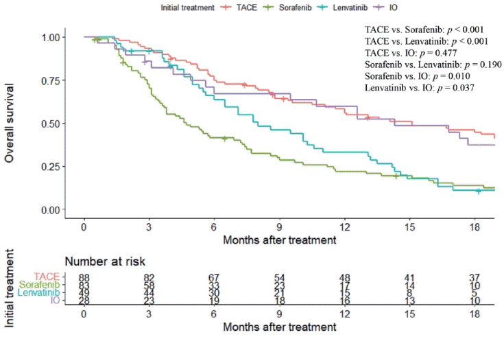
During the study period, TACE was associated with a median OS of 15.1 months (95% CI, 11.1-22.2), demonstrating statistical superiority over sorafenib and lenvatinib, which were associated with median OS values of 4.7 (95% CI, 3.7-7.3) and 8.0 (95% CI, 6.5-11.0) months, respectively, with P<0.001 for both comparisons (Fig. 1). Conversely, there was no significant survival difference between TACE and IO therapies (median OS, 15.1 months [95% CI, 11.1-22.2] vs. 14.3 months [95% CI, 9.5-27.0]; P=0.477). Upon direct comparisons among systemic therapies, we found that sorafenib therapy was associated with a lower median OS than IO (P=0.010). However, the difference between sorafenib and lenvatinib failed to reach statistical significance (P=0.190). IO elicited a significant yet modest OS advantage over lenvatinib (P=0.037).

Univariable and multivariable Cox regression analyses

According to the multivariable analysis, factors associated with worse survival included ALBI grade 3 (HR, 2.01; 95% CI, 1.25-3.25; P=0.004), elevated PIVKA-II (HR, 1.06; 95% CI, 1.00-1.13; P=0.045), and PV invasion (segmental/lobar HR, 1.76; 95% CI, 1.24-2.49; P=0.001; main/contralateral HR, 1.91; 95% CI, 1.25-2.91; P=0.003) (Table 2). Other clinical and demographic variables showed no significant effects. Upon comparing treatments, sorafenib was significantly associated with an increased risk of mortality (HR, 1.97; 95% CI, 1.36-2.85; P<0.001) when compared with TACE. Lenvatinib (HR, 1.21; 95% CI, 0.77-1.90; P=0.411) and IO (HR, 1.01; 95% CI, 0.61-1.67; P=0.973) did not differ significantly in survival outcomes.
Sequential multivariate models consistently revealed that sorafenib was inferior to TACE in terms of OS, with HRs favoring TACE for all adjustments (all P<0.001; Supplementary Table 1). Conversely, lenvatinib increased the HRs relative to TACE, which decreased with more comprehensive adjustments, losing statistical significance in fully adjusted model 4 (P=0.222). IO therapies showed no significant difference in HRs when compared with TACE in any model, suggesting that TACE offers a survival benefit comparable to that of lenvatinib and IO therapies when demographics, liver function, tumor markers, and tumor characteristics are considered.

Subgroup analysis

In the subgroup analysis comparing TACE with systemic therapies (lenvatinib and IO, excluding sorafenib), no significant differences were detected in survival in terms of most demographic and clinical characteristics, including age, sex, ALBI grade, etiology, and PV invasion (P>0.05) (Table 3). Nevertheless, the analysis detected a significant survival advantage in patients with smaller intrahepatic tumors (<5 cm) who received TACE when compared with those who received systemic therapy, with an adjusted HR of 5.05 (P=0.02). This finding suggested that TACE yielded a potential benefit in this subgroup. Overall, TACE had no significant survival benefit in any examined subgroup when compared with systemic therapy.

Comparison of Kaplan-Meier curves in PS-matched groups

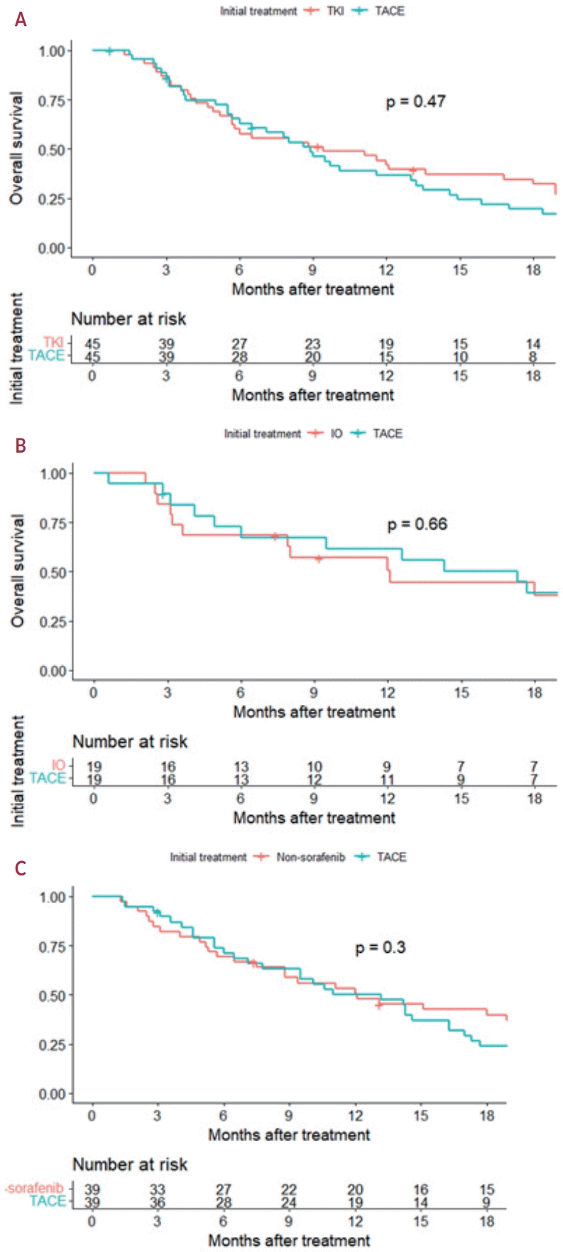
After PS matching to ensure group comparability based on key clinical factors (Supplementary Tables 2-4), Kaplan-Meier survival curves were compared using the log-rank test (Fig. 2). The analysis involved 45 matched pairs of TACE versus TKIs (sorafenib and lenvatinib), revealing no significant difference in survival (P=0.47). Similarly, no significant differences in survival were detected upon comparing 19 matched pairs treated with TACE and IO (P=0.66). Additionally, we detected no significant difference in survival upon comparing TACE and combined non-sorafenib systemic therapies (lenvatinib and IO) involving 39 matched pairs (P=0.3).

DISCUSSION

Herein, we reassessed the role of TACE in treating HCC patients with EHM in the current era of advanced systemic therapy. Our study showed that patients with HCC and EHM treated with TACE had an OS comparable to that of those treated with newer systemic therapies, such as IO.
In patients with advanced HCC, death is caused predominantly by liver failure owing to the progression of intrahepatic disease.18 Locoregional treatments, including TACE, are frequently performed palliatively in patients with EHM, with some studies suggesting a potential survival advantage.3,4,19 However, these studies typically involved highly selective patients with well-preserved liver function, limited metastatic spread to a single organ, and controlled intrahepatic lesions, leading to potential biases because of their retrospective nature and small sample sizes. The effectiveness of locoregional therapy in improving survival among patients with EHM remains uncertain owing to the lack of randomized controlled trials that directly compared TACE and systemic therapies in this context. Observational studies have reported mixed results, with some indicating survival benefits associated with TACE alone or in combination with sorafenib. However, heterogeneity in patient populations and treatment approaches complicates the establishment of definitive conclusions, particularly for advanced HCC, in which EHM may indicate more aggressive tumor behavior.20-24
In the current study, patients treated with systemic therapies had a higher incidence of unfavorable tumor characteristics than those treated with TACE. After adjustment, OS did not differ between patients treated with newer systemic agents and those who received TACE. This finding suggests that TACE is comparable to newer systemic agents in terms of survival outcomes after adjusting for tumor factors. Moreover, this finding underscores the continued relevance of TACE, even with the advent of advanced systemic therapies.
Herein, sorafenib-treated patients had significantly reduced OS when compared with those treated with TACE or other newer systemic agents, even after adjusting for demographic variables, liver function parameters, and tumor characteristics. This finding differs from a previous study conducted by our group, which found no statistical difference in survival between sorafenib- and TACE-treated patients after adjustment, suggesting that survival differences may be caused by indication bias rather than TACE being an independent prognostic factor.25 The result of this analysis, particularly the inferiority of sorafenib to that of other treatments, warrants a re-evaluation in the context of recent therapeutic advances. The study period of 2018-2020 coincides with the introduction and adoption of newer agents that may relegate sorafenib to a palliative option for patients with reduced liver function or those ineligible for newer treatments. This shift may explain the observed differences in survival and highlight the evolving therapeutic landscape and its effects on treatment efficacy.
Upon conducting a subgroup analysis excluding sorafenib and focusing on lenvatinib and IO, we detected no statistical difference in survival between TACE and combined newer systemic agents in several subgroups, including age, sex, liver function, etiology, and tumor characteristics. Conversely, TACE conferred a significant survival benefit over systemic therapy in patients with HCC and intrahepatic tumors <5 cm in size. Accordingly, TACE may be particularly beneficial in patients with smaller intrahepatic tumors, underscoring the need for a tailored treatment approach that leverages the strengths of locoregional and systemic therapies to optimize patient outcomes.
This observational study has several limitations. First, the inherent selection bias of observational studies may have impacted our results. The study did not account for subsequent treatments following the initial therapy, which possibly affected the OS. Second, this study was conducted at a single referral center, and almost all patients were Asians from an area endemic for hepatitis B. This specific demographic characteristic may limit the generalizability of the results. Third, the study only included a few patients treated with IO. Considering the growing use of IOs, the under-representation of patients receiving IOs may impact the generalizability of our findings to a broader population of patients with HCC currently receiving these agents. Fourth, our study did not examine PFS or ORR as secondary outcome measures. These outcomes would offer a comprehensive assessment of the therapeutic efficacy of TACE than that of systemic therapies. Finally, our retrospective study design did not allow the investigation of the safety profiles and tolerability of compared treatments. Acquiring this information can be informative and could influence treatment decisions.
In summary, the role of TACE in managing intrahepatic tumor control in patients with HCC with EHM remains valid in the era of new systemic agents. TACE could provide an OS comparable to that of newer systemic agents, suggesting that TACE remains a valuable strategy for patients with EHM. TACE was associated with superior survival benefits to newer systemic therapies for intrahepatic tumors <5 cm in size, highlighting its preferred role. However, further well-designed prospective studies are necessary to fully ascertain the effectiveness and applicability of TACE.

Notes

Conflicts of Interest

The authors have no conflicts of interest to disclose.

Ethics Statement

This study was conducted in accordance with the Declaration of Helsinki and approved by the Institutional Review Board of Samsung Medical Center (No. 2024-02-075).

Funding Statement

None.

Data Availability

The data that support the findings of this study are available from the authors upon reasonable request.

Author Contributions

Study concept and design: BGS, MSC

Data acquisition: BGS

Data analysis and interpretation: BGS

Manuscript draft: BGS

Data analysis plan and data management: BGS

Critical revision of manuscript: BGS, MJG, WK, DHS, GYG, YHP, MSC, JHL

Overall study supervision: MSC

All authors participated in the preparation of the manuscript and have seen and approved the final version.

References

1. Sung H, Ferlay J, Siegel RL, Laversanne M, Soerjomataram I, Jemal A, et al. Global cancer statistics 2020: GLOBOCAN estimates of incidence and mortality worldwide for 36 cancers in 185 countries. CA Cancer J Clin. 2021; 71:209–249.
2. Singal AG, Llovet JM, Yarchoan M, Mehta N, Heimbach JK, Dawson LA, et al. AASLD practice guidance on prevention, diagnosis, and treatment of hepatocellular carcinoma. Hepatology. 2023; 78:1922–1965.
crossref
3. Yoo DJ, Kim KM, Jin YJ, Shim JH, Ko GY, Yoon HK, et al. Clinical outcome of 251 patients with extrahepatic metastasis at initial diagnosis of hepatocellular carcinoma: does transarterial chemoembolization improve survival in these patients? J Gastroenterol Hepatol. 2011; 26:145–154.
crossref
4. Bettinger D, Spode R, Glaser N, Buettner N, Boettler T, Neumann-Haefelin C, et al. Survival benefit of transarterial chemoembolization in patients with metastatic hepatocellular carcinoma: a single center experience. BMC Gastroenterol. 2017; 17:98.
crossref
5. Aitcheson G, Pillai A, Dahman B, John BV. Recent advances in systemic therapies for advanced hepatocellular carcinoma. Curr Hepatol Rep. 2021; 20:23–33.
crossref
6. Goh MJ, Sinn DH, Kim JM, Lee MW, Hyun DH, Yu JI, et al. Clinical practice guideline and real-life practice in hepatocellular carcinoma: a Korean perspective. Clin Mol Hepatol. 2023; 29:197–205.
7. Yamashita T, Kudo M, Ikeda K, Izumi N, Tateishi R, Ikeda M, et al. REFLECT-a phase 3 trial comparing efficacy and safety of lenvatinib to sorafenib for the treatment of unresectable hepatocellular carcinoma: an analysis of Japanese subset. J Gastroenterol. 2020; 55:113–122.
crossref
8. Lee MMP, Chan LL, Chan SL. The role of lenvatinib in the era of immunotherapy of hepatocellular carcinoma. J Liver Cancer. 2023; 23:262–271.
crossref
9. Verset G, Borbath I, Karwal M, Verslype C, Van Vlierberghe H, Kardosh A, et al. Pembrolizumab monotherapy for previously untreated advanced hepatocellular carcinoma: data from the open-label, phase II KEYNOTE-224 trial. Clin Cancer Res. 2022; 28:2547–2554.
crossref
10. Yau T, Park JW, Finn RS, Cheng AL, Mathurin P, Edeline J, et al. Nivolumab versus sorafenib in advanced hepatocellular carcinoma (CheckMate 459): a randomised, multicentre, open-label, phase 3 trial. Lancet Oncol. 2022; 23:77–90.
crossref
11. Yau T, Kang YK, Kim TY, El-Khoueiry AB, Santoro A, Sangro B, et al. Efficacy and safety of nivolumab plus ipilimumab in patients with advanced hepatocellular carcinoma previously treated with sorafenib: the CheckMate 040 randomized clinical trial. JAMA Oncol. 2020; 6:e204564.
12. Finn RS, Qin S, Ikeda M, Galle PR, Ducreux M, Kim TY, et al. Atezolizumab plus bevacizumab in unresectable hepatocellular carcinoma. N Engl J Med. 2020; 382:1894–1905.
crossref
13. Han JW, Park SH. Advances in immune checkpoint inhibitors for hepatocellular carcinoma. J Liver Cancer. 2021; 21:139–145.
crossref
14. Sankar K, Gong J, Osipov A, Miles SA, Kosari K, Nissen NN, et al. Recent advances in the management of hepatocellular carcinoma. Clin Mol Hepatol. 2024; 30:1–15.
crossref
15. Lee HS. Management of patients with hepatocellular carcinoma and extrahepatic metastasis. Dig Dis. 2011; 29:333–338.
crossref
16. Uka K, Aikata H, Takaki S, Shirakawa H, Jeong SC, Yamashina K, et al. Clinical features and prognosis of patients with extrahepatic metastases from hepatocellular carcinoma. World J Gastroenterol. 2007; 13:414–420.
17. Korean Liver Cancer Association (KLCA); National Cancer Center (NCC) Korea. 2022 KLCA-NCC Korea practice guidelines for the management of hepatocellular carcinoma. Clin Mol Hepatol. 2022; 28:583–705.
18. Jung SM, Jang JW, You CR, Yoo SH, Kwon JH, Bae SH, et al. Role of intrahepatic tumor control in the prognosis of patients with hepatocellular carcinoma and extrahepatic metastases. J Gastroenterol Hepatol. 2012; 27:684–689.
crossref
19. Long HY, Huang TY, Xie XY, Long JT, Liu BX. Treatment strategies for hepatocellular carcinoma with extrahepatic metastasis. World J Clin Cases. 2021; 9:5754–5768.
crossref
20. Kirstein MM, Voigtländer T, Schweitzer N, Hinrichs JB, Marquardt J, Wörns MA, et al. Transarterial chemoembolization versus sorafenib in patients with hepatocellular carcinoma and extrahepatic disease. United European Gastroenterol J. 2018; 6:238–246.
crossref
21. Yoo JJ, Lee JH, Lee SH, Lee M, Lee DH, Cho Y, et al. Comparison of the effects of transarterial chemoembolization for advanced hepatocellular carcinoma between patients with and without extrahepatic metastases. PLoS One. 2014; 9:e113926.
crossref
22. Pinter M, Hucke F, Graziadei I, Vogel W, Maieron A, Königsberg R, et al. Advanced-stage hepatocellular carcinoma: transarterial chemoembolization versus sorafenib. Radiology. 2012; 263:590–599.
23. Nishikawa H, Osaki Y, Iguchi E, Takeda H, Nakajima J, Matsuda F, et al. Comparison of the efficacy of transcatheter arterial chemoembolization and sorafenib for advanced hepatocellular carcinoma. Exp Ther Med. 2012; 4:381–386.
crossref
24. Choi GH, Shim JH, Kim MJ, Ryu MH, Ryoo BY, Kang YK, et al. Sorafenib alone versus sorafenib combined with transarterial chemoembolization for advanced-stage hepatocellular carcinoma: results of propensity score analyses. Radiology. 2013; 269:603–611.
crossref
25. Kim J, Sinn DH, Choi MS, Kang W, Gwak GY, Paik YH, et al. Hepatocellular carcinoma with extrahepatic metastasis: are there still candidates for transarterial chemoembolization as an initial treatment? PLoS One. 2019; 14:e0213547.
crossref

Figure 1.
Overall survival according to initial treatment modality. Comparisons were performed using the log-rank test. Post-hoc pairwise comparisons of overall survival between treatment groups were performed using the log-rank test, with Bonferroni correction for multiple testing. TACE, transarterial chemoembolization; IO, immunotherapy.
jlc-2024-05-26f1.tif
Figure 2.
Comparison of Kaplan-Meier curves in propensity score matched groups. (A) TACE versus TKI, (B) TACE versus IO, and (C) TACE versus non-sorafenib systemic therapies (lenvatinib and IO). Comparisons were performed using the log-rank test. TKI, tyrosine kinase inhibitor; TACE, transarterial chemoembolization; IO, immunotherapy.
jlc-2024-05-26f2.tif
jlc-2024-05-26f3.tif
Table 1.
Baseline characteristics
Characteristic Treatment group
P-value
TACE (n=88) Sorafenib (n=83) Lenvatinib (n=49) IO (n=28)
Age (years) 58.6±11.1 59.8±11.0 56.4±11.0 58.2±11.5 0.383
Male 74 (84.1) 68 (81.9) 42 (85.7) 23 (82.1) 0.944
Etiology 0.500
 HBV 56 (63.6) 51 (61.4) 34 (69.4) 19 (67.9)
 HCV 8 (9.1) 5 (6.0) 0 (0.0) 2 (7.1)
 NBNC 24 (27.3) 27 (32.5) 15 (30.6) 7 (25.0)
ECOG performance status 0.477
 0 75 (85.2) 71 (85.5) 42 (85.7) 20 (71.4)
 1 12 (13.6) 11 (13.3) 7 (14.3) 7 (25.0)
 ≥2 1 (1.1) 1 (1.2) 0 (0.0) 1 (3.6)
CTP grade 0.089
 A 81 (92.0) 68 (81.9) 44 (89.8) 27 (96.4)
 B 7 (8.0) 15 (18.1) 5 (10.2) 1 (3.6)
ALBI grade <0.001
 1 66 (75.0) 45 (54.2) 23 (46.9) 21 (75.0)
 2 21 (23.9) 23 (27.7) 15 (30.6) 7 (25.0)
 3 1 (1.1) 15 (18.1) 11 (22.4) 0 (0.0)
AFP 177.3 (9.8-3,592.0) 2,618.5 (140.6-29,824.1) 3,361.0 (87.3-39,690.0) 484.0 (37.7-9,651.5) 0.001
PIVKA-II 1,381.0 (138.5-8,584.5) 7,976.0 (1,332.0-28,709.0) 6,835.0 (694.0-72,977.0) 1,686.0 (232.0-23,459.5) 0.001
Tumor number (intrahepatic) <0.001
1 36 (40.9) 9 (11.0) 6 (12.2) 3 (10.7)
2 12 (13.6) 10 (12.2) 4 (8.2) 2 (7.1)
3 9 (10.2) 2 (2.4) 2 (4.1) 2 (7.1)
≥4 31 (35.2) 56 (68.3) 32 (65.3) 20 (71.4)
Infiltrative 0 (0.0) 5 (6.1) 5 (10.2) 1 (3.6)
Tumor diameter (intrahepatic) 6.5 (3.8-9.6) 11.1 (7.8-13.5) 12.4 (10.0-15.0) 9.9 (6.8-12.1) <0.001
PV invasion <0.001
 None 52 (59.1) 20 (24.1) 13 (26.5) 12 (42.9)
 Segmental/lobar 28 (31.8) 39 (47.0) 18 (36.7) 11 (39.3)
 Main/contralateral 8 (9.1) 24 (28.9) 18 (36.7) 5 (17.9)
Bile duct invasion 10 (11.4) 8 (9.8) 5 (10.2) 3 (10.7) 0.989
Nodal metastasis 42 (47.7) 49 (59.0) 27 (55.1) 18 (64.3) 0.334
Distant metastasis 0.123
 Lung metastasis 25 (28.4) 36 (43.4) 18 (36.7) 12 (42.9)
 Bone metastasis 21 (23.9) 13 (15.7) 6 (12.2) 2 (7.1)

Values are presented as mean±standard deviation, number (%), or median (range).

TACE, transarterial chemoembolization; IO, immunotherapy; HBV, hepatitis B virus; HCV, hepatitis C virus; NBNC, non-B and non-C; ECOG, Eastern Cooperative Oncology Group; CTP, Child-Pugh; ALBI, albumin-bilirubin; AFP, alpha-fetoprotein; PIVKA-II, protein induced by vitamin K antagonist-II; PV, portal vein.

Table 2.
Univariable and multivariable Cox regression analysis for overall survival
Characteristic Univariable
Multivariable
HR (95% CI) P-value HR (95% CI) P-value
Age (years) 0.99 (0.98-1.01) 0.248
Male 0.83 (0.57-1.21) 0.325
ECOG PS ≥1 (vs. 0) 1.03 (0.71-1.51) 0.878
ALBI grade
 1 1 (reference) 1 (reference)
 2 1.33 (0.96-1.83) 0.089 1.28 (0.90-1.82) 0.163
 3 2.43 (1.56-3.77) <0.001 2.01 (1.25-3.25) 0.004
Etiology, viral (vs. non-viral) 0.92 (0.68-1.25) 0.596
Log (AFP) 1.07 (1.03-1.11) <0.001
Log (PIVKA-II) 1.12 (1.06-1.18) <0.001 1.06 (1.00-1.13) 0.045
Tumor number (intrahepatic) ≥3 1.36 (1.01-1.85) 0.045
Tumor size (intrahepatic) ≥5 cm 1.93 (1.31-2.84) <0.001
PV invasion
 None 1 (reference) 1 (reference)
 Segmental/lobar 2.03 (1.46-2.81) <0.001 1.76 (1.24-2.49) 0.001
 Main/contralateral 2.46 (1.68-3.61) <0.001 1.91 (1.25-2.91) 0.003
Lymph node metastasis 1.33 (1.00-1.77) 0.048
Lung metastasis 1.19 (0.89-1.59) 0.244
Bone metastasis 0.90 (0.63-1.30) 0.579
Treatment
 TACE 1 (reference) 1 (reference)
 Sorafenib 2.53 (1.79-3.57) <0.001 1.97 (1.36-2.85) <0.001
 Lenvatinib 1.98 (1.33-2.95) <0.001 1.21 (0.77-1.90) 0.411
 IO 1.18 (0.72-1.93) 0.521 1.01 (0.61-1.67) 0.973

HR, hazard ratio; CI, confidence interval; ECOG, Eastern Cooperative Oncology Group; PS, performance status; ALBI, albumin-bilirubin; AFP, alphafetoprotein; PIVKA-II, protein induced by vitamin K antagonist-II; PV, portal vein; TACE, transarterial chemoembolization; IO, immunotherapy.

Table 3.
Subgroup analysis of overall survival comparing transarterial chemoembolization with systemic therapies (lenvatinib and immunotherapy) adjusted for clinical and tumor factors
Characteristic Adjusted HR (95% CI)* P-value
Age (years)
 <60 (n=95) 1.09 (0.62-1.94) 0.76
 ≥60 (n=70) 1.83 (0.79-4.22) 0.16
Sex
 Male (n=139) 1.63 (0.97-2.73) 0.07
 Female (n=26) 0.72 (0.11-4.70) 0.74
ALBI grade
 1 (n=110) 1.30 (0.76-2.23) 0.33
 2 to 3 (n=55) 0.67 (0.28-1.59) 0.36
Etiology
 NBNC (n=46) 1.02 (0.35-3.00) 0.97
 Viral (n=119) 1.12 (0.66-1.92) 0.67
Tumor size (intrahepatic) (cm)
 <5 (n=36) 5.05 (1.34-19.0) 0.02
 ≥5 (n=129) 1.09 (0.66-1.79) 0.75
Tumor number (intrahepatic)
 <3 (n=63) 0.87 (0.34-2.20) 0.76
 ≥3 (n=102) 1.17 (0.66-2.08) 0.59
AFP (ng/mL)
 <400 (n=83) 1.32 (0.65-2.65) 0.44
 ≥400 (n=82) 1.49 (0.76-2.92) 0.25
PIVKA (ng/mL)
 <400 (n=50) 1.20 (0.40-3.62) 0.74
 ≥400 (n=115) 1.38 (0.78-2.41) 0.27
PV invasion
 None (n=77) 1.58 (0.80-3.11) 0.18
 Segmental/lobar (n=57) 0.89 (0.42-1.88) 0.76
 Main/bilateral (n=31) 1.05 (0.30-3.62) 0.94

HR, hazard ratio; CI, confidence interval; ALBI, albumin-bilirubin; NBNC, non-B and non-C; AFP, alpha-fetoprotein; PIVKA, protein induced by vitamin K antagonist; PV, portal vein.

* Adjusted for age, sex, ALBI grade, etiology, AFP, PIVKA, size, number, PV invasion except for grouping variables.

TOOLS
Similar articles