Case: A 64-year-old Korean man visited our hospital with sore throat and epigastric pain immediately after gastrointestinal endoscopy. He has had a history of high blood pressure and diabetes, but no other unusual medical history or surgical history. Before visiting our hospital, gastrointestinal endoscopy was performed at a local clinic (Fig. 1). During gastroscopy procedure, bleeding suddenly occurred at the pyriform sinus level, and the patient complained of sore throat and epigastric pain.
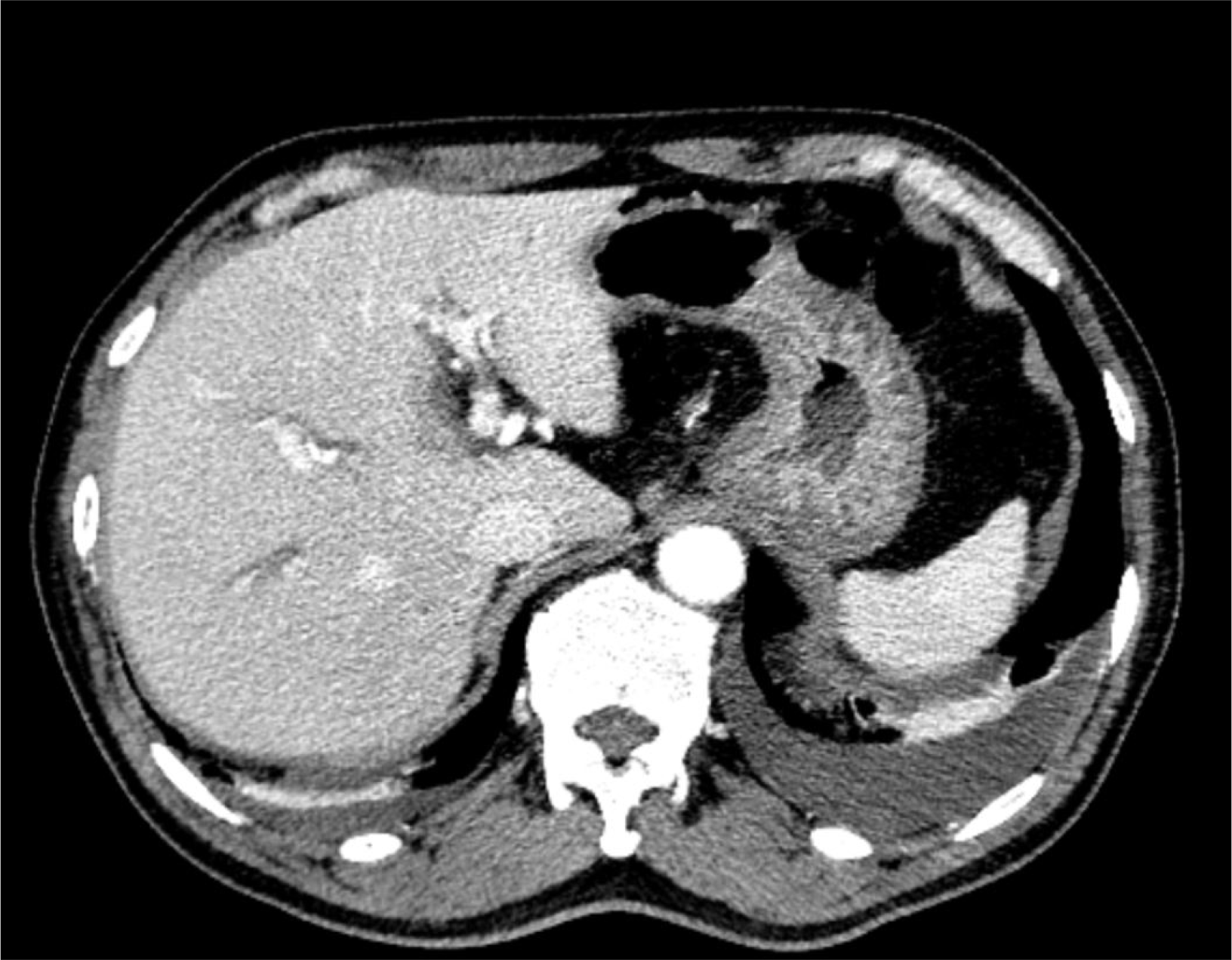
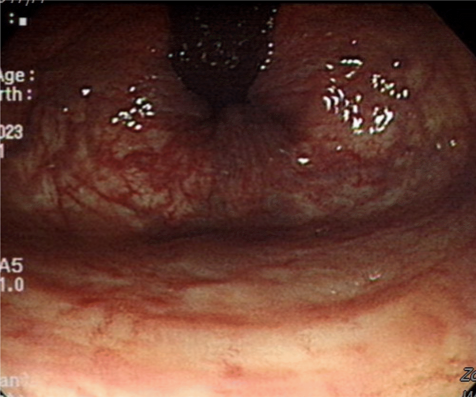
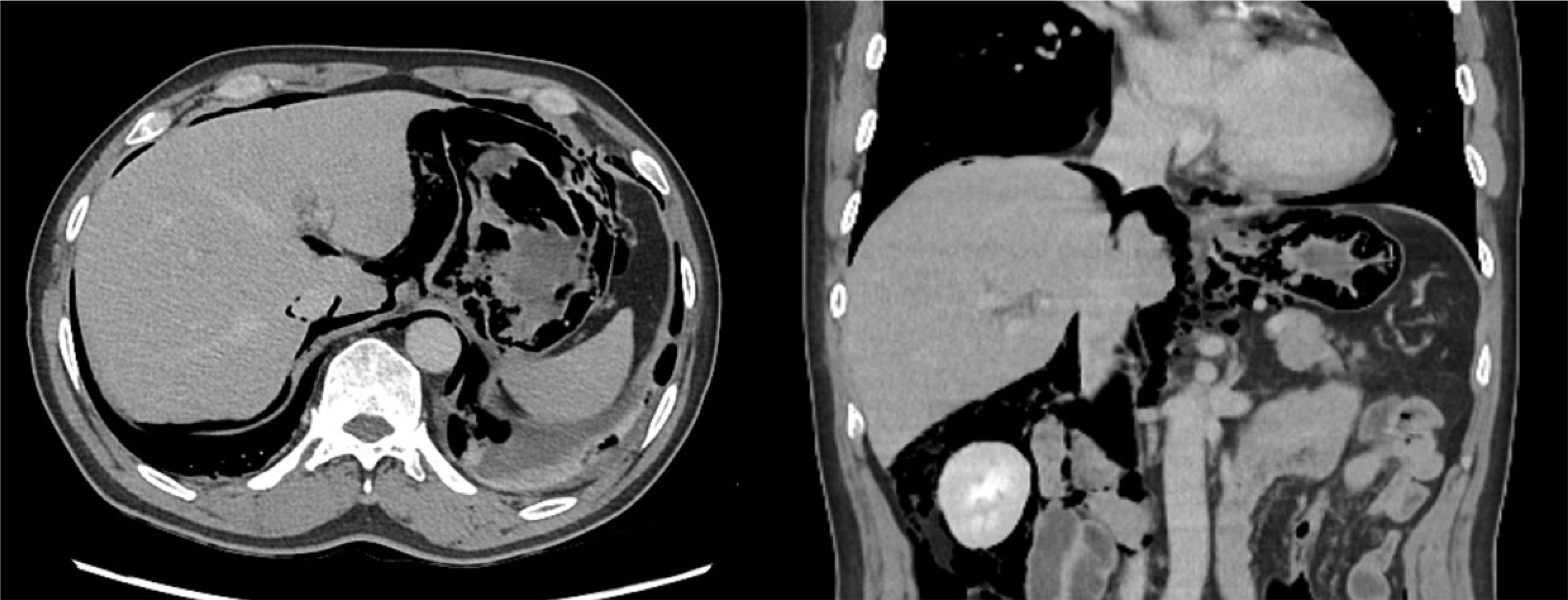
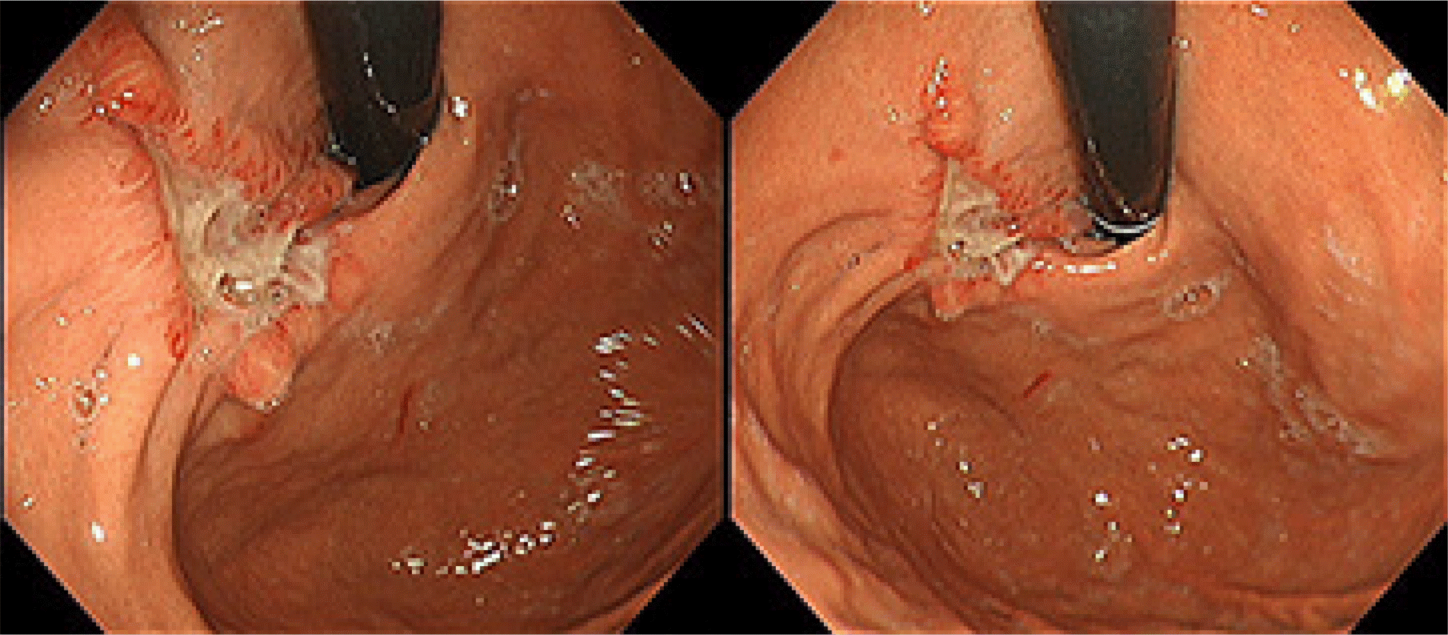
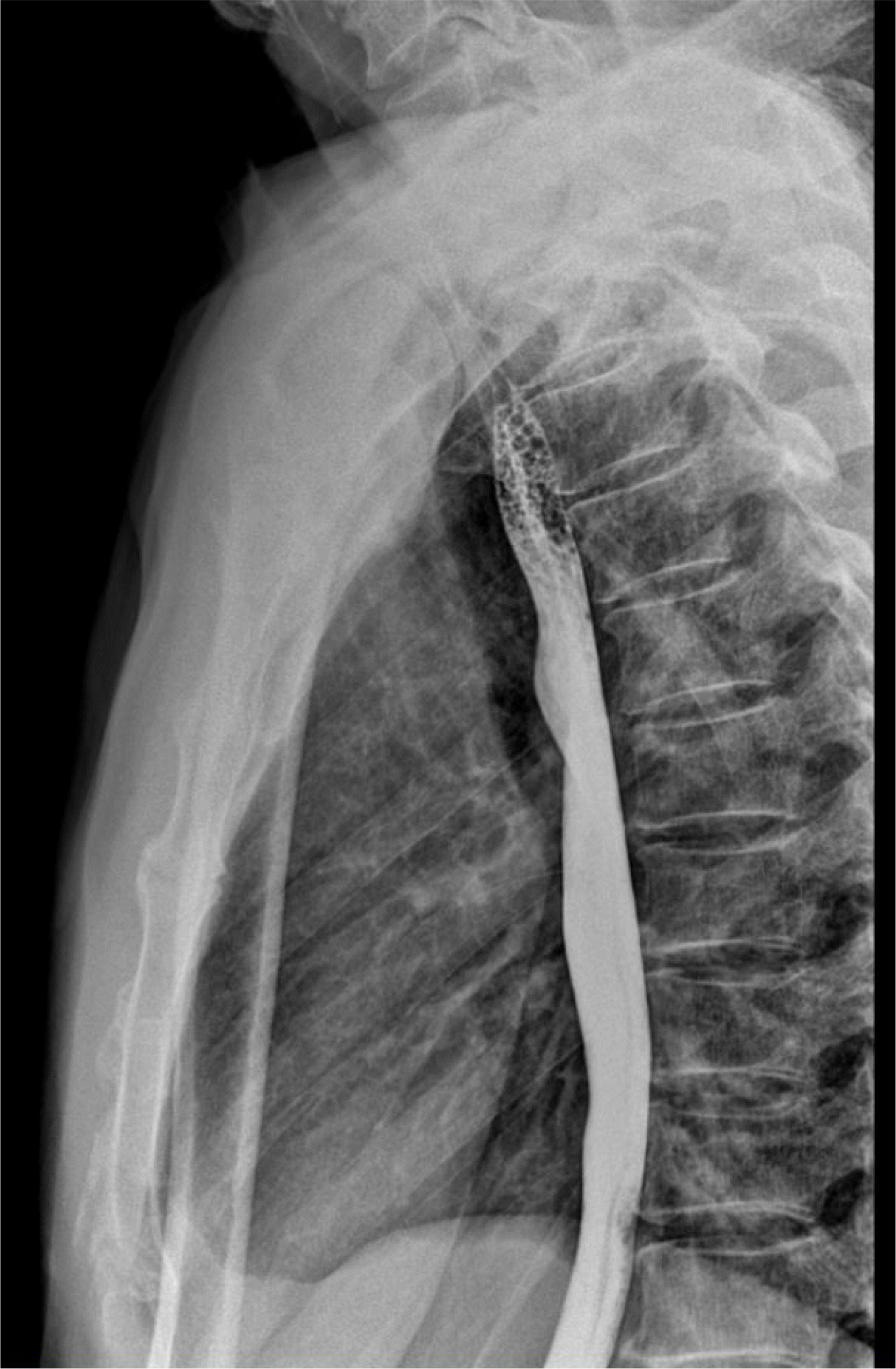
Afterwards, he was transferred to the hospital's emergency room, and his vital signs were: blood pressure 147/90, pulse 103, temperature 38.0, and respiration 20, accompanied by tachycardia and fever. On physical exam, in the neck and collarbone area, subcutaneous empysema was felt and epigastric tenderness was confirmed. A chest X-ray confirmed pneumomediastinum showing free air near the mediastinum, and so a computed tomography (CT) was performed. The Initial CT exam showed extensive pneumomediastinum extending to bilateral cervical spaces and diffuse pneumoperitoneum and extraperitoneal free air along the esophagus, stomach, perihepatic area, right retroperitoneal space, pelvic extraperitoneal space, right scrotum and bilateral adductor muscles (Fig. 2). No definite rupture of the cervical esophagus was seen. There was no evidence of prominent perforation such as fluid collection, and vital signs other than fever were relatively stable. After consulting with the surgeon, it was decided not to perform emergency surgery but to first hospitalize the patient in the internal medicine department for conservative treatment. The patient was admitted to the intensive care unit for vital sign monitoring, total non per oral & total parenteral nutrition were applied, and antibiotic treatment was administered. A second CT scan was performed the next day, and there was no significant change in the pneumoperitoneum, and there was no evidence of peritonitis. CT showed improvement, with partial resolving of the pneumomediastinum, no evidence of mediastinitis, and a thick esophagus wall, confirming the finding of esophageal inflammation rather than esophageal perforation (Fig. 3). Conservative treatment was maintained and intensive care unit care was performed. On the 8th day of hospitalization, a third CT was performed and it was confirmed that the existing emphysema, pneumomediastinum. pneumoperitoneum and esophagitis had significantly improved (Fig. 4). Gastroscopy was performed on the 9th day of hospitalization, and linear erosion from the upper esophageal sphincter to the cardia and ulcers near the cardia were confirmed, and there was no esophageal perforation (Fig. 5). Based on this endoscopic image, it appears that an ulcerated area of the cardia was the cause of the perforation. After the gastroscopy, liquid-soft diet was started, and after starting the diet, the patient was stable with no symptoms such as abdominal pain, and there were no special findings on lab or X-ray, so he was discharged on the 10th day of hospitalization. On the 5th day after discharge upper gastrointestinal (UGI) swallow radiographs was performed at an outpatient clinic, and no evidence of contrast leakage at esophagus was confirmed (Fig. 6).
Diagnosis: Pneumoperitoneum
There is a very rare chance of developing pneumoperitoneum or pneumomediastinum after UGI endoscopy.1 Pneumomediastinum is a condition in which air exists in the mediastinum due to perforation of adjacent organs, lung or esophageal disease. Pneumoperitoneum is a condition in which there is air in the abdomen and can usually occur when the abdomen is perforated. We present the case of a 64-year-old male patient who developed pneumoperitoneum and pneumomediastinum after UGI endoscopy. This patient showed no clear evidence of esophageal perforation on endoscopy, but perforation was suspected clinically. Generally, surgical intervention is considered necessary when gastrointestinal perforation occurs, but we would like to share this case because it improved without surgical intervention. Early identification and expeditious management of a perforation have been shown to decrease associated morbidity and mortality.2 In this case, we can suggest that pneumoperitoneum without clear evidence of esophageal perforation after gastroscopy can be improved with conservative care such as oxygen treatment, fasting, and antibiotics without surgical intervention.
REFERENCES
1. Krishnamurthy C, Hilden K, Peterson KA, Mattek N, Adler DG, Fang JC. 2012; Endoscopic findings in patients presenting with dysphagia: analysis of a national endoscopy database. Dysphagia. 27:101–105. DOI: 10.1007/s00455-011-9346-0. PMID: 21674194. PMCID: PMC5970000.
2. Lai CH, Lau WY. 2008; Management of endoscopic retrograde cholangiopancreatography-related perforation. Surgeon. 6:45–48. DOI: 10.1016/S1479-666X(08)80094-7. PMID: 18318088.
Fig. 3
Second computed tomography image showing partial resolving of the pneumomediastinum and esophageal inflammation with thickening of the esophageal wall.
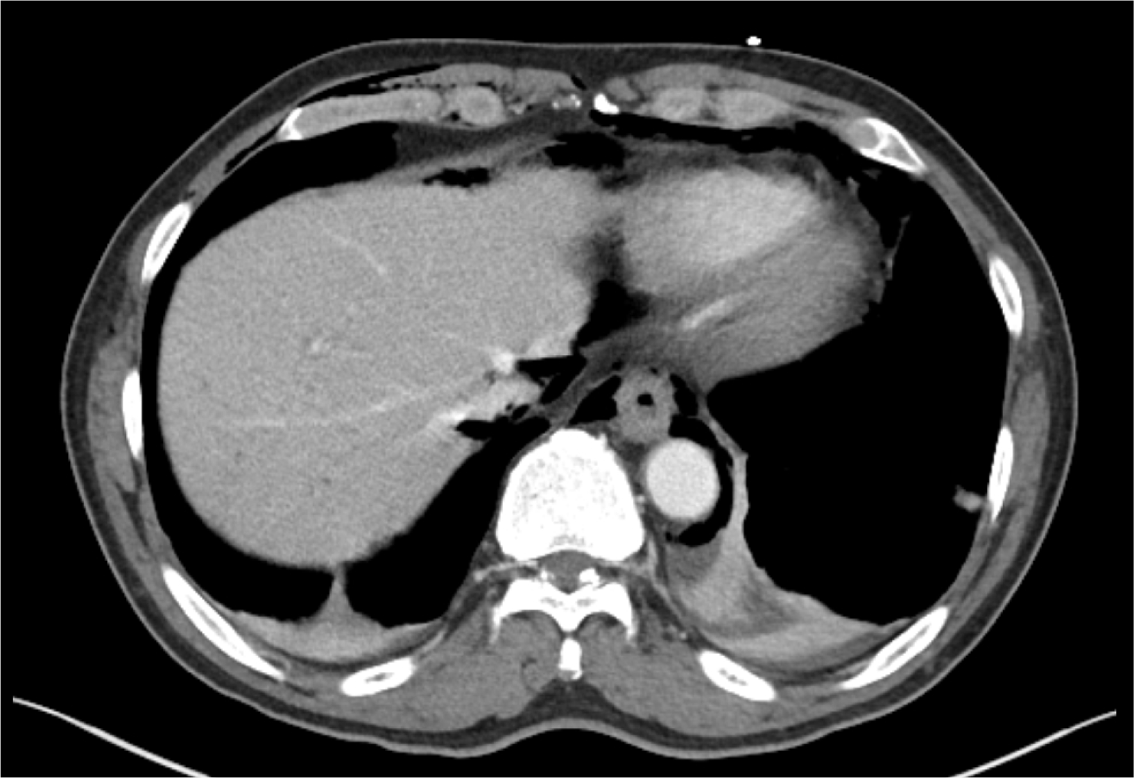
Fig. 4
Third Computed tomography image showing improved emphysema, pneumomediastinum, pneumoperitoneum and esophagitis.


 PDF
PDF Citation
 Print
Print


 XML Download