Journal List > J Stroke > v.26(3) > 1516088619

Feng, Chang, and Fu: Ruptured Peripheral Cerebral Aneurysms Associated With Moyamoya Disease: A Systematic Review

Abstract

Background and Purpose

A ruptured peripheral cerebral aneurysm (PPCA) associated with moyamoya disease (MMD) is a rare but potentially life-threatening condition with controversial management strategies. We aim to summarize the clinical characteristics, treatment strategies, and prognostic factors of PPCAs in MMD.

Methods

We reviewed studies published in PubMed between 1980 and 2023 and used logistic regression analysis to identify the risk factors for adverse outcomes.

Results

Of 425 identified studies, 48 eligible studies involving 121 participants were included in the current study. The mean age at diagnosis was 40.8±15.1 years, with a peak age of onset between 41 and 50 years. Among the identified participants, 59.6% were female, and 55.9% presented with impaired consciousness. Aneurysms were present in the posterior (35.5%) or anterior (30.6%) choroidal arteries in 66.1% of the cases, and 71.1% of the patients presented with intraventricular hemorrhage (IVH) with or without intracerebral hematoma (ICH). The treatment strategies were embolization (28.9%), direct surgery (21.5%), revascularization (22.3%), and conservation (27.3%). Favorable outcomes were achieved in 86.8% of all cases, with 97.1% for embolization, 65.4% for direct surgery, 96.3% for revascularization, and 84.8% for conservative treatment. Aneurysm rebleeding occurred in 11 (26.8%) of 41 patients managed conservatively, leading to worse outcomes in 7 patients (63.6%). Impaired consciousness (odds ratio [OR], 8.61; 95% confidence interval [CI], 2.06–36.00) and aneurysm rebleeding (OR, 16.54; 95% CI, 3.08–88.90) independently predicted poor outcomes.

Conclusion

PPCA should be considered in patients with hemorrhagic MMD, particularly those with IVH with or without ICH. Endovascular and bypass treatments are recommended as first-line options, with direct open surgery as an alternative in urgent hematoma evacuation cases. Detailed preoperative planning and intraoperative technical assistance are necessary to reduce procedure-related complications. Conservative management should be selected with caution because of the high risk of rebleeding and poor outcomes. Impaired consciousness and aneurysm rebleeding appeared to be independent risk factors for adverse prognoses. We emphasize that treatment selection should be personalized, and the potential benefits should be weighed against the associated risks.

Introduction

Cerebral aneurysms associated with moyamoya disease (MMD) remain a potential hemorrhagic factor, with a reported incidence of 3%–18% [1-3]. Based on the anatomic location, these aneurysms can be broadly classified as major trunk aneurysms located on the circle of Willis and peripheral cerebral aneurysms (PPCAs) originating from the choroidal, lenticulostriate, meningeal, or moyamoya arteries. MMD-associated aneurysms are more prone to bleeding because of their fragile aneurysmal wall and increased hemodynamic stress, which predisposes them to a poor prognosis [4-8].
Considering these risks, prompt and appropriate interventions are required to treat such aneurysms. Direct surgical or endovascular treatment is widely accepted for hemorrhagic aneurysms of the major trunk of the intracranial arteries. However, optimal treatment for ruptured PPCAs remains controversial. Some authors recommend direct treatment, including endovascular embolization or open surgery, whereas others advocate conservative observation or cerebral revascularization because of the tendency for spontaneous regression [2,5-51].
Notably, studies on ruptured PPCA associated with MMD are relatively few, and all of them are small, single-center, retrospective studies and case reports. To the best of our knowledge, there has been no comprehensive literature review on these studies. Consequently, we conducted a systematic review of MMD-related ruptured PPCAs published over the past 40 years to synthesize evidence on the clinical characteristics, treatment strategies, and prognostic factors, with the aim of improving the current understanding of this rare but important clinical entity.

Methods

Search strategies and study selection

A comprehensive literature search of the National Center for Biotechnology Information’s PubMed database was performed according to the Preferred Reporting Items for Systematic Reviews and Meta-Analyses (PRISMA) guidelines. The search term “(moyamoya [title/abstract]) AND (aneurysm [title/abstract])” was used to identify articles published between January 1980 and December 2023. The cited references were also searched to identify additional relevant studies.
Documents reporting intracranial aneurysms associated with MMD, regardless of the design, were considered eligible for inclusion. All the retrieved articles were screened based on their titles and abstracts. Studies were excluded based on the following criteria: irrelevant or non-English articles; reviews, letters to the editor, commentaries, abstracts, or book chapters; nonhuman studies; unruptured intracranial aneurysms; intracranial major artery aneurysms; and insufficient individual patient data.

Data extraction

We extracted the following information from each study: patient demographics; clinical presentation; hemorrhagic type; number of patients with acute hydrocephalus; aneurysm location; treatment modality; number of patients with aneurysm rebleeding; time of aneurysm rebleeding after the first ictus; time between treatment and aneurysm disappearance; and follow-up outcome.
Clinical outcomes, assessed using the Glasgow Outcome Scale (GOS) or the modified Rankin Scale (mRS), were dichotomized as good (GOS 4–5 or mRS 0–3) or poor (GOS 1–3 or mRS 4–6).
Two reviewers (ZF and YC) independently screened the full texts to identify potentially relevant studies and extracted the data. Discrepancies were resolved by consensus with a third reviewer (CF).

Statistical analyses

Continuous data are expressed as mean±standard deviation. Categorical variables were analyzed using Pearson’s chi-square test or Fisher’s exact test. Univariate and multivariate logistic regression analyses were used to identify the risk factors for poor outcomes. In the multivariate analysis, we performed pairwise comparisons between treatment modalities.
All statistical analyses were performed using SPSS version 19.0 (IBM Corp., Armonk, NY, USA). A two-sided P-value of less than 0.05 was considered statistically significant.

Results

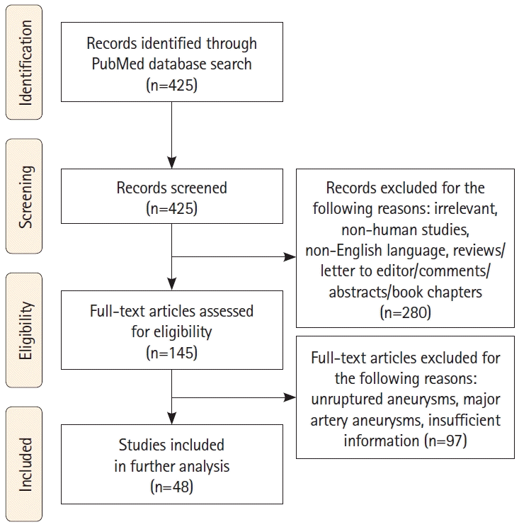
A total of 48 studies with 121 eligible patients were included in this systematic review (Figure 1). The characteristics of these publications are described in Supplementary Table 1. Regarding the bias concern in this systematic review of case reports, a JBI critical appraisal checklist for case reports (Joanna Briggs Institute, Adelaide, Australia; https://synthesismanual.jbi.global) is provided for the included articles (Supplementary Table 2).

Demographic and aneurysm characteristics

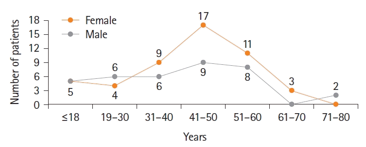
The general patient characteristics are summarized in Table 1. Regarding ethnicity, 41 (33.9%) were Chinese, 39 (32.2%) were Korean, 29 (24.0%) were Japanese, and 12 (9.9%) were Caucasian. Age data were available for 85 patients, and the mean age was 40.8±15.1 years (range, 7–74 years). Of these patients, eight were younger than 18 years, and the remaining 77 (90.6%) were 18 years or older. The peak age of onset was between 41 and 50 years in both male and female participants (Figure 2). Of the 104 patients with available sex data, 62 (59.6%) were female, resulting in a female-to-male ratio of 1.48:1. Admission presentation data were available for 68 patients, of whom 38 (55.9%) exhibited impaired consciousness, and 26 (38.2%) experienced headaches (with or without nausea/vomiting). The hemorrhagic types included subarachnoid hemorrhage (SAH), intraventricular hemorrhage (IVH), intracerebral hemorrhage (ICH), and their combinations. The most common type was IVH (61/121, 50.4%), followed by IVH+ICH (25/121, 20.7%) and ICH (18/121, 14.9%). Acute hydrocephalus was observed in 21 patients (17.4%). Aneurysms were frequently found in the posterior choroidal artery (PChA; 43/121, 35.5%) and anterior choroidal artery (AChA; 37/121, 30.6%).

Treatment modalities and clinical outcomes

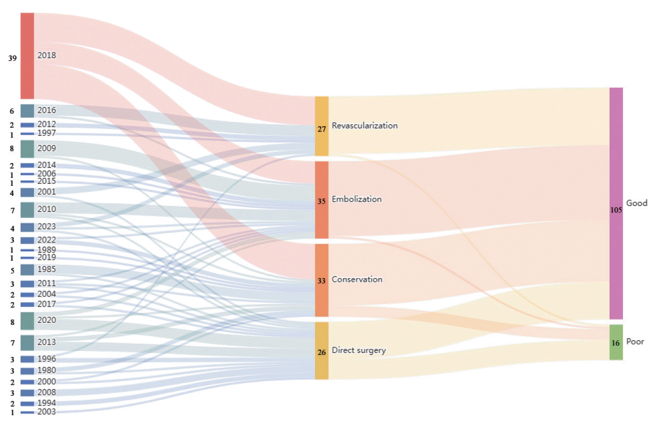
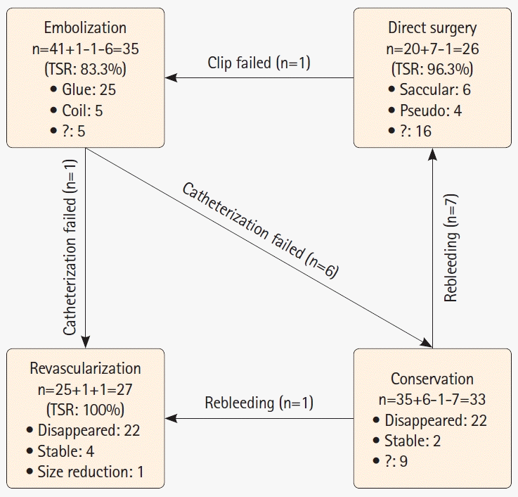
The relationships among the number of cases, year of report, treatment selection, and follow-up outcomes are presented in a Sankey diagram (Figure 3). Of the 121 patients with PPCA, 35 (28.9%) underwent embolization, 26 (21.5%) underwent direct surgery, 27 (22.3%) underwent revascularization, and 33 (27.3%) were managed conservatively (Figure 4). The technical success rates for targeted embolization, direct surgery, and cerebral revascularization were 83.3% (35/42), 96.3% (26/27), and 100% (27/27), respectively. Two patients underwent bypass surgery upon diagnosis of MMD before PPCA formation [26,40].
In the embolization group, aneurysms were embolized with or without sacrificing the parent arteries. Liquid adhesives were used for embolization in 25 patients (71.4%), and detachable coils were used in 5 patients (14.3%).
In the direct surgery group, pathological histology was examined in 10 (38.5%) of 26 patients; 6 patients (60%) had saccular aneurysms and 4 (40%) had pseudoaneurysms.
In the revascularization group, 22 (81.5%) of 27 PPCAs disappeared and 5 (18.5%) either decreased in size or remained stable during follow-up. The time from aneurysm diagnosis to disappearance was available for 7 patients, ranging from 1 to 24 months, with an average of 7 months.
In the conservative therapy group, follow-up angiographic imaging was available for 24 (72.7%) of 33 patients. Among these, 22 (91.7%) aneurysms disappeared, with time data available for 9 cases, ranging from 3 weeks to 12 months (mean, 6.1 months), and 2 aneurysms (8.3%) remained stable in size.
Overall, 105 (86.8%) of 121 patients achieved a favorable outcome, whereas the remaining 16 patients had unfavorable outcomes. The rates of good outcomes were 97.1% (34/35) in the embolization group, 65.4% (17/26) in the direct surgery group, 96.3% (26/27) in the revascularization group, and 84.8% (28/33) in the conservative therapy group.

Aneurysm rebleeding

Aneurysm re-rupture occurred in 11 (9.1%) of the 121 participants, all of whom underwent conservative treatment [10,12,14,16,26,32,35,40,47,50]. The rate of rebleeding in cases that received conservative treatment was 26.8% (11/41). The timing of rebleeding was available for 10 patients, which varied from 1 day to 7 years (mean, 12 months). Most rebleeding episodes (7/10, 70%) were observed within 35 days of the initial bleeding. Of the 11 patients, only four achieved a good outcome, and the remaining 7 (63.6%) had a poor outcome.

Prognostic factors

In the univariate logistic regression analysis, there were significant differences in the outcomes of impaired consciousness (P=0.042), treatment modality (P=0.001), and aneurysm rebleeding (P<0.001) between the two groups (Table 2). No significant correlation was found between the outcome and ethnicity, age, sex, hemorrhagic type, acute hydrocephalus, or aneurysm location.
Multivariate logistic regression analysis revealed that impaired consciousness (odds ratio [OR], 8.61; 95% confidence interval [CI], 2.06–36.00; P=0.003) and aneurysm rebleeding (OR, 16.54; 95% CI, 3.08–88.90; P=0.001) were independent risk factors for poor outcome, whereas treatment modality was not (Table 3).

Discussion

MMD-associated PPCA has been rarely reported. Only 121 ruptured PPCAs have been described in patients with MMD in the past 40 years. We hypothesized that these aneurysms may be underestimated in patients with MMD based on the following findings: this lesion often remains undiagnosed unless it ruptures; it tends to be missed by routine brain imaging, especially when the size is small. Therefore, catheter-based cerebral angiography is recommended for the detection of aneurysms, when possible.
The exact mechanisms contributing to the formation, growth, and rupture of such aneurysms have not been clearly elucidated. Several factors may be involved, including pathological vascular architecture, long-term hemodynamic stress, and aneurysm location [5-7,41]. Unilateral MMD is susceptible to the formation of anterior circulation aneurysms on the unaffected side, whereas bilateral MMD is prone to the formation of posterior circulation aneurysms [1]. Under normal circumstances, high Suzuki stages are often associated with uncoordinated distribution of the cerebral circulation and development of collateral compensation in the posterior circulation, thereby predisposing to the PPCA formation owing to the long-term hemodynamic burden on the fragile collateral vessels [1,52,53]. Recent clinical studies suggested the impact of choroidal anastomosis (so-called choroidal channel) as dangerous collaterals with a high bleeding risk among the variety of “periventricular anastomosis” in adult patients with hemorrhagic MMD [52-54]. Based on this observation, PPCAs at choroidal channel are considered to be at a high risk of rupture, which requires prompt management.
In terms of ethnicity, the largest number of participants were from Asia, particularly China, followed by Korea and Japan. Our study revealed that the mean age at diagnosis was 40.8±15.1 years, and there was a higher prevalence in females than in males, which is consistent with the findings of previous studies [55,56]. Compared with SAH in main trunk aneurysms, the most common hemorrhagic type in PPCAs was IVH with or without ICH. Such aneurysms occurred more frequently in the PChA and AChA. Therefore, PPCA should be considered as a potential cause of hemorrhagic MMD, particularly when manifested as IVH with or without ICH, and catheter-based angiography is essential to identify this underestimated but important lesion.
Theoretically, the presence of MMD-associated PPCA represents a potential hemorrhagic risk if not obliterated. Some studies show that hemorrhagic MMD has higher rates of rebleeding (28%–35%) and mortality (7%–10%) [4,5,41,57,58]. The rate of good outcome after the first rupture was 60%, whereas it decreased to 40% after further bleeding [4,57]. Rhim et al. [41] reported that the rate of rebleeding in aneurysm-related hemorrhagic MMD (8/19, 42.1%) was significantly higher than that of aneurysm-unrelated hemorrhagic MMD (13/58, 22.4%); all aneurysmal rebleeding events occurred at least 6 months after the initial hemorrhage. Iwama et al. [59] reported 7 participants with rebleeding at the same site, 4 with MMD-associated aneurysms, and most rebleeding episodes (6/7, 85.7%) were observed within 2 months after the first onset. In the present study, aneurysmal rebleeding occurred in 9.1% of the cases, and the rate of poor outcome was 63.6% among them; in terms of timing, 70% of aneurysmal rebleeding occurred within the first 35 days. Therefore, elimination of such aneurysms to prevent recurrent rupture is plausible but remains controversial. Several authors have preferred to treat this lesion with direct endovascular or surgical intervention, whereas others have opted for revascularization surgery or conservative management, because of its tendency to regress spontaneously [2,5-51,60].
Endovascular embolization is recommended as a safe, feasible, and effective treatment option for most PPCAs in patients with MMD [2,5,7,21,22,24,25,27,28,30,33,37-39,41,43,45,46,51]. Kim et al. [5] reported on a cohort of 8 ruptured PPCAs in MMD cases treated with endovascular therapy, 7 of which were uneventful. Endovascular treatment was attempted in 42 patients and was successful in 35. In contrast to the treatment of main trunk aneurysms, liquid adhesive embolization, rather than coiling with or without a stent-assisted technique, is the preferred option for aneurysms of the collateral vessels. In the cases we identified, 25 (71.4%) of the 35 patients who underwent successful endovascular treatment were treated with n-butyl cyanoacrylate, ethylene-vinyl alcohol copolymer (Onyx), or cyanoacrylate (Glubran). Typically, aneurysms and their adjacent parent arteries are occluded together. Some authors have explained the use of liquid embolic agents for aneurysm obliteration because the lesion and the collateral vessel tend to be small and fragile, and the delivery of the detachable coil or stent might predispose them to rupture or dangerous tearing [5,15]. In addition, accessible selective catheterization is critical to the technical success of embolization because the parent artery is usually thin and tortuous. Our current study showed that 7 (16.7%) of 42 PPCAs could not be embolized because of approach inaccessibility [5,9,26,28,36,39,47].
Direct surgery, such as neck clipping or aneurysm resection, has been considered too risky for the treatment of MMD-associated ruptured PPCAs for the following reasons: these aneurysms are generally small and deep-seated, making them difficult to localize and identify; surgical clipping is difficult to perform in some cases due to the lack of a defined aneurysm neck; these aneurysms are prone to re-rupture with significant morbidity and mortality during intraoperative dissection and retraction, given their fragile characteristics; there is a potential risk of injury to the abnormal, fragile collateral vessels during surgical exposure; patients with MMD might be more susceptible to cerebral ischemia because of the poor reserve capacity of hemodynamics [2,39,40,42]. Under such circumstances, direct surgical repair might be a significant challenge and is not recommended for these aneurysms. Nevertheless, this therapeutic strategy has unique advantages over the other three, including surgical hematoma evacuation and aneurysm acquisition for further histopathological examination. The pathological features of MMD-associated PPCAs remain unclear owing to limited histopathological data and because they are typically considered as pseudoaneurysms based on their anatomic location and tendency for early morphologic change [2,25,48,49,51]. However, in this review, of the 10 PPCAs with available histopathological diagnosis, 6 (60%) were saccular aneurysms and the other 4 (40%) were pseudoaneurysms [1,6,10-12,14,17,18,29].
Cerebral revascularization has been reported as an alternative treatment to eliminate MMD-associated PPCAs [2,15,16,19,31,39,48,49]. Ni et al. [2] treated 13 MMD-related PPCAs with pure revascularization (superficial temporal artery–middle cerebral artery bypass combined with encephalo-duro-myo-synangiosis) because of microcatheter inaccessibility, of which 12 were obliterated and 1 remained stable during the follow-up period. Liu et al. [39] performed bilateral encephalo-duro-arterio-synangiosis to treat 5 pediatric PPCAs, 3 of which regressed spontaneously and the other 2 were stable at follow-up. Here, we observed 27 ruptured PPCAs treated with revascularization, of which 22 disappeared, 4 were stable, and 1 decreased in size. The exact mechanism underlying the spontaneous resolution of this lesion after bypass surgery is currently unknown. However, the reduction of the hemodynamic burden on the aneurysm and its parent artery by bypass surgery explains this phenomenon to some extent [48,49]. In particular, Lee et al. [49] found dynamic changes in diameter associated with altered hemodynamics in the parent artery (from 2.42 mm initially to 5.33 mm and finally to 1.51 mm), which poses a risk of aneurysm rupture. Moreover, there is a potential risk of PPCA formation and rupture if hemodynamic stress is not effectively reduced, even after surgical revascularization, as shown in two reported cases [26,40]. The authors suggested that impaired collateral circulation after bypass could result in aneurysm formation, and diminished collateral circulation during the follow-up period might be a sign of aggravated hemodynamic stress leading to aneurysm formation. Notably, de novo aneurysm formation around the anastomosis site or within the collateral vascular network after revascularization surgery cannot be ignored [61-67]. This postoperative complication has been rarely reported but may be clinically underestimated because it is often asymptomatic and undiagnosed [62]. Regarding the mechanism of aneurysm formation after intracranial anastomotic surgery, the following hypotheses have been proposed: disruption of the internal elastic layer and tunica media of the arterial anastomosis during the revascularization procedure might lead to traumatic aneurysm formation at the suture line; the jet blood flow from the donor arteries could directly impact the artificial T-bifurcation, causing wall stress shear and leading to spindle-like dilatation around the site of anastomosis, predisposing to the aneurysm formation under the influence of increased hemodynamic stress; a patient with MMD with the genetic variant RNF213 c.14576G>A might carry a higher risk of repeated de novo aneurysm formation. The time from bypass surgery to aneurysm diagnosis in the reported cases ranged from 6 months to 20 years, suggesting that aneurysm formation is probably time-dependent [61-67]. Therefore, long-term surveillance after cerebral revascularization seems to be essential.
Conservative therapy was selected as a viable alternative for MMD-related hemorrhagic PPCA in the following situations: high-risk surgical procedures; inaccessible endovascular approach; and the potential tendency of the aneurysm to resolve spontaneously [5,7-9,11-13,17,28,34-36,41,43]. Our review of the literature showed that 33 ruptured PPCAs were treated with conservative observation, of which 22 (66.7%) spontaneously regressed and 2 (6.1%) remained stable. The mean time to aneurysm disappearance was 6.1 months, ranging from 3 weeks to 1 year. Some authors have speculated that cerebral vasospasm and secondary thrombosis might explain the phenomenon of spontaneous aneurysm regression in the setting of conservative treatment [8,36]. Considering that such aneurysms usually originate from small-caliber collateral branches, vasospasm after aneurysmal hemorrhage might lead to the retention of blood flowing into the aneurysm sac, thereby resulting in thrombosis obliteration. In addition, some authors have suggested spontaneous regression as the natural history of pseudoaneurysms [68,69]. Notably, conservatively managed MMD-associated ruptured PPCAs are at a high risk of recurrent hemorrhage, which correlates with high morbidity and mortality. In the present review, we identified 11 cases of aneurysm re-rupture; all rebleeding occurred during the observation period [10,12,14,16,26,32,35,40,47,50]. Most rebleeding events (70%) occurred within the first 35 days after ictus. The rates of rebleeding and the associated poor prognosis in these patients were as high as 26.8% and 63.6%, respectively, suggesting that aggressive intervention should be undertaken to prevent future rupture.
The overall outcomes of these patients were favorable (86.8%). Good outcomes were achieved in 97.1%, 65.4%, 96.3%, and 84.8% of the cases treated with endovascular embolization, direct surgery, cerebral revascularization, and conservative observation, respectively. These results indicate that the therapeutic effect of embolization may be comparable to that of revascularization, all of which are superior to conservative observations. The treatment outcomes of direct open surgery were the worst. However, it should be emphasized that most participants in the open surgery group were treated 15 years ago, when knowledge about this lesion was relatively inadequate and the surgical procedures were potentially more invasive. Studies have reported that the advancement and application of stereotaxy, neuronavigation, and neuroendoscopic techniques have made open surgery safer, more effective, and less invasive [10,23,29,42]. However, the prognostic factors for patients with MMD-associated PPCAs remain unclear. Our review identified the independent risk factors for poor outcomes in these patients, including impaired consciousness and aneurysm rebleeding. Patients with impaired consciousness or aneurysm rebleeding showed an 8.6- and 16.5-fold increased risk of adverse prognosis, respectively, compared with those without symptoms.
This systematic review has several limitations. First, some eligible records may have been excluded because only PubMed was searched. Second, we only included articles published in English; thus, articles from high-incidence regions in other languages, such as Chinese, Japanese, and Korean, were excluded. Third, there is the potential for study heterogeneity, and a statistical bias exists. Fourth, our results were based on retrospective case series and single-case reports, which are considered lower-quality evidence. Fifth, the reliability of our study is weakened by the small sample size. Nevertheless, this review provides preliminary evidence for the selection of appropriate treatment options for such aneurysms.

Conclusions

MMD-associated ruptured PPCAs are more prevalent in females, with a peak age of onset at 41–50 years. These aneurysms most commonly arise from the choroidal arteries and frequently present with IVH with or without ICH. When hemorrhagic MMD is encountered, PPCA should be considered, and catheter-based angiography is essential to identify such lesions. Endovascular intervention or bypass surgery may be a first-line therapeutic option. In certain cases, particularly when urgent hematoma evacuation is required, direct open surgery may be an alternative. However, detailed preoperative planning and intraoperative technical assistance are required to reduce procedure-related complications. Conservative observation should be chosen with caution because of the high risk of aneurysm rebleeding and the associated worse outcomes, unless individuals are deemed unsuitable for surgical or endovascular treatment. The overall outcome seemed to be favorable, and impaired consciousness and aneurysm rebleeding may be independent risk factors for an unfavorable prognosis. We emphasize the importance of personalized treatment plans to balance the benefits and risks. Further studies with larger sample sizes or randomized controlled trials are warranted to facilitate a better understanding of this rare but clinically important condition.

Supplementary materials

Supplementary materials related to this article can be found online at https://doi.org/10.5853/jos.2024.02061.
Supplementary Table 1.
Literature review of ruptured PPCAs associated with MMD (1980–2023)
jos-2024-02061-Supplementary-Table-1.pdf
Supplementary Table 2.
Risk of bias assessment criteria for included case reports (JBI critical appraisal checklist)
jos-2024-02061-Supplementary-Table-2.pdf

Notes

Funding statement

None

Conflicts of interest

The authors have no financial conflicts of interest.

Author contribution

Conceptualization: ZF, CF. Study design: ZF, CF. Methodology: all authors. Data collection: all authors. Investigation: all authors. Statistical analysis: CF. Writing—original draft: ZF, CF. Writing—review & editing: CF. Approval of final manuscript: all authors.

References

1. Larson AS, Rinaldo L, Brinjikji W, Lanzino G. Location-based treatment of intracranial aneurysms in moyamoya disease: a systematic review and descriptive analysis. Neurosurg Rev. 2021; 44:1127–1139.
crossref
2. Ni W, Jiang H, Xu B, Lei Y, Yang H, Su J, et al. Treatment of aneurysms in patients with moyamoya disease: a 10-year single-center experience. J Neurosurg. 2018; 128:1813–1822.
crossref
3. Kim S, Jang CK, Park EK, Shim KW, Kim DS, Chung J, et al. Clinical features and outcomes of intracranial aneurysm associated with moyamoya disease. J Clin Neurol. 2020; 16:624–632.
crossref
4. Yoshida Y, Yoshimoto T, Shirane R, Sakurai Y. Clinical course, surgical management, and long-term outcome of moyamoya patients with rebleeding after an episode of intracerebral hemorrhage: an extensive follow-up study. Stroke. 1999; 30:2272–2276.
5. Kim SH, Kwon OK, Jung CK, Kang HS, Oh CW, Han MH, et al. Endovascular treatment of ruptured aneurysms or pseudoaneurysms on the collateral vessels in patients with moyamoya disease. Neurosurgery. 2009; 65:1000–1004.
crossref
6. Okawa M, Abe H, Ueba T, Higashi T, Inoue T. Surgical management of a ruptured posterior choroidal intraventricular aneurysm associated with moyamoya disease. Cent Eur J Med. 2013; 8:818–821.
crossref
7. Kim JH, Kwon TH, Kim JH, Chong K, Yoon W. Intracranial aneurysms in adult moyamoya disease. World Neurosurg. 2018; 109:e175–e182.
crossref
8. Yamada H, Saga I, Kojima A, Horiguchi T. Short-term spontaneous resolution of ruptured peripheral aneurysm in moyamoya disease. World Neurosurg. 2019; 126:247–251.
crossref
9. Arai Y, Matsuda K, Isozaki M, Nakajima T, Kikuta K. Ruptured intracranial aneurysms associated with moyamoya disease: three case reports. Neurol Med Chir (Tokyo). 2011; 51:774–776.
10. Ali MJ, Bendok BR, Getch CC, Gottardi-Littell NR, Mindea S, Batjer HH. Surgical management of a ruptured posterior choroidal intraventricular aneurysm associated with moyamoya disease using frameless stereotaxy: case report and review of the literature. Neurosurgery. 2004; 54:1019–1024.
crossref
11. Tanaka Y, Takeuchi K, Akai K. Intracranial ruptured aneurysm accompanying moyamoya phenomenon. Acta Neurochir (Wien). 1980; 52:35–43.
crossref
12. Konishi Y, Kadowaki C, Hara M, Takeuchi K. Aneurysms associated with moyamoya disease. Neurosurgery. 1985; 16:484–491.
crossref
13. Grabel JC, Levine M, Hollis P, Ragland R. Moyamoya-like disease associated with a lenticulostriate region aneurysm: case report. J Neurosurg. 1989; 70:802–803.
crossref
14. Hamada J, Hashimoto N, Tsukahara T. Moyamoya disease with repeated intraventricular hemorrhage due to aneurysm rupture: report of two cases. J Neurosurg. 1994; 80:328–331.
15. Kawaguchi S, Sakaki T, Morimoto T, Kakizaki T, Kamada K. Characteristics of intracranial aneurysms associated with moyamoya disease: a review of 111 cases. Acta Neurochir (Wien). 1996; 138:1287–1294.
crossref
16. Kawai K, Narita K, Nakayama H, Tamura A. Ventricular hemorrhage at an early stage of moyamoya disease—case report. Neurol Med Chir (Tokyo). 1997; 37:184–187.
crossref
17. Miyake H, Ohta T, Kajimoto Y, Ogawa R, Deguchi J. Intraventricular aneurysms—three case reports. Neurol Med Chir (Tokyo). 2000; 40:55–60.
18. Lee JK, Lee JH, Kim SH, Lee MC. Distal anterior choroidal artery aneurysm in a patient with moyamoya disease: case report. Neurosurgery. 2001; 48:222–225.
crossref
19. Kuroda S, Houkin K, Kamiyama H, Abe H. Effects of surgical revascularization on peripheral artery aneurysms in moyamoya disease: report of three cases. Neurosurgery. 2001; 49:463–467. discussion 467-468.
20. Wong GK, Boet R, Poon WS. Ruptured distal anterior choroidal artery aneurysm presenting with right intracerebral haematoma: clipping aided by subpial uncal resection. J Clin Neurosci. 2003; 10:689–691.
21. Koebbe CJ, Horowitz MB. A rare case of a ruptured middle meningeal aneurysm causing intracerebral hematoma in a patient with moyamoya disease. AJNR Am J Neuroradiol. 2004; 25:574–576.
22. Baik SK, Kim YS, Lee HJ, Kim IS, Kang DS. Endovascular management of peripheral lateral posterior choroidal artery aneurysm associated with moyamoya disease: case report and review of the literature. Neurointervention. 2006; 1:44–49.
23. Gandhi CD, Gilad R, Patel AB, Haridas A, Bederson JB. Treatment of ruptured lenticulostriate artery aneurysms. J Neurosurg. 2008; 109:28–37.
24. Choulakian A, Drazin D, Alexander MJ. NBCA embolization of a ruptured intraventricular distal anterior choroidal artery aneurysm in a patient with moyamoya disease. J Neurointerv Surg. 2010; 2:368–370.
25. Park YS, Suk JS, Kwon JT. Repeated rupture of a middle meningeal artery aneurysm in moyamoya disease: case report. J Neurosurg. 2010; 113:749–752.
crossref
26. Leung GK, Lee R, Lui WM, Hung KN. Thalamo-perforating artery aneurysm in moyamoya disease–case report. Br J Neurosurg. 2010; 24:479–481.
27. Yang S, Yu JL, Wang HL, Wang B, Luo Q. Endovascular embolization of distal anterior choroidal artery aneurysms associated with moyamoya disease. A report of two cases and a literature review. Interv Neuroradiol. 2010; 16:433–441.
crossref
28. Yu JL, Wang HL, Xu K, Li Y, Luo Q. Endovascular treatment of intracranial aneurysms associated with moyamoya disease or moyamoya syndrome. Interv Neuroradiol. 2010; 16:240–248.
crossref
29. Lévêque M, McLaughlin N, Laroche M, Bojanowski MW. Endoscopic treatment of distal choroidal artery aneurysm. J Neurosurg. 2011; 114:116–119.
30. Xu HW, Li MH, Guan S, Sun SL. Two different methods of endovascular treatment for ruptured intracranial aneurysm associated with moyamoya disease and review of the literature. Life Sci J. 2011; 8:386–389.
31. Ni W, Xu F, Xu B, Liao Y, Gu Y, Song D. Disappearance of aneurysms associated with moyamoya disease after STA-MCA anastomosis with encephaloduro myosynangiosis. J Clin Neurosci. 2012; 19:485–487.
crossref
32. Hayashi K, Horie N, Nagata I. A case of unilateral moyamoya disease suffered from intracerebral hemorrhage due to the rupture of cerebral aneurysm, which appeared seven years later. Surg Neurol Int. 2013; 4:17.
crossref
33. Chalouhi N, Tjoumakaris S, Gonzalez LF, Dumont AS, Shah Q, Gordon D, et al. Onyx embolization of a ruptured lenticulostriate artery aneurysm in a patient with moyamoya disease. World Neurosurg. 2013; 80:436.e7–436.e10.
crossref
34. Okamura K, Higuchi T, Izumo T, Takahira R, Sadakata E, Yoshida M, et al. Ruptured basilar artery perforator aneurysm: a novel mechanism of pure subarachnoid hemorrhage in moyamoya disease. Illustrative case. J Neurosurg Case Lessons. 2022; 4:CASE22238.
35. He K, Zhu W, Chen L, Mao Y. Management of distal choroidal artery aneurysms in patients with moyamoya disease: report of three cases and review of the literature. World J Surg Oncol. 2013; 11:187.
crossref
36. Yuan Z, Woha Z, Weiming X. Intraventricular aneurysms: case reports and review of the literature. Clin Neurol Neurosurg. 2013; 115:57–64.
crossref
37. Hwang K, Hwang G, Kwon OK. Endovascular embolization of a ruptured distal lenticulostriate artery aneurysm in patients with moyamoya disease. J Korean Neurosurg Soc. 2014; 56:492–495.
crossref
38. Daou B, Chalouhi N, Tjoumakaris S, Rosenwasser RH, Jabbour P. Onyx embolization of a ruptured aneurysm in a patient with moyamoya disease. J Clin Neurosci. 2015; 22:1693–1696.
crossref
39. Liu P, Lv XL, Liu AH, Chen C, Ge HJ, Jin HW, et al. Intracranial aneurysms associated with moyamoya disease in children: clinical features and long-term surgical outcome. World Neurosurg. 2016; 94:513–520.
40. Kim YS, Joo SP, Lee GJ, Park JY, Kim SD, Kim TS. Ruptured choroidal artery aneurysms in patients with moyamoya disease: two case series and review of the literatures. J Clin Neurosci. 2017; 44:236–239.
crossref
41. Rhim JK, Cho YD, Jeon JP, Yoo DH, Cho WS, Kang HS, et al. Ruptured aneurysms of collateral vessels in adult onset moyamoya disease with hemorrhagic presentation. Clin Neuroradiol. 2018; 28:191–199.
crossref
42. Tokairin K, Kazumata K, Gotoh S, Sugiyama T, Kobayashi H. Neuroendoscope-assisted aneurysm trapping for ruptured intraventricular aneurysms in moyamoya disease patients. World Neurosurg. 2020; 141:278–283.
crossref
43. Zhao X, Wang X, Wang M, Meng Q, Wang C. Treatment strategies of ruptured intracranial aneurysms associated with moyamoya disease. Br J Neurosurg. 2021; 35:209–215.
crossref
44. Larson A, Rinaldo L, Brinjikji W, Meyer F, Lanzino G. Intracranial aneurysms in white patients with moyamoya disease: a U.S. single-center case series and review. World Neurosurg. 2020; 138:e749–e758.
45. Goto Y, Oka H, Hino A. Managing intervention for severe intraventricular hemorrhage casting in moyamoya disease: report of two cases. Int J Surg Case Rep. 2020; 73:271–276.
crossref
46. Byeon Y, Kim HB, You SH, Yang K. A ruptured lenticulostriate artery aneurysm in moyamoya disease treated with Onyx embolization. J Cerebrovasc Endovasc Neurosurg. 2022; 24:154–159.
crossref
47. Fu C, Jiang P, Zhao Y, Li Y. Recurrent artery of heubner aneurysm masquerading as caudate hemorrhage without subarachnoid hemorrhage in moyamoya disease: a case report and literature review. Curr Med Imaging. 2022; 18:429–431.
crossref
48. Kusakabe T, Oda K, Kobayashi H, Kawano D, Yoshinaga S, Fukumoto H, et al. Disappearance of a moyamoya-related distal anterior cerebral artery aneurysm after target bypass revascularization: illustrative case. J Neurosurg Case Lessons. 2023; 6:CASE23200.
crossref
49. Lee GY, Cho BK, Hwang SH, Roh H, Kim JH. Hydration-induced rapid growth and regression after indirect revascularization of an anterior choroidal artery aneurysm associated with moyamoya disease: a case report. J Cerebrovasc Endovasc Neurosurg. 2023; 25:75–80.
50. Ding W, Zhao Y, Liu L, Wang P, Qiu W, Ren H, et al. Moyamoya disease with distal anterior choroidal artery aneurysm resected via transcallosal approach: a case report and review. Medicine (Baltimore). 2023; 102:e33973.
crossref
51. Ando M, Maki Y, Hojo M, Hatano T. Ruptured saccular aneurysm of the lenticulostriate artery embolized without parent artery occlusion in a case of moyamoya disease. Neuroradiol J. 2023; 36:108–111.
crossref
52. Funaki T, Takahashi JC, Houkin K, Kuroda S, Takeuchi S, Fujimura M, et al. Angiographic features of hemorrhagic moyamoya disease with high recurrence risk: a supplementary analysis of the Japan adult moyamoya trial. J Neurosurg. 2018; 128:777–784.
crossref
53. Fujimura M, Funaki T, Houkin K, Takahashi JC, Kuroda S, Tomata Y, et al. Intrinsic development of choroidal and thalamic collaterals in hemorrhagic-onset moyamoya disease: casecontrol study of the Japan adult moyamoya trial. J Neurosurg. 2019; 130:1453–1459.
crossref
54. Wiedmann MKH, Davidoff C, Lo Presti A, Ni W, Rhim JK, Simons M, et al. Treatment of ruptured aneurysms of the choroidal collateral system in moyamoya disease: a systematic review and data analysis. J Neurosurg. 2021; 136:637–646.
55. Sato Y, Kazumata K, Nakatani E, Houkin K, Kanatani Y. Characteristics of moyamoya disease based on national registry data in Japan. Stroke. 2019; 50:1973–1980.
crossref
56. Ge P, Ye X, Zhang Q, Liu X, Deng X, Zhao M, et al. Clinical features, surgical treatment, and outcome of intracranial aneurysms associated with moyamoya disease. J Clin Neurosci. 2020; 80:274–279.
crossref
57. Saeki N, Nakazaki S, Kubota M, Yamaura A, Hoshi S, Sunada S, et al. Hemorrhagic type moyamoya disease. Clin Neurol Neurosurg. 1997; 99(Suppl 2):S196–S201.
crossref
58. Kobayashi E, Saeki N, Oishi H, Hirai S, Yamaura A. Long-term natural history of hemorrhagic moyamoya disease in 42 patients. J Neurosurg. 2000; 93:976–980.
crossref
59. Iwama T, Morimoto M, Hashimoto N, Goto Y, Todaka T, Sawada M. Mechanism of intracranial rebleeding in moyamoya disease. Clin Neurol Neurosurg. 1997; 99(Suppl 2):S187–S190.
60. Yan J, Lv M, Zeng E, Tang B, Xie S, Hong T. Clinical features and prognostic analysis of moyamoya disease associated with intracranial aneurysms. Neurol Res. 2020; 42:767–772.
crossref
61. Eguchi H, Arai K, Kawamata T. Aneurysm appearing at the anastomosis site 11 years after superficial temporal arterymiddle cerebral artery bypass surgery: moyamoya disease with a rapidly growing aneurysm. Illustrative case. J Neurosurg Case Lessons. 2023; 6:CASE23529.
crossref
62. Kondo K, Hara S, Kaneoka A, Inaji M, Tanaka Y, Nariai T, et al. Spontaneous occlusion of ruptured microaneurysm formed on postoperative transosseous anastomosis after indirect revascularization in a patient with moyamoya disease. Acta Neurochir (Wien). 2024; 166:206.
crossref
63. Nishimoto T, Yuki K, Sasaki T, Murakami T, Kodama Y, Kurisu K. A ruptured middle cerebral artery aneurysm originating from the site of anastomosis 20 years after extracranial-intracranial bypass for moyamoya disease: case report. Surg Neurol. 2005; 64:261–265.
crossref
64. Eom KS, Kim DW, Kang SD. Intracerebral hemorrhage caused by rupture of a giant aneurysm complicating superficial temporal artery-middle cerebral artery anastomosis for moyamoya disease. Acta Neurochir (Wien). 2010; 152:1069–1073.
65. Fukushima Y, Miyawaki S, Inoue T, Shimizu S, Yoshikawa G, Imai H, et al. Repeated de novo aneurysm formation after anastomotic surgery: potential risk of genetic variant RNF213 c.14576G>A. Surg Neurol Int. 2015; 6:41.
crossref
66. Yokota H, Yokoyama K, Noguchi H. De novo aneurysm associated with superficial temporal artery to middle cerebral artery bypass: report of two cases and review of literature. World Neurosurg. 2016; 92:583.e7–583.e12.
crossref
67. Aburakawa D, Fujimura M, Niizuma K, Sakata H, Endo H, Tominaga T. Navigation-guided clipping of a de novo aneurysm associated with superficial temporal artery-middle cerebral artery bypass combined with indirect pial synangiosis in a patient with moyamoya disease. Neurosurg Rev. 2017; 40:517–521.
crossref
68. Takahashi H, Baba Y, Usui R, Ohkuchi A, Kijima S, Matsubara S. Spontaneous resolution of post-delivery or post-abortion uterine artery pseudoaneurysm: a report of three cases. J Obstet Gynaecol Res. 2016; 42:730–733.
crossref
69. Muroya T, Ogura H, Shimizu K, Tasaki O, Kuwagata Y, Fuse T, et al. Delayed formation of splenic pseudoaneurysm following nonoperative management in blunt splenic injury: multiinstitutional study in Osaka, Japan. J Trauma Acute Care Surg. 2013; 75:417–420.

Figure 1.
Systematic literature review flowchart.
jos-2024-02061f1.tif
Figure 2.
Frequency of onset age in different sex groups.
jos-2024-02061f2.tif
Figure 3.
Relationship between number of cases and published year, treatment strategy, and follow-up outcome in the Sankey diagram.
jos-2024-02061f3.tif
Figure 4.
Treatment flowchart. TSR, technical success rate.
jos-2024-02061f4.tif
Table 1.
Patient demographics and aneurysm characteristics
Characteristics Value (n=121)
Ethnicity
 Chinese 41 (33.9)
 Korean 39 (32.2)
 Japanese 29 (24.0)
 Caucasian 12 (9.9)
Age, yr (n=85) 40.8±15.1 [7–74]
 ﹤18 11.5±3.0 [7–16]
 ≥18 43.8±12.8 [18–74]
Sex (n=104)
 Female 62 (59.6)
 Male 42 (40.4)
Admission presentation (n=68)
 Impaired consciousness 38 (55.9)
 Headache with or without nausea/vomiting 26 (38.2)
Hemorrhagic type
 IVH 61 (50.4)
 IVH+ICH 25 (20.7)
 ICH 18 (14.9)
 SAH 11 (9.1)
 IVH+SAH 4 (3.3)
 ICH+SAH 2 (1.7)
Acute hydrocephalus 21 (17.4)
Aneurysm location
 PChA 43 (35.5)
 AChA 37 (30.6)
 LSA 15 (12.4)
 Distal PCA 7 (5.8)
 Moyamoya vessel 6 (5.0)
 Distal ACA 5 (4.1)
 Distal MCA 3 (2.5)
 MMA 3 (2.5)
 Basilar perforator 1 (0.8)
 RAH 1 (0.8)
Treatment modality
 Embolization 35 (28.9)
 Conservation 33 (27.3)
 Revascularization 27 (22.3)
 Direct surgery 26 (21.5)
Aneurysm rebleeding 11 (9.1)
Follow-up outcome
 Good 105 (86.8)
 Poor 16 (13.2)

Values are presented as n (%) or mean±standard deviation [range].

n, available for analysis; IVH, intraventricular hemorrhage; ICH, intracerebral hemorrhage; SAH, subarachnoid hemorrhage; PChA, posterior choroidal artery; AChA, anterior choroidal artery; LSA, lenticulostriate artery; PCA, posterior cerebral artery; ACA, anterior cerebral artery; MCA, middle cerebral artery; MMA, middle meningeal artery; RAH, recurrent artery of Heubner.

Table 2.
Univariate logistic regression analysis of risk factors predicting outcome
Variable Poor outcome Good outcome P
Ethnicity 0.076
 Chinese 3 38
 Korean 3 36
 Japanese 7 22
 Caucasian 3 9
Age, yr (total n=85) 0.342
 ﹤18 0 11.5±3.0
 ≥18 46.8±14.0 43.0±12.4
Sex (total n=104) 0.581
 Female 11 51
 Male 5 37
Impaired consciousness (total n=68) 0.042
 Yes 12 26
 No 3 27
Headache (total n=68) 0.136
 Yes 3 23
 No 12 30
Hemorrhagic type 0.225
 IVH 8 53
 IVH+ICH 5 20
 ICH 0 18
 SAH 3 8
 IVH+SAH 0 4
 ICH+SAH 0 2
Acute hydrocephalus 0.152
 Yes 5 16
 No 11 89
Aneurysm location 0.242
 PChA 4 39
 AChA 9 28
 LSA 1 14
 Distal PCA 0 7
 Moyamoya vessel 1 5
 Distal ACA 0 5
 Distal MCA 0 3
 MMA 0 3
 Basilar perforator 0 1
 RAH 1 0
Treatment modality 0.001
 Embolization 1 34
 Conservation 5 28
 Revascularization 1 26
 Direct surgery 9 17
Aneurysm rebleeding ﹤0.001
 Yes 7 4
 No 9 101

n, available for analysis; IVH, intraventricular hemorrhage; ICH, intracerebral hemorrhage; SAH, subarachnoid hemorrhage; PChA, posterior choroidal artery; AChA, anterior choroidal artery; LSA, lenticulostriate artery; PCA, posterior cerebral artery; ACA, anterior cerebral artery; MCA, middle cerebral artery; MMA, middle meningeal artery; RAH, recurrent artery of Heubner.

Table 3.
Independent risk factors for poor outcome
Variable OR (95% CI) P
Impaired consciousness 8.61 (2.06–36.00) 0.003
Treatment modality
 Conservation compare to direct surgery 0.34 (0.10–1.18) 0.088
 Conservation compare to embolization 6.07 (0.67–55.04) 0.109
 Conservation compare to revascularization 4.64 (0.51–42.42) 0.174
Aneurysm rebleeding 16.54 (3.08–88.90) 0.001

OR, odds ratio; CI, confidence interval.

TOOLS
Similar articles